Changes in Pulmonary Function after Surgical Treatment of
Congenital Heart Disease with Pulmonary Hyperflow
Lilian Goraieb
1, Ulisses Alexandre Croti
1, Suzana Renata Perez Orrico
1, Omar Yesid Prieto Rincon
2, Domingo
Marcolino Braile
1Hospital de Base da Fundação Regional de Medicina (FUNFARME / FAMERP), São José do Rio Preto, São Paulo1, Instituto do Coração (INCOR), Brasília, DF 2 - Brazil
Mailing address: Lilian Goraieb •
Rua Duarte Pacheco, 1401/Casa 32, Higienópolis, 15.085-140, São José do Rio Preto, SP - Brazil
E-mail: [email protected]
Manuscript received November 01, 2007; revised manuscript received December 04, 2007; accepted December 26, 2007.
Summary
Background: Analysis of pulmonary status of pediatric patients in the postoperative phase of cardiac surgery.
Objective: To assess pulmonary compliance and airway resistance in patients with congenital heart disease and pulmonary hyperflow submitted to surgical treatment with the use of extracorporeal circulation.
Methods: Thirty-five patients were evaluated during surgery with measurements of static compliance and airway resistance at four different timepoints. Pulmonary measurements were performed non-invasively using end-inspiratory airway occlusion and specific mathematical formulas. The variables examined and related to pulmonary changes were: preoperative - age, weight, and relationship between systemic and pulmonary blood flow; intraoperative - perfusion times, anoxia times and minimum temperature; postoperative - time on mechanical ventilation and length of stay in the intensive care unit.
Results: Pulmonary compliance in all patients had an immediate and significant increase (P<0.001) at the end of surgery. Patients older than 30 months experienced a greater increase (P=0.0004). Those with more than 10kg also had a greater increase (P=0.0006). In patients on extracorporeal circulation for more than 50 minutes, the increase in pulmonary compliance took longer to occur (P=0.04). Airway resistance was not significantly changed at the end of surgery (P=0.393).
Conclusion: All patients experienced improved pulmonary compliance at the end of surgery, and this was significantly influenced by age, weight and time on extracorporeal circulation. Airway resistance, however, was not changed. (Arq Bras Cardiol 2008;91(2):70-76)
Key words: Heart defects, congenital; lung compliance; extracorporeal circulation.
The need for extracorporeal circulation (ECC), when lungs are not ventilated and remain collapsed for a period of time, causes pulmonary edema and results in decreased Pc and increased Awr3.
Therefore, we tried to assess changes in pulmonary function by measuring lung compliance and airway resistance in patients with congenital heart disease and pulmonary hyperflow who had undergone surgical treatment assisted by extracorporeal circulation.
Methods
This study was carried out at the Hospital de Base da Fundação Faculdade Regional de Medicina (FUNFARME) in São José do Rio Preto, state of São Paulo.
From February 2004 to February 2005, 35 patients with congenital heart disease and pulmonary hyperflow, who had undergone corrective surgery with extracorporeal circulation, were prospectively observed. Thirteen patients (37.1%) were males, and 22 (62.9%) were females. Age varied from 4 to 115 months (average 59.23 ± 43.01 months), with a median
Introduction
Pulmonary hyperflow in patients with congenital heart disease may lead to changes in pulmonary mechanics, increased respiratory work and oxygen consumption, thus worsening heart failure1.
Pulmonary function may be compromised, with changes in pulmonary compliance (Pc) and airway resistance (Awr)2.
Correction of the defect, with consequent normalization of flow, must be performed at an appropriate time for each patient, depending on the clinical repercussions and magnitude of the defect.
Original Article
Goraieb et al Pulmonary function in surgery for congenital heart disease
of 63 months. Weight varied from 3.9kg to 36.2kg (average 17.45 ± 10.38kg), with a median of 17kg. The relationship between pulmonary and systemic flow (Qp/Qs) varied from 1 to 7 (average 2.92 ± 1.29).
Pre-, intra- and postoperative variables were analyzed. The study was approved by the Research Ethics Committee of the Faculdade de Medicina de São José do Rio Preto, state of São Paulo, according to Expert Opinion # 012/2004, on February 16 2004.
Patients included were those diagnosed with congenital heart disease and pulmonary hyperflow who had partial or total interventricular communication (IVC), interatrial communication (IAC) or atrioventricular septal defects (AVSD), were less than 10 years of age, and needed the assistance of extracorporeal circulation (ECC).
Echocardiograms were performed up to 30 days before surgery to confirm diagnosis and calculate Qp/Qs.
Patients with other congenital or acquired heart diseases were systematically excluded, as well as preterm babies, patients with prior chronic pulmonary diseases, genetic syndromes, prior cardiovascular or pulmonary surgery, and allergic reactions during anesthesia induction.
The anesthetic and ventilatory technique used was similar for all patients, using combined and balanced general anesthesia associated with rachianesthesia.
Intravenous preanesthetic medication consisted of midazolam 0.1 mg/kg, followed by inhalatory induction with sevoflurane through facial mask (5% and 95% O2). After loss of consciousness, propofol, atracurium and isoflurane were administered according to the patient’s need.
Manual mask ventilation was followed by endotracheal intubation with a tube of appropriate diameter. Controlled mechanical ventilation (CMV) with 10ml/kg tidal volume and age-appropriate respiratory rate maintained an inspiratory-to-expiratory time ratio of 1:1 with a 0.6 fraction of inspired oxygen (FiO2) and zero positive end-expiratory pressure (PEEP).
Rachianesthesia was performed using 0.5 mg/kg isobaric bupivacaine and 1mcg/kg sufentanyl.
After surgery, all patients were started on dobutamine and/or milrinone immediately after release from ECC.
According to the specific protocol, data were colleted at three different timepoints: pre-, intra- and postoperatively.
Before surgery, data on the following variables were collected: age, gender, weight and echocardiographic data, such as diagnosis and Qp/Qs.
During the procedure, the following variables were analyzed: perfusion time, myocardial ischemia time, and minimum temperature, besides the ventilatory data needed for the calculation of Pc and Awr.
Mechanical ventilation was performed with a DATEX OHMEDA AESTIVA 3.000TM device. Parameters were adjusted for volume-controlled ventilation, 10ml/kgm tidal volume, square-wave flow and age-appropriate respiratory rate.
Airway occlusion was performed following an end-inspiration pause, during which tracheal pressure is reduced until a plateau and a constant flow equal to zero are reached.
Ventilatory data on maximal inspiratory pressure, plateau pressure, tidal volume, FiO2 and PEEP were obtained from the ventilator panel. The ventilator had to be adjusted to check plateau pressure. With the device programmed for the volume mode, configuration was set through the main menu to adjust for the inspiratory pause. Aiming at maintaining a 1:3 inspiratory-to-expiratory time ratio, the inspiratory pause was adjusted at 25% of the inspiratory time. Thus, when returning to the initial screen, at each respiratory cycle the plateau pressure appears on the screen right below the maximal pressure.
These ventilatory data were observed and measured at four different well-defined timepoints.
The first measurement was made with the child sedated, curarized, intubated and under MV, after draping; the second, following sternotomy, pericardiotomy and placement of the retractors; the third, five minutes after the end of ECC with the retractors still in place; and the fourth, after total chest closure.
All patients underwent ECC with a membrane oxygenator (Braile BiomédicaTM, Brazil) and a roller pump. At the end of surgery, patients under manual ventilation were transferred to the intensive care unit (ICU) and later, MV was started. Depending on the severity of the disease, characteristics of the ECC, and evaluation by the anesthesiologist, some patients were extubated in the operating room.
Postoperative variables observed in the ICU were duration of MV and length of stay in the ICU.
To calculate Pc and Awr, the value of Spc, which had been calculated using the formula in Figure 1, was considered4,5.
Awr expressed in centimeters of water (cm/H2O) was calculated using the formula in Figure 24,5.
For the statistical analysis of the data, Spc and Awr values were translated into compliance rate (CR) and resistance rate (RR), respectively. Rates were obtained by dividing the values of compliance and resistance by body surface, which allowed the comparison among different individuals. These rates were related to pre-, intra- and postoperative variables.
Results
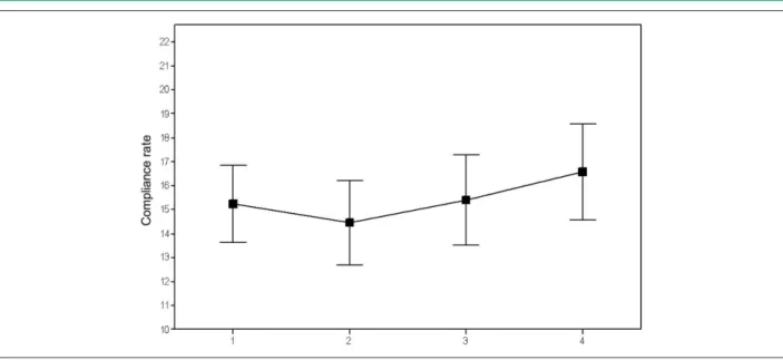
Table 1 displays calculations associated with the CR for the four timepoints, also showing a small change in mean CR values from one timepoint to the next during the intraoperative period. CR values are less dispersed at the first timepoint and reach the greatest variation at the fourth timepoint, with the lowest mean value at the second timepoint. By applying a two-factor mean comparison test (Two-way ANOVA), significant differences were found (p<0.001).
The quantitative variables examined in the univariate analysis of the compliance rate relationship were: age, weight, Qp/Qs, perfusion time, anoxia time, minimum temperature, MV time and length of stay in the ICU.
The variables most strongly associated with CR values for the different timepoints were age, weight and perfusion time. The other variables showed no relationship pattern.
Mann-Figure 1 -Compliance rate variation at the four timepoints.
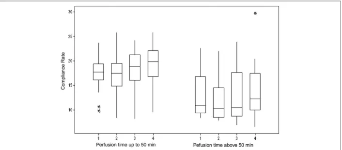
Figure 2 -Boxplot for compliance rate data according to weight subgroups.
Table 1 - CR variations at the four timepoints
CR N Mean SD Median Extremities Quartiles (25% – 75%)
Multiple comparisons*
Timepoint 1 35 15.25 4.7 16.8 8.4 – 23.7 10.4 – 18.5 A
Timepoint 2 35 14.45 5.11 15.1 7.9 – 25.8 9.4 – 18.9 A
Timepoint 3 35 15.40 5.52 16.5 7.0 – 24.2 10.2 – 20.3 A
Timepoint 4 35 16.59 5.86 16.9 6.6 – 29.8 10.3 – 20.4 B
P value <0.001
CR - compliance rate; SD - standard deviation; N - number of patients; * different letters indicate timepoints when CR means were statistically different.
Whitney test in comparisons of two stratified samples by age in the four CR measurements.
Original Article
Goraieb et al Pulmonary function in surgery for congenital heart disease
Table 2 - Results of the Mann-Whitney test for comparison of independent samples
CR Age ≤ 30 months Age > 30 months
N Median N Median
Timepoint 1 13 9.8 22 17.7
Timepoint 2 13 8.8 22 17.45
Timepoint 3 13 8.8 22 18.95
Timepoint 4 13 10.2 22 19.85
P value 0.0165 0.0004
CR - compliance rate; N - number of patients.
Figure 3 shows that by relating two variables, weight and age, we found that most patients up to 30 months of age weighed up to 10kg.
As to the relationship between CR and perfusion time, sample stratification, according to the “Tperf_50” categorical variable, indicated a statistically significant difference between the median values at each timepoint of CR measurement as
Figure 3 -Relationship between age and weight.
Figure 4 -Boxplot for compliance rate according to perfusion time subgroups.
per the Boxplot shown in Figure 4.
Figure 5 shows the percentage variation of the compliance rate according to the perfusion time.
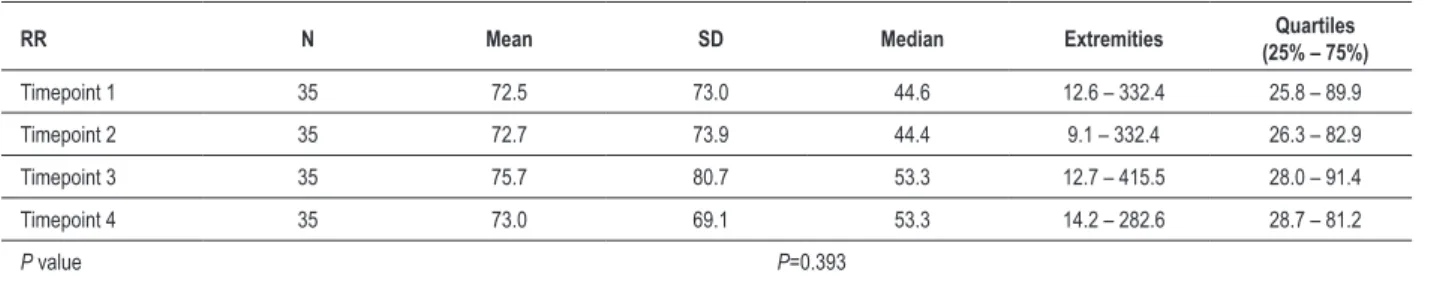
Table 3 shows calculations associated with RR for the four measurement timepoints.
Discussion
Pulmonary changes following pediatric cardiac surgery with ECC often account for the increase in morbidity and mortality3,6.
In children with pulmonary hyperflow, we noticed changes in pulmonary mechanics during the postoperative phase with ECC.
The mean compliance value obtained at the fourth measurement is statistically different from the mean obtained at the other timepoints. That is, CR had a statistically significant increase at the end of the surgery.
Diseases that result in abnormal flows of blood in the lungs may cause changes in pulmonary mechanics, increasing respiratory work and oxygen consumption, and worsening the cardiac condition1. The improvement in pulmonary compliance at the end of the surgery is probably due to the correction of pulmonary hyperflow, with immediate improvement of pulmonary mechanics. This improvement was translated by the increased CR, i.e, the larger it is, the more distensible is the lung and greater is the variation in volume during inspiration; consequently, the smaller it is, the more rigid is the lung, and smaller is the volume7.
Changes resulting from the use of ECC, hypothermia, absence of [mechanical] ventilation during surgery, and by the surgical procedure itself would be enough to impair pulmonary function after surgery. These changes often account for patient morbidity8.
Figure 5 -Percentage variation of the compliance rate related to the perfusion time.
Table 3 - Descriptive statistics for resistance rate per timepoint during the intraoperative period
RR N Mean SD Median Extremities Quartiles
(25% – 75%)
Timepoint 1 35 72.5 73.0 44.6 12.6 – 332.4 25.8 – 89.9
Timepoint 2 35 72.7 73.9 44.4 9.1 – 332.4 26.3 – 82.9
Timepoint 3 35 75.7 80.7 53.3 12.7 – 415.5 28.0 – 91.4
Timepoint 4 35 73.0 69.1 53.3 14.2 – 282.6 28.7 – 81.2
P value P=0.393
RR - resistance rate; N - number of patients; SD - standard deviation.
This confirms the fact that the benefits obtained from hyperflow correction supersede potential pulmonary complications resulting from ECC and the surgical procedure itself.
In a study conducted with 106 children under the age of 1 year, Satyer et al9, also observed that in those patients with pulmonary hyperflow, surgical correction improves pulmonary function. Despite the similarities in the results, that study assessed the dynamic Pc, whereas ours measured SPc.
In a study conducted with 23 children undergoing cardiac surgery, aged 2 months to 10 years, analogous to our findings, Lanteri et al10 observed abnormalities in pulmonary dynamic and static compliances, as well as an increase in respiratory resistance among patients with pulmonary blood hyperflow. After surgery, these values were normal.
Takeuchi et al11 included in their study only children weighing less than 10 kg and also observed an improvement in Pc after surgery for pulmonary hyperflow correction.
CR analysis performed with study variables showed that only age, weight and perfusion time displayed some pattern of correlation with CR.
Patients up to 30 months of age had lower, more concentrated RC values at the four timepoints. Above 30 months of age, dispersion of RC values increases.
In the subgroup of patients aged ≤30 months, the
percentage increase in CR is only 4.1% (p=0.0165), whereas in the subgroup of patients older than 30 months, this increase is 12.1% (p=0.0004).
The fact that children under 30 months of age had lower Pc values for all timepoints compared to those older than 30 months, and that weight was similar in children under 10 kg, with lower Pc means at all timepoints, is probably due to pulmonary maturation.
Taking into consideration the growth table used worldwide by the National Center of Health Statistics (NCHS), one can see that children under 30 months of age are those who weigh up to approximately 10kg. Therefore, our findings as to weight and age variables were consistent and similar to those of younger children who weighed less, as well as for those older ones who weighed more.
Original Article
Goraieb et al Pulmonary function in surgery for congenital heart disease
contributes to greater Pc values only after approximately eight years of age. Other authors who have analyzed pulmonary maturation show that after 4 months of age there is an increase in the complexity of alveolar shape, and the number of alveoli multiplies more rapidly over the first 3 years of life12,13.This certainly contributes to a higher Pc with increasing age.
Older children showed a 12% improvement in Pc from timepoint one to timepoint four. Children under 30 months of age and weighing less than 10kg also showed an improvement in lung compliance at the end of surgery, but only in the order of 4.1%.
Thus, older children experienced a significant improvement in Pc at the end of surgery. The younger patients experienced less CR variation at all timepoints, with values concentrated within a narrower interval, whereas in older children CR values showed greater variation.
Therefore, we can infer more accurately that in the group of younger children, static pulmonary compliance (Spc) will be lower at all timepoints when compared with children older than 30 months.
It is noteworthy that the increase in early postoperative Pc did not show any relationship pattern with the pulmonary hyperflow, i.e., when only Qp/Qs is related to the level of improvement in postoperative pulmonary function, no positive correlation is observed.
The lack of relationship between Qp/Qs and pulmonary development suggests that the level of pulmonary flow does not interfere with the pulmonary capacity in patients with pulmonary hyperflow.
Some authors have suggested that only pulmonary hyperflow with increased PAP (PAP >18 mmHg) would reduce compliance after surgical correction of the hyperflow. That is, the improvement is inversely proportional to the existing PAP in the preoperative stage. The greater the preoperative PAP, the smaller the improvement in Pc11.
As to time under extracorporeal circulation, in both the
group of children with perfusion time ≤ 50 minutes and the
group of children with perfusion time > 50 minutes, there was an improvement in Pc. However, in the group that remained less time under extracorporeal circulation, an improvement in Pc was recorded as of timepoint three, when the thorax was still open and the retractors were in place. As to the group under ECC for a longer period, the improvement in Pc was observed only at the end of the surgery, when the thorax had already been closed. In fact, Pc in these patients even worsened after ECC weaning (timepoint three).
ECC duration is reported in literature as one of the key factors that make MV weaning difficult in patients who have undergone cardiac surgery, due to the significant physiological disorder caused by the inflammatory reaction. The change in the membranes of pulmonary capillaries negatively affects gas exchange, increasing surgical risk14.
Brown et al14, in a study conducted to identify risk factors due to a longer stay in the ICU following pediatric cardiac surgery, identified time under ECC longer than 30 minutes as a determinant factor for worsening of pulmonary mechanics and, consequently, requiring a longer stay in the ICU regardless
of the subsequent postoperative events.
Several adverse effects have been associated with the use of ECC in children. The increase in capillary permeability results in edema formation, which consequently reduces pulmonary compliance and gas exchange.
During the intra-operative phase, efforts to reduce the adverse effects of ECC as well as the deleterious effects of post-perfusion capillary leakage syndrome include, besides a reduction of the volume of circuits and diuretic and anti-inflammatory therapy, the ultra filtration technique. Studies conducted on the use of hemoconcentration in cardiac surgery with ECC have concluded that the main advantages are removal of the macromolecules that mediate the systemic inflammatory reaction, reduction of circulating endotoxins, elimination of excess body water and pulmonary hypertension15,16.
An interesting item of data found refers to the correlation between ECC duration and age. Upon analysis of the inter-group data, children who had spent less time under ECC showed significantly higher Pc values at all timepoints, as compared to those with longer periods under ECC. We observed that the mean age of children in the first group was greater than in the second group. The older children remained less time under ECC than the younger ones. The justification for this may lie in the fact that indication for surgery at an earlier stage is due to more complex defects with greater hemodynamic repercussions, which explains the longer time under ECC.
In this study, Awr showed no significant change at the end of the surgery (P=0.393). Di Carlo et al17 reported A
wr values recorded immediately after surgery with successful or unsuccessful extubation, and concluded that Awr values over 75 cm H2O identify the possibility of difficult extubation due to respiratory failure. The authors also believe that the increase in Awr is probably due to pulmonary fluid build-up. That study was conducted with children undergoing several types of cardiac surgery and not exclusively for hyperflow correction, as was the case in our study17.
Our study did not identify any relationship between the change in Awr and any pre-, intra- or postoperative variable. Thus, there was no relationship between Awr and MV time, although the mean Awr at the end of surgery was less than 75 cm H2O.
Awr is greater in smaller pulmonary volumes. This seems to be related to the fact that changes take place in the diameters of airways with different pulmonary volumes4. This explains what our results have shown, i.e., that the greatest resistance was found in the group of children younger than 30 months and who weighed less than 10 kilos.
Awr variations were very small in both groups during the intraoperative period, and the changes at the end of surgery were not significant.
On the other hand, studies have shown that neonates suffered a 25% decrease in respiratory resistance after surgery, whereas older children experienced a reduction of only 16%9.
References
1. Lister G, Pitt BR. Cardiopulmonary interactions in the infant with congenital cardiac disease. Clin Chest Med. 1983; 4 (2): 219-32.
2. Reid LM. Lung growth in health and disease. Br J Dis Chest. 1984; 78: 113-24.
3. Babik B, Astalos T, Peták F, Deák ZI, Hantos Z. Changes in respiratory mechanics during cardiac surgery. Anesth Analg. 2003; 96: 1280-7.
4. Vieira SRR, Plotnik R, Fialkow L. Monitorização da mecânica respiratória durante a ventilação mecânica. In: Carvalho CRR (ed). Ventilação mecânica. São Paulo: Atheneu; 2000. p. 215-52.
5. Carvalho AP, Ayres FC. Circulação e respiração: fundamentos da biofísica e fisiologia. 3a ed. Rio de Janeiro: Cultura Médica; 1983. p.175.
6. Barnas GM, Watson RJ, Green MD, Sequeira AJ, Gilbert TB, Kent J, et al. Lung and chest wall mechanical properties before and after cardiac surgery with cardiopulmonary bypass. J Appl Physiol. 1994; 76: 166-75.
7. Zin WA, Rocco PRM. Mecânica respiratória nomal. In: Auler Junior JOC, Gomide do Amaral RV (eds). Assistência ventilatória mecânica. São Paulo: Atheneu; 1995. p. 3-24.
8. Schulze-Neick I, Li J, Penny DJ, Redington AN. Pulmonary vascular resistance after cardiopulmonary bypass in infants: effect on postoperative recovery. J Thorac Cardiovasc Surg. 2001; 121: 1033-9.
9. Stayer SA, Diaz LK, East DL, Gouvion JN, Vencill TL, McKenzie ED, et al. Changes in respiratory mechanics among infants undergoing heart surgery. Anesth Analg. 2004; 98: 49-55.
10. Lanteri CJ, Kano S, Duncan AW, Sly PD. Changes in respiratory mechanics in
children undergoing cardiopulmonary bypass. Am J Respir Crit Care Med. 1995; 152 (Pt 1): 1893-900.
11. Takeuchi M, Kinouchi K, Fukumitsu K, Kishimoto H, Kitamura S. Postbypass pulmonary artery pressure influences respiratory system compliance after ventricular septal defect closure. Pediatr Anaesth. 2000; 10: 407-11.
12. Davies G, Reid L. Growth of the alveoli and pulmonary arteries in childhood. Thorax. 1970; 25: 669-81.
13. Hislop A, Reid LM. Pulmonary arterial development during childhood: branching pattern and structure. Thorax. 1973; 28: 129-35.
14. Brown KL, Ridout DA, Goldman AP, Hoskote A, Penny DJ. Risk factors for long intensive care unit stay after cardiopulmonary bypass in children. Crit Care Med. 2002; 31: 1.
15. Souza DD, Braile DM. Assessment of a new technique of hemoconcentration and the necessities of blood derivates for transfusion in patients submitted to heart surgery using cardiopulmonay bypass. Braz J Cardiovasc Surg. 2004; 19 (3): 287-94.
16. Elliot MJ. Ultrafiltration and modified ultrafiltration in pediatric open heart operations. Ann Thorac Surg. 1993; 56 (6): 1518-22.
17. Di Carlo JV, Raphaely RC, Steven JM, Norwood WI, Costarino AT. Pulmonary mechanics in infants after cardiac surgery. Crit Care Med. 1992; 20 (1): 22-7.
18. Martins F. Monitorização no pós-operatório de cirurgia cardíaca. In: Auler Jr JOC, Oliveira AS (eds). Pós-operatório de cirurgia torácica e cardiovascular. Porto Alegre: Artmed; 2004. p.18-42.
but also enable a better functional assessment of the patient. Thus, the noninvasive measurement of Spc should be routinely used in clinical practice and especially during the postoperative phase of cardiac surgery. This measurement is appropriate for the evaluation of pulmonary parenchyma since the cardiovascular and respiratory systems are closely related, both anatomically and functionally.
In our study, the main findings were:children showed improvement in CR after the surgical correction of pulmonary hyperflow; in younger children and, therefore, with lower weights, this improvement was minor; those who spent more time under ECC took longer to show improvement in the CR. CRshowed no significant changes at the end of surgery.
Potential Conflict of Interest
No potential conflict of interest relevant to this article was reported.
Sources of Funding
There were no external funding sources for this study.
Study Association