https://doi.org/10.1590/0004-282X20170066
TRICK OF THE TRADE
Discordant clinical outcomes of congenital
Zika virus infection in twin pregnancies
Evolução clínica discordante da infecção congénita do vírus Zika em gestação gemelar
Vanessa van der Linden
1,2, Hélio van der Linden Junior
3, Mariana de Carvalho Leal
4,5,
Epitacio Leite Rolim Filho
2,4, Ana van der Linden
6, Maria de Fátima Viana Vasco Aragão
7,8,
Alessandra Mertens Brainer-Lima
9, Danielle Di Cavalcanti Sousa Cruz
6, Liana O. Ventura
10,11,
Telma Lúcia Tabosa Florêncio
12, Marli Tenório Cordeiro
13, Silvio da Silva Caudas Neto
4, Regina Coeli Ramos
141Hospital Barão de Lucena, Recife PE, Brasil;
2Associação de Assistência à Criança Deiciente (AACD), Recife PE, Brasil; 3Centro de Reabilitação Dr. Henrique Santillo, Goiania GO, Brasil; 4Universidade Federal de Pernambuco, Recife PE, Brasil; 5Hospital Agamenon Magalhães, Recife PE, Brasil;
6Instituto de Medicina Integral Professor Fernando Figueira, Recife PE, Brasil; 7Centro Diagnóstico Multimagem, Recife PE, Brasil;
8Universidade Mauricio de Nassau, Recife PE, Brasil; 9Universidade de Pernambuco, Recife PE, Brasil; 10Fundação Altino Ventura, Recife PE, Brasil;
11Hospital de Olhos de Pernambuco (HOPE), Recife PE, Brasil; 12UniVision, Recife PE, Brasil;
13Centro de Pesquisas Aggeu Magalhães-Fiocruz, Recife PE, Brasil; 14Hospital Universitário Oswaldo Cruz, Recife PE, Brasil;
Correspondence: Vanessa van der Linden; AACD Pernambuco, Av. Adv José Paulo Cavalcanti, 155; 50080-810 Recife PE, Brasil; E-mail: [email protected]
Conflict of interest: There is no conlict of interest to declare.
Received 05 February 2017; Received in inal form 27 February 2017; Accepted 06 March 2017.
ABSTRACT
Congenital Zika syndrome is an emergent cause of a congenital infectious disorder, resulting in severe damage to the central nervous
system and microcephaly. Despite advances in understanding the pathophysiology of the disease, we still do not know all the mechanisms
enrolled in the vertical transmission of the virus. As has already been reported in other types of congenital infectious disorders in dizygotic
twin pregnancies, it is possible that the virus affects only one of the fetuses. In this article, we report on two cases of twin pregnancies
exposed to the Zika virus, but with only one of the fetuses affected with microcephaly and brain damage. This indicates the urgent need
for more studies regarding the pathophysiology of viral infection and the mechanisms involved in the natural protection against the virus.
Keywords:
Zika virus; Zika virus infection; twins, dizygotic.
RESUMO
A síndrome congênita do Zika vírus é uma causa de infecção congênita emergente, resultando em graves danos ao sistema nervoso central
e microcefalia. Apesar dos avanços na compreensão da isiopatologia da doença, ainda não conhecemos todo o mecanismo envolvido na
transmissão vertical do vírus. Como já foi relatado em outros tipos de infecções congênitas em gestações gemelares dizigóticas, é possível
que apenas um dos fetos seja afetado pelo vírus. Este artigo descreve 2 casos de gestações gemelares expostas ao vírus Zika, onde apenas
um dos fetos foi afetado, com microcefalia associado a graves danos no sistema nervoso central. Isso indica a necessidade urgente de mais
estudos sobre a isiopatologia da infecção viral e os mecanismo envolvidos na proteção natural contra o vírus.
Palavras-chave:
Zika virus; infecção pelo Zika virus; gêmeos dizigóticos;
Gestational Zika virus (ZIKV) infection has been robustly
associated with a well-delimited congenital syndrome
includ-ing microcephaly and speciic neuroradiological
abnormali-ties, deining the congenital Zika syndrome (CZS)
1,2,3. Other
symptoms have also been described in association with the
syndrome, such as ophthalmologic lesions, hearing loss and
arthrogryposis
4,5,6. Like other congenital viral infections, only
in which only one fetus was born with CZS.
METHODS
Data collection
A standard form was used to collect demographic and
clin-ical data, including the recollection of a rash during pregnancy.
All investigations described were conducted as part of
the clinical protocol or clinical indication; no investigations
were conducted for research reasons. Informed consent
was obtained from the participants or their legally
autho-rized representatives.
Laboratory tests
Serologic tests were performed on both mother and
new-borns to exclude the main diferential diagnoses of CZS
(i.e.
,
other congenital infections that lead to brain calciications
and microcephaly), which are cytomegalovirus,
toxoplas-mosis, rubella, syphilis and HIV. When cytomegalovirus IgG
was present in both mother and child, real-time polymerase
chain reaction was performed in urine or blood.
he cerebrospinal luid (CSF) of patients was tested
for IgM antibody capture enzyme-linked immunosorbent
assay (ELISA) for ZIKV, following the Center for Disease
Control and Prevention (CDC) protocol, as described by
Martin et al.
9Laboratory conirmation was considered as
a positive ZIKV-speciic IgM in CSF with capture ELISA,
according to the CDC Emergency Use Authorization
proto-col with reagents by Robert Lanciotti (CDC, Fort Collins, CO,
USA), as recently published by Cordeiro et al.
10Clinical evaluation
For clinical evaluation, microcephaly was deined as
a head circumference two standard deviations below the
mean for gestational age and sex, according to the Fetal
International and Newborn Growth Consortium for the
21st Century (Intergrowth-21st) for newborns and the
World Health Organization child growth standards for
infants
11,12. Birth weight was classiied as appropriate,
small or large for gestational age and sex according to the
Intergrowth-21st curve
11.
All the infants underwent neurologic, orthopedic,
ophthal-mological, and hearing evaluations, including clinical
exami-nation and ancillary exams. he infant patients had brain
imaging by non-contrasted computerized tomography (CT)
and simple radiography of the hips. Clinical assessment of
dys-phagia was made by a speech therapist. Audiometric screening
was carried out by auditory brainstem response audiometry,
and conirmed by diagnostic tests (conirmatory
frequency-speciic auditory brainstem response with tone burst stimuli
and behavioral audiometry) using the routine recommended
CASE REPORTS
Patient 1
A boy, born in 2015 in Recife, State of Pernambuco, from a
dizygotic twin pregnancy. His mother had an episode of skin
rash associated with itching and fever in the irst month of
gestation. At the 25th week of pregnancy, microcephaly was
diagnosed by obstetric ultrasonography, but no anomaly
was detected by this method in the twin brother. Delivery
occurred at a gestational age of 37 weeks, with the newborn
weighing 2,100 g; the head circumference was 28 cm (3 SD
from the gestational age and sex, classiied as severe
micro-cephaly). he patient also presented with craniofacial
disportion, closed anterior fontanelle, exuberant occipital
pro-tuberance, redundant scalp skin and a right clubfoot. Brain
CT revealed difuse bilateral reduction of cerebral
paren-chyma, ventriculomegaly, cortical underdevelopment,
mul-tiple calciications predominantly in the basal ganglia and
cortical/subcortical white matter regions, and hypoplasia of
the brainstem and cerebellum (Figure 1).
First eye exam (13 days after birth): clear cornea, depth
anterior chamber, phakic, isocoria, pathologic red relex
test both eyes. High myopia (-12.00 OD and -9.00 OS),
vitre-ous haze, bilateral staphyloma chorioretinal lesions (sharply
demarcated atrophy) involving the posterior pole and optic
disc OD and only macula in OS. After three months of age,
developed congenital glaucoma OD and underwent surgery.
Last exam (one year old): ocular pressure under control,
clear cornea, stable myopia, fundus indings were the same,
clear vitreous.
Auditory evaluation by frequency-speciic auditory
brain-stem response and behavioral audiometry detected bilateral
profound hearing loss. After an unsuccessful attempt at
reha-bilitation with hearing aids and speech therapy, a cochlear
implant was scheduled for this child.
At seven months of age, he started presenting with
spasms in clusters. he pattern of EEG was focal, with
dis-charge in the frontal lobe at irst, and at 12 months of age, the
EEG presented with multifocal discharge. he seizures were
controlled with valproate. At the age of 12 months he
pre-sented with neurodevelopmental arrest, with no interaction
with the environment and no head control. An X-ray of the
hips showed left hip dysplasia.
he development and neurological evaluation of his
brother was normal at 10 months of age.
Patient 2
was diagnosed at 32 weeks of pregnancy by ultrasonography in
the female twin. After delivery at 35 weeks, the female newborn
had a head circumference of 26 cm (below 3 SD for the
gesta-tional age and sex) and weighed 1,750 g. he same craniofacial
indings observed in Patient 1 were present. Brain CT revealed
difuse bilateral reduction of cerebral parenchyma,
ventriculo-megaly, malformation of cortical development, multiple
calci-ications in cortical/subcortical white matter regions and mild
hypoplasia of the brainstem and cerebellum (Figure 1). he
ante-rior and posteante-rior segments of the eye were normal in the
oph-thalmological assessment. he auditory evaluation was normal.
Epilepsy was diagnosed at six months of age, with the
same seizure pattern, but we are still waiting for the EEG. Her
seizures were reduced with levetiracetam. At the last
evalua-tion, at seven months of age, she presented with severe
neu-rodevelopmental delay, with no interaction with the
environ-ment and no head control. he X-ray of the hips was normal.
he development and neurological evaluation of her
brother were normal at seven months of age.
In these two cases, screening tests for the most common
causes of congenital infection (toxoplasmosis,
cytomegalovi-rus, rubella, syphilis and HIV) were negative. he CSF
sam-ple was tested by IgM antibody capture ELISA for ZIKV and
it was positive. And, in both cases, the unafected twin was
normal on clinical examination, and blood and CSF tests for
ZIKV were negative.
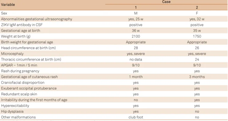
Tables 1 and 2 summarize the main indings of the two
newborns.
DISCUSSION
his article describes two twin siblings exposed to the
Zika virus during pregnancy, but only one of the siblings
pre-sented with a typical picture of CZS. he craniofacial aspects
of our patients (Figure 2) were described by Russel et al. as a
fetal brain disruption sequence
14. he fetal brain disruption
sequence phenotype is hypothesized to be a result of loss in
brain volume and decrease in intracranial pressure, and it is
not speciic to the etiologic agent. Moore et al. described the
fetal brain disruption sequence phenotype related to CZS
15.
he brain imaging of the two patients, showing
calciica-tions predominantly in the subcortical region, with
abnor-malities of cortical development, was consistent with the
pattern fully described by Aragão et al. for CZS
3. he
diagno-sis of this syndrome was based on the neuroimaging indings,
exclusion of other congenital infections and the presence of
positive IgM in the CSF of the two afected children.
Only one patient presented with ophthalmic and auditory
abnormalities. Congenital infection due to presumed ZIKV
expo-sure has been shown to be associated with vision-threatening
indings, such as the bilateral macular and optic nerve
abnor-malities seen in the irst patient of this study. Glaucoma has
been described in one infant from Bahia, Brazil
5,16.
Congenital infection by ZIKV is a new condition and
the real risk of mother-fetus transmission of this virus
is still unknown. Nishiura et al. tried to estimate a
theo-retical risk of microcephaly occurrence from congenital
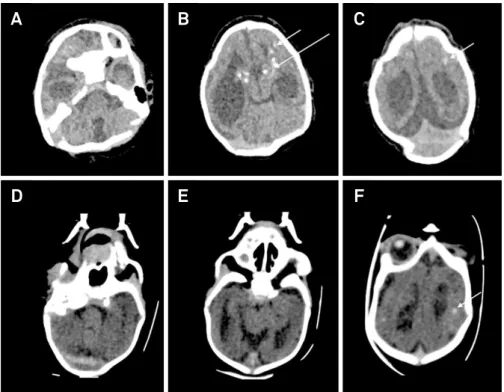
Figure 1.
A, B and C are brain computerized tomography from Patient 1 and D, E and F are from Patient 2. A and D show the
brainstem and cerebellum hypoplasia; B, C, E and F show diffuse bilateral reduction of cerebral parenchyma, ventriculomegaly
and cortical underdevelopment; B, C and F show calciications in the basal ganglia (long arrow) and cortical/subcortical white
matter regions (short arrow).
A
D
B
E
C
ZIKV and assumed that it could be of at least 14.0%
17For
Ellington et al.
18, following the Puerto Rico outbreak, the
risk of microcephaly ranged from 1% to 13% for maternal
infection in the irst trimester, up to 0.7% in the second
tri-mester, and up to 0.2% in the third trimester.
here are many reports in the literature about
congeni-tal infections in twin pregnancy afecting only one sibling,
most of them dizygotic
7,8,19,20,21,22. Maternal factors related to
immunologic competency may explain why some mothers
transmit the virus to the fetus, causing neurologic damage, and
others do not. However, this cannot explain the diferences in
the outcomes of the children in twin pregnancies. One
possi-ble explanation for this is the diferences in fetal susceptibility.
Lazzarotto et al. showed that twin fetuses may react diferently
to primary maternal cytomegalovirus infection, in spite of
being exposed to the same maternal inluences
7. A few reports
Variable
1
2
Sex
M
F
Abnormalities gestational ultrasonography
yes, 25 w
yes, 32 w
ZIKV IgM antibody in CSF
positive
positive
Gestational age at birth
36 w
35 w
Weight at birth (g)
2100
1750
Birth weight for gestational age
Appropriate
Appropriate
Head circumference at birth (cm)
28
26
Microcephaly
yes, severe
yes, severe
Thoracic circumference at birth (cm)
no data
24
APGAR – 1min / 5 min
9/10
9/10
Rash during pregnancy
yes
yes
Gestational age of cutaneous rash
1 month
3 months
Craniofacial disproportion
yes
yes
Exuberant occipital protuberance
yes
yes
Redundant scalp skin
yes
yes
Irritability during the irst months of age
no
yes
Hyperexcitability
yes
yes
Hip dysplasia
yes
no
Other malformations
club foot
no
M: male; F: female; w: weeks; ZIKV: Zika virus; CSF: cerebrospinal luid; min: minutes.
Table 2.
Neurological indings.
Variable
Patient
1
2
Age of testing
12 mo
7 mo
Corrected age
11 months
6 months
Head circumference
38 cm
34 cm
Microcephaly
yes
yes
Weight
8020g
Visual ixation and pursuit
no
no
Interaction with the environment
no interaction
no interaction
Strabismus
yes
yes
Nystagmus
yes
yes
Social smile
no
no
Head control
no
no
Sitting without support
no
no
Grasp
grasping relex
grasping relex
Asymmetric tonic neck relex
present
present
Muscle tone
Limb hypertonia with pyramidal
and extrapyramidal signs
Limb hypertonia with pyramidal
and extrapyramidal signs
Dysphagia
yes, moderate
yes, moderate
of diferent outcomes in monozygotic twins in
cytomegalovi-rus-afected pregnancies corroborate this theory
23,24.
Another explanation is the diferences in placental
func-tion in dizygotic twin pregnancies. Although the placenta
acts as a portal for mother-fetus transmission of viral
dis-eases, it is not completely permeable, as demonstrated by
Fowler et al., who found fetal contamination in only 40% of
cases of gestational cytomegalovirus
25.his placental barrier
function can be explained by several mechanisms, but it is
still not completely understood how, in various cases of twin
pregnancies with viral infection, only one of the fetuses is
afected
7,26. In the patients presented, the placentas were not
studied. he comparison of the placentas from the normal
and the afected child could throw some light onto the
ques-tion of distinct outcomes.
Despite the advances in understanding the
pathophysi-ology of the disease, we still do not know all the mechanisms
enrolled in vertical transmission of the ZIKV virus. As has
already been reported in other types of congenital infectious
disorders in dizygotic twin pregnancies, it is possible for the
virus to afect only one of the fetuses. his indicates the urgent
need for more studies regarding the pathophysiology of the viral
infection. Other possible variants, like genetic factors, viral
tro-pism and the placenta barrier could inluence the
pathophysi-ology of the CZS and must be investigated in further studies.
here was no pathology study of the placentas in this
patient series. his could be important to deine whether the
virus compromised the placenta of the non-afected child or
not. his information is crucial to understand whether the
pla-centa is the most important barrier against the virus invasion,
or if intrinsic fetal factors are more important for this
partic-ular protection. here is lack of evidence on how exactly the
virus disseminates through the pregnant body, how it reaches
the fetus, and which type of barrier could inluence this
mech-anism. Further genetic studies may elucidate if there are genes
involved in speciic protection against external agents.
References
1. Mlakar J, Korva M, Tul N, Popović M, Poljšak-Prijatelj M, Mraz J N, Popović M, Poljšak-Prijatelj M, Mraz J et al. Zika virus associated with microcephaly. N Engl J Med. 2016;374(10):951-8. https://doi.org/10.1056/NEJMoa1600651
2. Rasmussen SA, Jamieson DJ, Honein MA, Petersen LR. Zika virus and birth defects: reviewing the evidence for casuality. N Engl J Med. 2016;374(20):1981-7. https://doi.org/10.1056/NEJMsr1604338 3. Aragão MFVV, Linden V, Brainer-Lima AM, Coeli RR, Rocha MA, Silva PS et al.
Clinical features and neuroimaging (CT and MRI) indings in presumed Zika virus related congenital infection and microcephaly: retrospective case series study. BMJ. 2016;353:i1901. https://doi.org/10.1136/bmj.i1901 4. Linden V, Rolim Filho EL, Lins OG, Linden A, Viana MF, Aragão MFVV et al.
Congenital Zika syndrome with arthrogryposis: retrospective case series study. BMJ. 2016;354:i3899. https://doi.org/10.1136/bmj.i3899
5. Ventura CV, Maia M, Ventura BV, Linden VV, Araújo EB,
Ramos RC et al. Ophthalmological indings in infants with microcephaly and presumable intra-uterus Zika virus infection. Arq Bras Oftalmol. 2016;79(1):1-3. https://doi.org/10.5935/0004-2749.20160002 6. Leal MC, Muniz LF, Ferreira TSA, Santos CM, Almeida LC,
Linden V et al. Hearing loss in infants with microcephaly and evidence of congenital Zika virus infection — Brazil, November 2015–May 2016. MMWR Morb Mortal Wkly Rep. 2016;65(34):917-9. https://doi.org/10.15585/mmwr.mm6534e3
7. Lazzarotto T, Gabrielli L, Foschini MP, Lanari M,
Guerra B, Eusebi V et al. Congenital cytomegalovirus infection in twin pregnancies: viral load in the amniotic fluid and pregnancy outcome. Pediatrics. 2003;112(2):e153-7. https://doi.org/10.1542/peds.112.2.e153
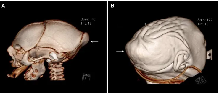
Figure 2.
A and B show the computed tomography of the skull with reconstruction, of Patient 1, with the typical phenotype of fetal
brain disruption sequence characterized by microcephaly with exuberant occipital protuberance (short arrow) and redundant skin
on the scalp (long arrow).
Spin: -78
Tilt: 16
Spin: 122
Tilt: 18
https://doi.org/10.1007/s12098-009-0208-9
9. Martin DA, Muth DA, Brown T, Johnson AJ, Karabatsos N, Roehrig JT. Standardization of immunoglobulin M capture enzyme-linked immunosorbent assays for routine diagnosis of arboviral infections. J Clin Microbiol. 2000;38(5):1823-6.
10. Cordeiro MT, Pena LJ, Brito CA, Gil LH, Marques ET. Positive IgM for Zika virus in the cerebrospinal luid of 30 neonates with microcephaly in Brazil. Lancet. 2016;387(10030):1811-2. https://doi.org/10.1016/S0140-6736(16)30253-7
11. Villar J, Cheikh Ismail L, Victora CG, Ohuma EO, Bertino E, Altman DG et al. International Fetal and Newborn Growth
Consortium for the 21st Century (INTERGROWTH-21st). International standards for newborn weight, length, and head circumference by gestational age and sex: the Newborn Cross-Sectional Study of the INTERGROWTH-21st Project. Lancet. 2014;384(9946):857-68. https://doi.org/10.1016/S0140-6736(14)60932-6
12. World Health Organization – WHO, Department of Nutrition for Helath and Development. WHO child growth standards: Head circumference-for-age, arm circumference-for-age, triceps skinfold-for-age and subscapular skinfold-for-age: methods and development. Geneva: World Health Organization; 2007. 13. American Academy of Pediatrics, Joint Committee on Infant
Hearing. Year 2007 position statement: principles and guidelines for early hearing detection and intervention programs. Pediatrics. 2007;120(4):898-921. https://doi.org/10.1542/peds.2007-2333 14. Russell LJ, Weaver DD, Bull MJ, Weinbaum M, Opitz JM.
In utero brain destruction resulting in collapse of the fetal skull, microcephaly, scalp rugae, and neurologic impairment: the fetal brain disruption sequence. Am J Med Genet. 1984;17(2):509-21. https://doi.org/10.1002/ajmg.1320170213
15. Moore CA, Staples JE, Dobyns WB, Pessoa A, Ventura CV, Fonseca EB et al. Characterizing the pattern of anomalies in congenital Zika syndrome for pediatric clinicians. JAMA Pediatr. 2017;171(3):288-95. https://doi.org/10.1001/jamapediatrics.2016.3982
16. Freitas BP, Ko AI, Khouri R, Mayoral M, Henriques DF,
Maia M et al. Glaucoma and congenital Zika syndrome. Ophthalmology. 2017;124(3):407-8. https://doi.org/10.1016/j.ophtha.2016.10.004
during pregnancy with Zika virus infection. Epidemics. 2016;15:66-70. https://doi.org/10.1016/j.epidem.2016.03.001 18. Ellington SR, Devine O, Bertolli J, Martinez Quiñones A,
Shapiro-Mendoza CK, Perez-Padilla J et al. Estimating the number of pregnant women infected with Zika virus and expected infants with microcephaly following the Zika virus outbreak in Puerto Rico, 2016. JAMA Pediatr. 2016;170(10):940-5. https://doi.org/10.1001/jamapediatrics.2016.2974
19. Martino M, Tovo PA, Galli L, Caselli D, Gabiano C,
Mazzoni PL et al. HIV-I infection in perinatally exposed siblings and twins: the Italian register for HIV infection in children. Arch Dis Child. 1991;66(10):1235-8. https://doi.org/10.1136/adc.66.10.1235 20. Barlow KM, Mok JY. Dizygotic twins discordant for HIV
and hepatitis C virus. Arch Dis Child. 1993;68(4):507. https://doi.org/10.1136/adc.68.4.507
21. Egaña-Ugrinovic G, Goncé A, García L, Marcos MA, López M, Nadal A et al. Congenital cytomegalovirus infection among twin pairs. J Matern Fetal Neonatal Med. 2016;29(21):3439-44. https://doi.org/10.3109/14767058.2015.1130818
22. Tomasik T, Zawilińska B, Pawlik D, Ferek J, Ferek J, Wójtowicz A et al. [Congenital cytomegaly in one twin: a case report]. Med Wieku Rozwoj. 2012;16(3):252-60. Polish.
23. Seguin J, Cho CT. Congenital cytomegalovirus infection in one monozygotic twin. JAMA. 1988;260(22):3277. https://doi.org/10.1001/jama.1988.03410220061021 24. Wu HY, Huang SC, Huang HC, Hsu TY, Lan KC. Cytomegalovirus
infection and fetal death in one monozygotic twin. Taiwan J Obstet Gynecol. 2011;50(2):230-2. https://doi.org/10.1016/j.tjog.2011.01.008 25. Fowler KB, Stagno S, Pass RF, Britt WJ, Boll TJ, Alford CA. The
outcome of congenital cytomegalovirus infection in relation to maternal antibody status. N Engl J Med. 1992;326(10):663-7. https://doi.org/10.1056/NEJM199203053261003