Maternal-Fetal Outcome and Prognosis of Cardiac Surgery During
Pregnancy
Walkiria Samuel Ávila
1, Ana Maria Milani Gouveia
1, Pablo Pomerantzeff
1, Maria Rita Lemos Bortolotto
2, Max Grinberg
1,
Noedir Stolf
1, Marcelo Zugaib
2Instituto do Coração do Hospital das Clínicas da Faculdade de Medicina da Universidade de São Paulo1; Departamento de Obstetrícia e Ginecologia do Hospital das Clínicas da Faculdade de Medicina da Universidade de São Paulo2, São Paulo, SP - Brazil
Summary
Background: Cardiac surgery improves the maternal prognosis in cases refractory to medical therapy. However, it is associated with risks to the fetus when performed during pregnancy.
Objectives: To analyze maternal-fetal outcome and prognosis related to cardiac surgery performed during pregnancy and puerperium.
Methods: The outcome of 4 gestations of women undergoing cardiac surgery during pregnancy and puerperium was studied. Fetal cardiotocography was performed throughout the procedure in patients with gestational age above 20 weeks.
Results: Mean maternal age was 27.8 ± 7.6 years; there was a predominance of patients with rheumatic valve disease (87.8%), of whom 5 (4.6%) underwent reoperation due to prosthetic valve dysfunction. Mean extracorporeal circulation time was 87.4± 43.6min and hypothermia was used in 27 (67.5%) cases. Thirteen (3.7%) mothers experienced no events and gave birth to live healthy newborns. Postoperative outcome of the remaining 28 (68.3%) pregnancies showed: 7 (4.5%) maternal complications and three (7.3%) deaths; 2 (29.2%) fetal losses, and four (0%) cases of neurological malformation, two of which progressed to late death. One patient was lost to follow-up after surgery. Nine (2.9%) patients underwent emergency surgery, and this variable was correlated with maternal prognosis (p<0.00).
Conclusion: Cardiac surgery during pregnancy allowed survival of 92.7% of the mothers, and 56.0% of the patients who presented cardiac complications refractory to medical therapy gave birth to healthy children. Worse maternal prognosis was correlated with emergency surgery. (Arq Bras Cardiol 2009;93():8-3)
Key Words: Thoracic surgery; pregnancy; clinical evaluation; prognosis; maternal-fetal relations.
Mailing address: Walkiria Samuel Ávila •
Rua Martiniano de Carvalho 864 cj 1107/1108 - Bela Vista - 01321-000– São Paulo, SP, Brazil
E-mail: [email protected], [email protected]
Manuscript received July 01, 2008; revised manuscript received September 23, 2008; accepted September 23, 2008.
Introduction
Heart diseases are estimated to occur in 0.5 to 1.0% of pregnancies, and they contribute to increase the maternal-fetal morbidity and mortality rate1. In Brazil, among other
types of heart diseases, rheumatic heart disease is still the most frequent in women during pregnancy and is associated with high complication rates2-4.
The functional deterioration that results from the physiological circulatory overload observed in the gestational period5,6 in patients with a reduced functional reserve is
frequently refractory to medical therapy.
Cardiac surgery has provided a favorable maternal prognosis7,8 in these cases. However, this procedure is
associated with high obstetric and fetal risks, with emphasis on the impact of extracorporeal circulation (ECC) and anesthetic
agents. Thus, surgical treatment of heart diseases during pregnancy and puerperium is performed only in selected cases.
We present our experience with cardiac surgery performed after it was considered the only therapeutic method able to revert the poor prognosis in a specific clinical circumstance.
Methods
From 1986 to 2007, 41 patients followed up by the Department of Heart Disease and Pregnancy of the Heart Valve Diseases Team of Instituto do Coração (InCor) underwent surgery during pregnancy and puerperium.
During surgery, cardiotocography continuously recorded the uterine contractions and fetal heart beats in patients with gestational age above 20 weeks. Surgical and anesthetic techniques followed the recommendations established for cardiac surgery. Additional care in relation to conventional operations was taken regarding the patient’s position, heparinization, ECC priming content, degree of hypothermia, and myocardial protection9.
Tabela 1 - Maternal heart disease
Maternal heart disease Absolute frequency
Relative percentage
Mitral valve disease
Prosthetic valve dysfunction 11 26.82 Mitral stenosis 10 24.39 Mitral regurgitation 6 14.63
Aortic valve disease
Prosthetic valve dysfunction 2 4.87 Aortic stenosis 3 7.31 Aortic regurgitation 2 4.87 Mitral + aortic prostheses
dysfunction 2 4.87 Aneurysm + coarctation of
the aorta 1 2.43 Aortic aneurysm + Marfan
syndrome 1 2.43 Aortic dissection in normal
vessel 1 2.43 Complete transposition of the
great arteries 1 2.43
Acute coronary insuficiency 1 2.43
Total 41 100
preoperative variables, including age, heart disease, gestational age at the time of operation, and reason for surgical indication; b) intraoperative variables, including type of procedure, ECC duration (in minutes) and temperature; and c) postoperative variables, including cardiac complications, hemorrhage, infection and death.
We also analyzed the occurrence of fetal losses, which included spontaneous abortion (up to 20 weeks of gestation), stillbirth or neonatal death (up to 28 days after birth), and prematurity (birth up to 37 weeks of gestation); birth weight; incidence of congenital malformation; and late death, defined as those occurring after the first year of life.
Prevention of premature labor was made using natural progesterone (50-mg capsules every 12 hours in the intra and postoperative periods) in place of indometacin due to the risk of the latter causing intrauterine ductus arteriosus closure.
Descriptive statistical analysis of maternal and newborn results was performed using the mean and standard deviation after performing the Shapiro-Wilk normality test. The analysis of the independent effects of maternal characteristics on the chance of complications was carried out using the Wolf method to obtain the confidence interval.
Results
Maternal age ranged between 15 and 44 years (mean of 27.8 ± 7.6 years) and, among the underlying cardiac lesions, there was a predominance of rheumatic heart valve disease, corresponding to 36 (90%) cases. Of these, 17 (42.5%) presented prosthetic valve dysfunction (15 biological and two mechanical valves) (Table 1).
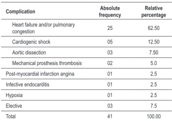
Among the clinical conditions accounting for surgical
indication, heart failure and/or pulmonary congestion were the major causes, present in 25 (62.5%) cases; cardiogenic shock in five (12.5%) cases; and aortic dissection in three (7.3%) patients, of whom one had coarctation of the aorta, another had Marfan syndrome, and the third had a structurally normal heart (Table 2).
Thirty seven of the 41 operations were performed within four and 34 (mean of 21.3 ± 9.3) weeks of gestation, and four were performed during puerperium (mean 10.2±8 days after delivery). Reoperation was indicated in 15 (37.5%) cases due to prosthetic valve dysfunction, followed by 14 (34.1%) cases of heart valve prosthesis implantation, seven (17.07%) mitral commissurotomies, three (7.31%) cases of correction of aortic aneurysm, one (2.43%) case of coronary artery bypass grafting, and one (2.43%) case of Blalock-Taussig procedure, which was the only intervention performed off-pump in this case series. Emergency surgery was performed in nine (21.9%) patients.
ECC time ranged from 16 to 203 minutes (mean of 87.4± 43.6). Hypothermia was used during ECC in 27 cases and ranged from 13.00 C to 32.20C(30.10 C ± 4.30 C), being
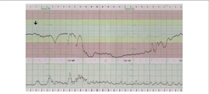
classified as mild (11), moderate (11) and deep (five cases). During ECC, fetal bradycardia was recorded with a mean of 60 beats/minute (bpm), which reverted spontaneously to normal heart rate, mean of 140 bpm, after ECC (Figure 1).
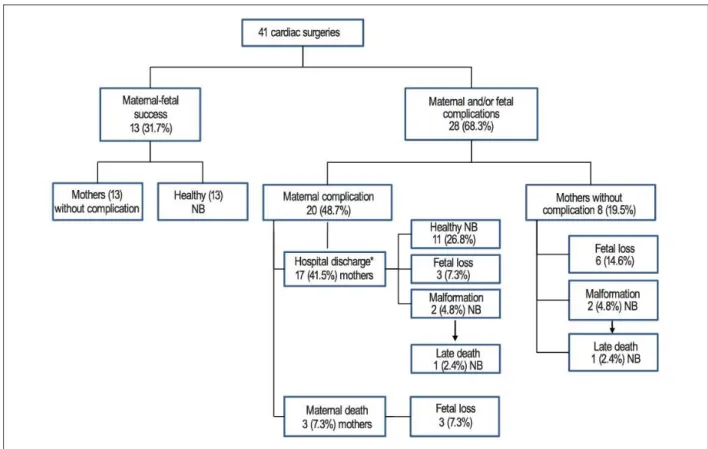
Thirteen gestations (31.7%) progressed with no maternal or fetal complications (Figure 2). The outcome of the remaining 28 (68.3%) gestations showed: 17 (41.4%) maternal complications and three (7.31%) deaths in the postoperative period (Table 3); 12 (29.2%) fetal losses; and four (10%) cases of neurological malformation, two of which progressed to late death. One patient was lost to obstetric follow-up after discharge in the postoperative period of the cardiac surgery.
The mean weight of 27 (63.4%) live births was 2118.1 ± 875.1g; there were 14 (35%) premature deliveries, among which eight deaths occurred and four (10%) had neurological complications (cerebral anoxia associated with tetraparesis, cortical atrophy, hydrocephalus and mental retardation). These four malformations were verified in two cases of biological valve prosthesis implantation (one in the aortic position and
Table 2 - Clinical indications for surgery
Complication Absolute
frequency
Relative percentage
Heart failure and/or pulmonary
congestion 25 62.50
Cardiogenic shock 05 12.50 Aortic dissection 03 7.50 Mechanical prosthesis thrombosis 02 5.0 Post-myocardial infarction angina 01 2.5 Infective endocarditis 01 2.5
Hypoxia 01 2.5
Elective 03 7.5
Figure 1 - Cardiotocographic record during aortic valve replacement on the 28th week of gestation. The upper tracing shows fetal heart beats, and the lower tracing
shows uterine contraction recording. The arrow (↓) shows the beginning of extracorporeal circulation. Table 3 - Case distribution according to maternal outcome
Complication Absolute
frequency
Relative percentage
Death 03 7.3(1)
Complications 17 41.5(2)
Heart failure 07 41.2
Stroke 02 11.7
Bleeding 03 17.6
Infective endocarditis 02 11.7 Cardiac arrhythmia* 03 17.6
Total 41 100.00
1 - Relative to the total case series; 2 - Relative to the total complications that did not progress to death; Arrhythmia (atrial ibrillation in 3 cases and atrioventricular block in 2).
the other in the mitral aortic position), and in two cases of reoperation for replacement of a biological valve prosthesis in the aortic and mitral positions, respectively.
The emergency nature of surgical indication was the only variable that significantly correlated maternal characteristics with postoperative complications (Table 4).
Discussion
Results of the worldwide experience on cardiac surgery during pregnancy are controversial10-13. Studies are
predominantly retrospective and the procedures used are heterogeneous; additionally, it is difficult to standardize the surgical techniques. Thus, judicious analysis of prognostic variables and their effects on the management of these patients during pregnancy is impaired.
The present study included 41 patients followed up by the same multidisciplinary team during pregnancy, according to a standardized clinical, surgical and obstetric management.
The predominance of mitral valve disease, 27 (65.8%) cases, particularly stenotic lesions (24.3% in native valves and 26.8% in biological prostheses), is one of the consequences of rheumatic disease which has not yet been eradicated in our country and affects mainly the mitral valve of female individuals, with clinical manifestations during childbearing age.
The progressive increase of cardiac output in the beginning of the second trimester of pregnancy facilitates New York Heart Association (NYHA) functional class (FC) progression from I/II to III/IV, which is common in patients with heart valve diseases and demands constant clinical reassessment and treatment of occasional complications including surgical intervention in refractory cases.
In Arnoni et al14 study, the major cause for indication of
surgery during pregnancy in 49 (84.4%) cases of heart valve disease was progressive hemodynamic deterioration, acute pulmonary edema, and persistence of FC III/IV, despite medical treatment.
Another relevant aspect is the gestational age at which cardiac surgery is performed. The earlier the onset of complications in patients with severe lesions, the greater the tendency to indicate surgery, even in the cases in which there is immediate favorable response to medical treatment, because the likelihood of progression to hemodynamic deterioration throughout pregnancy and during delivery and puerperium is potentially very high, thus favoring recurrence of complications and maternal death. This reasoning justifies the mean gestational age of 20 weeks found in our study.
Figure 2 -Cardiac surgery during pregnancy. Maternal-fetal outcomes. * NB - newborn; * Lost to follow-up”1(2,4%) NB.
Table 4 - Correlation of maternal characteristics with the odds of occurrence of complications
Maternal characteristic Endpoint p OR(2) IC(OR;95%)(3)
Maternal complication(1)
Age (years) 0.879 1.01 (0.92 – 1.10)
Gestational age (weeks) 0.398 1.03 (0.96 – 1.12)
ECC (minutes) 0.261 1.01 (0.99 – 1.04)
ECC temperature (oCelsius) 0.622 1.05 (0.87 – 1.27)
Emergency care 0.046 9.82 (1.04 – 92.78)
Fetal loss
Age (years) 0.325 0.95 (0.86 – 1.05)
Gestational age (weeks) 0.639 0.98 (0.90 – 1.07)
ECC (minutes) 0.996 1.00 (0.98 – 1.02)
ECC temperature (oCelsius) 0.945 1.01 (0.80 – 1.27)
Emergency care 0.549 1.64 (0.32 – 8.33)
NB death(4)
Age (years) 0.301 0.95 (0.87 – 1.04)
Gestational age (weeks) 0.939 1.00 (0.93 – 1.09)
ECC (minutes) 0.546 1.01 (0.99 – 1.02)
ECC temperature (oCelsius) 0.794 1.03 (0.84 – 1.26)
Emergency care 0.544 1.60 (0.35 – 7.30)
dysfunction during pregnancy, whether due to calcification in biological prostheses or to thrombosis in mechanical prostheses.
These findings are consistent with those of Salazar et al15,
who showed that among 15 cases of cardiac surgery during pregnancy, 13 (86.6%) had rheumatic heart valve disease, of which eight (61.5%) presented prosthetic valve dysfunction. Therefore, prosthetic valve dysfunction should be regarded as a maternal risk during pregnancy with a potentially high need for reoperation due to hemodynamic deterioration which is sometimes irreversible.
In turn, aortic dissection, which was the reason for emergency surgery in three cases, is a less common but a high risk event during pregnancy16. Difficulties in the diagnosis,
misinterpretation of symptoms (which may be mistaken for normal pregnancy) and limitations to invasive investigation (due to the potential risk for the concept) result in delayed treatment and increased mortality rate for the disease.
In our study, aortic dissection occurred in a woman with Marfan syndrome and in another with coarctation of the aorta; this condition has been already documented and is known to be related to pregnancy13. However, its occurrence in healthy
pregnant women is unexpected, as was described in the third patient of our case series, thus leading to the hypothesis of a strong correlation between pregnancy and aortic dissection as a result of structural vascular changes of gestation17.
Surgical indication for dissection was based on the chances of maternal and fetal survival, type of dissection, maternal clinical conditions and gestational age. In the two cases in which dissection occurred with a viable fetus (32 and 33 weeks of gestation), the cesarean section that preceded the surgical repair of the aneurysm was aimed at preserving the life of the fetus, and prevented the risk of spontaneous labor in the postoperative period18.
It is assumed thatthe risk of maternal death due to cardiac surgery is not modified by pregnancy. However, the 7.5% maternal death rate found in our study, along with that of 8.6% reported by Arnoni et al14, and of 13.3% described by Salazar
et al15 are relatively high when compared to those found for
surgical treatment of heart valve disease in this age range. According to the authors, these percentages were due to the emergency nature of the surgical indication which, in the present study, was the only variable that correlated with complications and maternal death in the postoperative period.
As regards the pregnancy outcomes, the percentages of 25% of fetal losses and 35% of prematurity recorded in our study are higher than those found in the population of pregnant women with heart diseases, estimated at 12% and 15%, respectively - and probably result from variations in blood flow and in arterial oxygen saturation consequent to anesthesia and ECC.
Hyperventilation during anesthesia results in a reduction of the uterine flow by 25% and reduction of maternal venous return and cardiac output due to the mechanical effects of the
positive pressure19. Maternal hyperventilation and respiratory
alkalosis also lead to decreased fetal arterial oxygen tension and facilitate shift of the maternal oxyhemoglobin dissociation curve20.
It is assumed that ECC leads to the formation of placental microemboli due to piling up of red cells and of microbubbles which, together with maternal blood pressure variations and hypothermia, aggravate hypoxia in the uteroplacental circulation and trigger uterine contractions and labor21.
These effects also explain the fetal bradycardia that may be attributed to placental flow changes which, during ECC, turn from pulsatile to nonpulsatile and laminar, and also to hemodilution and hypothermia22,23.
Teratogenic risks that could result from anesthesia and ECC in the embryogenic phase were not observed in the three cases of elective surgery performed in the first trimester of pregnancy. However, there were four (11.4%) cases of neurologic malformation in fetuses of patients who underwent surgery after 20 weeks of gestation, probably as a consequence of intrauterine hypoxia.
Blood flow reduction in the umbilical arteries causes hypoxemia and vasoconstriction in the fetal circulation which, when persistent and prolonged, exhaust the compensatory mechanism of vasodilatation and of diastolic blood flow increase in the middle cerebral artery, thus resulting in cerebral ischemia24,25.
This justifies the use of Doppler-flowmetry for monitoring flow in the uterine and umbilical arteries as a guide for the perfusionist to control the hemodynamic pattern during ECC26,27.
In conclusion, cardiac surgery performed during pregnancy and puerperium permitted maternal survival in 92.7% of the patients, and birth of healthy infants in 56% of the pregnant women who presented cardiac complications refractory to other therapies. The emergency nature of the indication of surgery was significantly correlated with a worse postoperative maternal prognosis.
Acknowledgements
To Dr. Maeve de Barros Correia for the orthographic review.
Potential Conflict of Interest
No potential conflict of interest relevant to this article was reported.
Sources of Funding
There were no external funding sources for this study.
Study Association
References
1. Feitosa HN, Moron AF, Born D, Almeida PAM. Mortalidade materna por cardiopatia. Rev Saude Pública. 1991; 25: 443-51.
2. Ávila WS, Rossi EG, Ramires JA, Grinberg M, Bortolotto MR, Zugaib M, et al. Pregnancy and heart disease: experience with 1000 cases. Clin Cardiol. 2003; 26 (3): 135-42.
3. Andrade J, Ciari Jr C, Marcus RER, Almeida PAM, Meneghello Z, Siqueira AF, et al. Evolução da gravidez em pacientes cardíacas com prótese de dura mater. Arq Bras Cardiol. 1979; 32 (3 sup.I): 31.
4. Born D, Martinez EE, Almeida PAM, Santos DV, Carvalho AC, Moron AF, et al. Pregnancy in patients with prosthetic heart valves: effects of anticoagulation on mother, fetus, and neonate. Am Heart J. 1992; 124 (2): 413-7.
5. Grinberg M, Avila WS, Amaral FMC. Modificações hemodinâmicas da gravidez. In Andrade J, Avila WS (eds). Doença cardiovascular, gravidez e planejamento familiar. São Paulo: Atheneu; 2003. p. 11-20.
6. Ueland K, Metcalfe J. Circulatory changes in pregnancy. Clin Obstet Gynaecol. 1975; 18: 41-8.
7. Weiss BM, Von Segesser LK, Alon E, Seifert B, Turina MI. Outcome of cardiovascular surgery and pregnancy: a systematic review of the period 1984-1996. Am J Obstet Gynecol. 1998; 179: 1643-53.
8. Bernal JM, Miralles PJ. Cardiac surgery with cardiopulmonary bypass during pregnancy. Obstet Gynecol Surv. 1986; 41: 1-6.
9. Pomini F, Mercogliano D, Cavalletti C, Caruso A, Pomini P. Cardiopulmonary bypass in pregnancy. Ann Thorac Surg. 1996; 61: 259-68.
10. Méier MA, Feldman J, Maia JC, Jazbik W, Pernambuco P, et al. Cirurgia cardíaca durante a gravidez: doze casos operados com circulação extracorpórea e hemodiluição. Arq Bras Cardiol. 1968; 21 (2): 73-86.
11. Born D, Massonetto JC, Almeida PAM, Moron AF, Buffolo E, Gomes WJ, et al. Cirurgia cardíaca com circulação extracorpórea em gestantes: análise da evolução materno-fetal. Arq Bras Cardiol. 1995; 64: (3): 207-11.
12. Pomerantzeff PMA, Benício A, Brandão CMAAvila WS, Bueno PC, Grinberg M, et al. Cirurgia valvar em gestantes: experiência de oito casos. Arq Bras Cardiol. 1998; 70 (6): 403-8.
13. Rossouw GJ, Knott-Craig CJ, Barnard PM, Macgregor LA, Van Zyl WP. Intracardiac operation in seven pregnant women. Ann Thorac Surg. 1993; 55: 1172-4.
14. Arnoni RT, Arnoni AS, Bonini RCA, de Almeida AF, Neto CA, Dinkhuysen JJ, et
al. Risk factors associated with cardiac surgery during pregnancy. Ann Thorac Surg. 2003; 76: 1605-8.
15. Salazar E, Espinola E, Molina FJ, Reyes A, Barragán R. Heart surgery with cardiopulmonary bypass in pregnant woman. Arq Cardiol Mexico. 2001; 71 (1): 20-7.
16. Zeebregts CJ, Schepens MA, Hameeteman TM, Morshuis WJ, de la Riviere AB. Acute aortic dissection complicating pregnancy. Ann Thorac Surg. 1997; 64: 1345-8.
17. Kelly BA, Bond BC, Poston L. Aortic adaptation to pregnancy: elevated expression of matrix metalloproteinases-2 and -3 in rat gestation. Mol Hum Reprod. 2004; 10: 331-7.
18. Wahlers T, Laas J, Alken A, Borst HG. Repair of acute type A aortic dissection after cesarean section in the thirty-nine week of pregnancy. J Thorac Cardiovasc Surg. 1994; 107: 314-5.
19. Strickland RA, Oliver WC, Chantigian RC, Ney JA, Danielson GK. Subject review: anesthesia, cardiopulmonary bypass, and the pregnant patient. Mayo Clin Proc. 1991; 66: 411-29.
20. Agarwal RC, Bhattacharya PK Bhattacharya L, Jain RK. Pregnancy and cardiopulmonary bypass. Indian J Anaesth. 2004; 48 (4): 259-63.
21. Chambers CE, Clark SL. Cardiac surgery during pregnancy. Clin Obstet Gynecol. 1994; 37: 316-23.
22. Hawkins JA, Paape KL, Adkins TP, Shaddy RE, Gay WA Jr. Extracorporeal circulation in the fetal lamb: effect of hypothermia and perfusion rate. J Cardiovasc Surg. 1991; 32: 295-300.
23. Parry AJ, Westaby S. Cardiopulmonary bypass during pregnancy. Ann Thorac Surg. 1996; 61: 1865-9.
24. Levy DL, Warringer RA, Burgess GE. Fetal response to cardiopulmonary bypass. Obstet Gynecol. 1980; 56: 112-5.
25. Zugaib M, Miyadahira S, Nomura RMY, Francisco RPV. Vitalidade fetal: propedêutica e avaliação. São Paulo: Atheneu; 2000. p. 9-15.
26. Lamb MP, Ross K, Johnstone AM, Manners JM. Fetal heart monitoring open heart surgery: two case reports. Brit J Obstet Gynaecol. 1991; 88: 669-74.