Case Report
ST-Segment Elevation in Pulmonary Thromboembolism
Fernando Santiago Montenegro, Valmir Barzan, Andrea Rocha De Lorenzo, Felipe José Monassa Pittella, Antônio
Sérgio Cordeiro da Rocha
Instituto Nacional de Cardiologia de Laranjeiras, Rio de Janeiro, RJ – Brazil
Introduction
In addition to acute myocardial infarction (AMI), there are several causes of ST-segment elevation,1 such as early repolarization, variation of the normal pattern (male pattern), left ventricular hypertrophy, complete left bundle branch block, acute pericarditis, myocarditis, Brugada syndrome, post-cardioversion, hyperkalemia, and pulmonary thromboembolism (PTE). However, the distinction between those conditions and AMI is clinically relevant, because of the benefit provided by early reperfusion in the presence of AMI with ST-segment elevation. This case report is about a patient diagnosed with PTE, whose electrocardiogram (ECG) mimicked AMI.
Case report
The patient is a 55-year-old, black, obese (BMI = 31 kg/m2) female, who was admitted to the emergency unit of another institution after four episodes of syncope accompanied by retrosternal pain and dyspnea. The first syncope occurred when she was walking, and the others when resting. The syncope episodes were followed by a retrosternal pressure sensation, with no radiation, and dyspnea. There was no history of diabetes or arterial hypertension, but she reported being an ex-smoker (tobacco load of 60 packs per year) and undergoing radiotherapy for uterus cancer 12 years before.
The report of her admission was as follows: tachycardia
(150 bpm); blood pressure of 132/74 mmHg; and O2
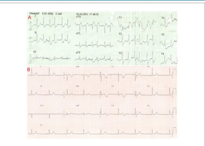
saturation of 91% at room air. Her heart rhythm was irregular, with neither accessory heart sounds nor murmurs. The respiratory sounds were reduced in the lower third of both hemithoraces. The ECG on admission showed atrial fibrillation with ventricular response of 150 bpm, S1Q3 pattern, incomplete right bundle-branch block (RBBB), ST-segment elevation in the leads aVR and V1, and ST-segment depression in the leads D1, D2, aVF, and V2-V6 (Figure 1A). The markers of myocardial necrosis were positive and the troponin I peak was 4.42. Two days after starting the treatment for acute coronary syndrome (ACS), the patient underwent coronary
angiography at our institution, which showed no coronary artery obstruction. Eleven days after the initial episode, after being transferred to our institution, still undergoing treatment for ACS (double platelet antiaggregation with prophylactic anticoagulation), painful and asymmetric swelling of the left leg was evidenced, with neither calf changes nor inflammatory signs. A transthoracic echocardiogram revealed normal left and right ventricular (RV) function, while venous Doppler echography of the lower limb evidenced recent thrombosis of the popliteal and small saphenous veins.
Computed tomographic angiography of the pulmonary arteries showed thrombi in both main pulmonary arteries (Figure 2). An ECG performed ten days after the initial episode showed disappearance of the S1Q3 pattern and of the RBBB, in addition to T wave inversion in the leads V1-V3 (Figure 1B). Because of clinical stability, the patient was initially treated with venous anticoagulation, later maintained on oral anticoagulation, and referred to an oncology service to investigate possible recurrence of the neoplasia.
Discussion
The classical ECG changes of PTE are as follows: T wave inversion on the right precordial leads; simultaneous T wave inversion and ST-segment elevation on the anteroseptal and inferior leads; S1Q3T3 pattern; complete or incomplete RBBB; and sinus tachycardia. The presence of atrial arrhythmias, RBBB, and ST-segment elevation or depression is associated with RV failure and poor prognosis2.
Livaditis et als.3 have reported the case of a patient with massive PTE and ST-segment elevation in the leads aVR and V1-V3, which, with thrombolytic treatment, reverted completely3. Although our patient did not undergo thrombolytic treatment, she improved clinically and electrocardiographically with the anticoagulant treatment.
Another study has reported three patients with ST-segment elevation in aVR and elevated troponin, of whom only one showed acute occlusion of the left main coronary artery. Considering the two other patients with normal coronary arteries, in one the ST-segment elevation in aVR was attributed to paroxysmal junctional tachycardia, while, in the other, to PTE, confirmed on computed tomographic angiography 4.
Lin et als.5 have reported a case of PTE, in which the patient had syncope, chest pain and dyspnea, similarly to the findings of the present report, and the initial ECG, in addition to ST-segment elevation from leads V1 to V4, showed incomplete RBBB, S waves in D1, V5, and V6, and Q wave in D3. Similarly to the case presently reported, the ECG changes normalized with treatment, and the initial diagnosis was AMI5.
Mailing Address: Fernando Santiago Montenegro •
Avenida Epitácio Pessoa, 2330/906, Lagoa. Postal Code 22411-071, Rio de Janeiro, RJ – Brazil
E-mail: [email protected]
Manuscript received August 22, 2011; manuscript received August 22, 2011; accepted December 19, 2012.
Keywords
Pulmonary embolism; electrocardiography/methods
Case Report
Montenegro et al. ST elevation in pulmonary thromboembolismArq Bras Cardiol 2012;99(3):e131-e133
Figure 1A - 12-lead electrocardiogram performed at the emergency room; 1B - 12-lead electrocardiogram performed at the ward
Figure 2 - Computed tomographic angiography of the pulmonary arteries showing illing failures (arrows) due to thrombi in both pulmonary arteries.
Case Report
Montenegro et al.ST elevation in pulmonary thromboembolism
1. Wang K, Asinger RW, Marriott HJL. ST-segment elevation in conditions other than acute myocardial infarction. N Engl J Med. 2003;349(22):2128-35. 2. Geibel A, Zehender M, Kasper W, Olschewski M, Klima C, Konstantinides
SV. Prognostic value of the ECG on admission in patients with acute major pulmonary embolism. Eur Respir J. 2005; 25(5): 843–8.
3. Livaditis IG, Paraschos M, Dimopoulos K. Massive pulmonary embolism with ST elevation in leads V1-V3 and successful thrombolysis with tenecteplase. Heart. 2004;90(7):e41.
4. Beygui F., Tran H., Montalescot G. Chest pain, ST-segment elevation in aVR lead, and high troponina levels. Arch Cardiovasc Dis. 2009;102(1):79-80. 5. Lin JF, Li YC, Yang PL. A case of massive pulmonary embolism with ST
elevation in leads V1-4. Circ J. 2009;73(6):1157–9.
6. Torbicki A, Perrier A, Konstantinides S, Agnelli G, Galie N, Pruszczyk P, et al. Guidelines on the diagnosis and management of acute pulmonary embolism. The Task Force for the Diagnosis and Management of Acute Pulmonary Embolism of the European Society of Cardiology (ESC). Eur Heart J. 2008;29(18):2276–315.
7. Rodger M, Makropoulos D, Turek M, Quevillon J, Raimond F, Rasuli P, et al. Diagnostic value of the electrocardiogram in suspected pulmonary embolism. Am J Cardiol. 2000;86(7):807–9.
8. Kosuge M, Ebina T, Kiuoshi H, Morita S, Endo M, Maejima N, et al. An early and simple predictor of severe left main and/or three-vessel disease in patients with non-ST-segment elevation acute coronary syndrome. Am J Cardiol. 2011;107(4):495-500.
References
Arq Bras Cardiol 2012;99(3):e131-e133
The ECG changes of PTE are attributed to changes in the cardiac hemodynamics. Volume overload increases the parietal tension and the RV muscle work. The ventricular septum bulging into the left ventricle reduces cardiac output, decreasing coronary flow, which, associated with hypoxemia and the increased heart work, contributes to myocardial ischemia6, justifying the elevation of myocardial necrosis markers.
Although the ST-segment elevation is associated with massive PTE, being, thus, an important prognostic marker, it is a rare alteration with few reports in the literature. A study with 246 patients, assessing the value of the ECG for the diagnosis of PTE, has reported that only 9.3% of the patients with the confirmed diagnosis had a change in the ST segment7.
According to her Wells score (4.5 points), the patient here reported had an intermediate probability of PTE: 3 points for the clinical symptoms of deep venous thrombosis (asymmetric swelling with tenderness on palpation) and 1.5 points for heart rate greater than 100 bpm6. Adding the ECG changes found increased the probability of PTE. However, the ST-segment elevation in the leads aVR and V1 could also indicate severe damage to the left main coronary artery and/or proximal lesion of the anterior descending artery8. Thus, the presence of the ST-segment elevation in the leads aVR and V1 could mean both massive PTE and severe coronary artery lesion.
This case report illustrates the difficulty of differentiating between both syndromes, especially at the emergency room, where decisions should be made rapidly aiming at saving patients’ lives. In the present case, the emergency room team chose to treat the most prevalent cause, ACS. In that circumstance, echocardiography availability would be an important tool to help in the differential diagnosis and in guiding the treatment, because it could demonstrate changes in pulmonary artery pressures or RV function, or the presence of a thrombus in the pulmonary trunk or pulmonary artery main branches occurring in massive PTE.
Potential Conflict of Interest
No potential conflict of interest relevant to this article was reported.
Sources of Funding
There were no external funding sources for this study.
Study Association
This study is not associated with any post-graduation program.