AR
TIGO ORIGINAL / ORIGINAL AR
TICLE
INTRODUCTION
Inlammatory bowel disease (IBD) is a term that describes different pathologies, such as ulcerative coli-tis and Crohn`s disease. Inlammation is thought to develop due to the breakdown of the intestinal epithe-lial barrier, which is accompanied by the subsequent exposition of luminal antigens to the mucosal immune system, resulting in the recruitment of inlammatory cells, such as neutrophils and monocytes. The cellular recruitment and local active inlammation reduce the availability of oxygen, leading to hypoxia(6).
Hypoxia triggers an adaptive response in which the expression of hypoxia-inducible factors (HIFs) plays a central role. HIFs are believed to confer a protective response through the HIF-dependent induction of barrier-protective genes in the epithe-lium(10). Under hypoxic conditions, genes that are related to angiogenesis, such as vascular endothelial growth factor (VEGF), or to cellular proliferation, such as apelin, are induced(11). The apoptotic effects of tumor necrosis factor (TNF-a) were also inhibited
NITRIC OXIDE INTERFERES WITH
HYPOXIA SIGNALING DURING COLONIC
INFLAMMATION
Cintia Rabelo e Paiva
CARIA
, Camila Henrique
MOSCATO
, Renata Bortolin Guerra
TOMÉ
,
José
PEDRAZZOLI Jr
, Marcelo Lima
RIBEIRO
and Alessandra
GAMBERO
ABSTRACT - Context - Intestinal inlammation can induce a local reduction in oxygen levels that triggers an adaptive response cen-tered on the expression of hypoxia-inducible factors (HIFs). Nitric oxide, a well-described inlammatory mediator, may interfere with hypoxia signaling. Objectives - We aimed to evaluate the role of nitric oxide in hypoxia signaling during colonic inlammation. Method - Colitis was induced by single (acute) or repeated (reactivated colitis) trinitrobenzenosulfonic acid administration in rats. In addition, one group of rats with reactivated colitis was also treated with Nw-Nitro-L-arginine methyl ester hydrochloride to block nitric oxide synthase. Colitis was assessed by macroscopic score and myeloperoxidase activity in the colon samples. Hypoxia was determined using the oxygen-dependent probe, pimonidazole. The expression of HIF-1a and HIF-induced factors (vascular endothelial growth factor - VEGF and apelin) was assessed using Western blotting. Results - The single or repeated administration
of trinitrobenzenosulfonic acid to rats induced colitis which was characterized by a high macroscopic score and myeloperoxidase activity. Hypoxia was observed with both protocols. During acute colitis, HIF-1a expression was not increased, but VEGF and apelin were increased. HIF-1a expression was inhibited during reactivated colitis, and VEGF and apelin were not increased. Nw-Nitro-L-arginine methyl ester hydrochloride blockade during reactivated colitis restored HIF-1a, VEGF and apelin expression.
Conclusions - Nitric oxide could interfere with hypoxia signaling during reactivated colitis inlammation modifying the expression of proteins regulated by HIF-1a.
HEADINGS – Colitis. Hypoxia-inducible factor 1. Nitric oxide. NG-Nitroarginine methyl ester. Endothelial growth factors.
Declared conflict of interest of all authors: none
Financial support: Fundação de Amparo à Pesquisa do Estado de São Paulo (FAPESP 2010/02991-6).
Research performed at: Unidade Integrada de Farmacologia e Gastroenterologia (UNIFAG), Faculdade de Medicina da Universidade São Francisco, SP, Brasil. Correspondence: Alessandra Gambero. Unidade Integrada de Farmacologia e Gastroenterologia (UNIFAG), Faculdade de Medicina da Universidade São Francisco.
by hypoxia(13). However, hypoxia and inlammation are interdependent phenomena; hypoxia can elicit local inflammation, and inflammation can induce local hypoxia, as mentioned previously.
Decreases in oxygen need to be detected by cells in the hypoxic environment to induce the appropriate response. When oxygen levels are suficient, prolyl hydroxylases (PHDs) hydroxylate and degrade the
a subunit of HIF via ubiquitylation(14). Nitric oxide (NO) displays many similarities to oxygen and may interfere with oxygen sensing mechanisms, as well as with hypoxia signaling(19). The increased synthesis of NO via inducible nitric oxide synthase (iNOS) is a well-described event that occurs during intestinal inlammation(20).
used in the literature but, repeated administration of TNBS provides a better correlation between experimental and clin-ical indings because of its ability to replicate relapse and re-mission periods that are frequent in Cronh’s disease patients.
METHOD
Animals
Speciic pathogen-free male Wistar rats (200-250 g, 6-8 weeks old) were obtained from the Multidisciplinary Cen-ter for Biological Research (State University of Campinas, Campinas, SP, Brazil). The experiments were performed in accordance with the principles outlined by the National Council for the Control of Animal Experimentation (CON-CEA, Brazil), and received approval from the Ethics Com-mittee of São Francisco University, Bragança Paulista, SP, Brazil (Protocol 002.09.09). The rats were maintained in a room with controlled humidity and temperature in collective cages and were exposed to 12 hour light-dark cycles. Twelve hours prior to an experiment, the animals were deprived of food (standard chow), but not water. The studies were carried out using 5-7 rats per group and in a irst round four groups were performed: control of acute colitis, control of reacti-vated colitis, acute colitis and reactireacti-vated colitis. In a second round of experiments another three groups were performed: control of reactivated colitis treated with saline, reactivated colitis treated with saline and reactivated colitis treated with Nw-Nitro-L-arginine methyl ester hydrochloride (L-NAME) with ive rats per group.
TNBS-induced colitis
The animals were anesthetized using ketamine/xylazine (1:1 v/v), and colitis was induced by the intracolonic instilla-tion of TNBS, either 10 mg on day 0 (acute colitis group) or 3 mg on days 0, 14 and 28 (reactivated colitis group), dissolved in 0.3 mL of 50% ethanol (Sigma, St. Louis, MO, USA). The solution was injected into the colon 8 cm proximal to the anus using a catheter. The instillation procedure lasted only a few seconds, and the rats were maintained in a vertical position until they had recovered from the anesthesia. The rats from the acute colitis group were sacriiced on the 7th day, and the rats from the reactivated colitis group were sacriiced on the 35th day under overdose of ketamine/xylazine (1:1 v/v). The control rats received saline via the same route of administration (control group).
L-NAME treatment
An additional group of TNBS-induced colitic rats with reactivation was treated with Nw-Nitro-L-arginine methyl ester hydrochloride (L-NAME; 10 mg/kg/day) intraperi-toneally during the last 7 days of the protocol described above. Healthy and colitis controls were carried out and, rats received saline instead of L-NAME as described.
Colitis characterization by macroscopic damage and myeloperoxidase activity
The colons were immediately removed from the animals,
opened longitudinally and evaluated for macroscopically visible damage by two observers who were unaware of the experimental groups. The criteria for the assessment of the macroscopic colonic damage of each animal were as fol-lows: no damage (no points), hyperemia without any ulcers (1 point), linear ulcer with no signiicant inlammation (2 points), linear ulcer with inlammation at one site (3 points), two or more sites of ulceration/inlammation (4 points) and two or more major sites of ulceration and inlammation or one site of ulceration/inlammation extending 1 cm along the length of the colon (5 points). If the damage extended 2 cm along the length of the colon, the score was increased by 1 point for each additional centimeter of involvement (6-10 points).
Colon samples obtained longitudinally from a site of macroscopically detectable inlammation (or a corresponding site in the tissue with no macroscopically detectable inlam-mation) were homogenized in 0.5% (w/v) hexadecyltrimeth-ylammonium bromide in 50 mM potassium phosphate buffer, pH 6.0. For the myeloperoxidase (MPO) assay, 50 mL of each sample was added to 200 mL of o-dianisidine solution (0.167 mg/mL o-dianisidine dihydrochloride and 0.0005% hydrogen peroxide in 50 mM phosphate buffer, pH 6.0) immediately prior to reading the change in absorbance at 460 nm over a 5 min period using a microplate reader (Multiscan MS, Labsystems, Joensuu, Finland).
Pimonidazole staining
To assess colonic hypoxia during colitis induced TNBS administration, rats were treated intraperitoneally with 60 mg/kg pimonidazole 1 h prior to sacriice. Following the manufacturer’s recommended instructions, colonic pimo-nidazole distribution was visualized on parafin-embedded sections using a Hypoxyprobe Omni Kit (Natural Pharmacia International Inc., Massachusetts, USA).
TNF-a measurements and Western blot analysis
To measure TNF-a using an enzymatic assay (EIA), colon samples were collected as previously described, excised and immediately homogenized in solubilizing buffer at 4°C (1% Triton X-100, 100 mM Tris-HCl, 100 mM sodium pyrophos-phate, 100 mM sodium luoride, 10 mM EDTA, 10 mM so-dium orthovanadate, 2 mM PMSF and 0.1 mg of aprotinin/ ml). The insoluble material was removed by centrifugation for 20 min at 9,000 g and 4°C. The protein concentrations of the supernatants were determined using the Biuret method. TNF-a was quantiied using a commercially available kit (R&D Systems, Minneapolis, MN, USA).
a Tris buffered solution. The membranes were then incubated overnight with anti-iNOS (sc-8310), anti-VEGF (sc-507), anti-HIF-1a (sc-10790) and anti-apelin (sc-33804) antibodies at dilutions of 1:1000 (Santa Cruz Biotechnology, Inc., CA, USA). The nitrocellulose membranes were developed using commercially available chemiluminescence kits (GE Health-care, UK). The band intensities were quantiied by the optical densitometry (Scion Image software, ScionCorp, Frederick, MD) of the developed autoradiography.
Statistical analysis
All of the data are expressed as the means ± Standard Error of the Mean (SEM). Non-parametric data were ex-pressed as the medians. The comparisons between groups of data were performed using unpaired Student’s test. Statistical
analysis were performed using GraphPad InStat (GraphPad Software, La Jolla, CA, USA). An associated probability (P
value) of less than 5% was considered signiicant.
RESULTS
Colitis assessments
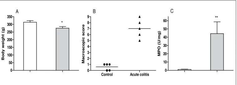
The single or repeated administration of TNBS in alcoholic vehicle inluenced both hyperemia and the number of small ulcers that were observed macroscopically, resulting in a higher macroscopic score (Figures 1B and 2B). Colonic inlammation decreased body weight gain during acute or reactivated colitis, as shown in Figures 1A and 2A, respectively. Myeloperoxidase (MPO) activity, a biochemical marker of neutrophil iniltration, was also higher in the colons of colitic rats (Figures 1C and 2C).
FIGURE 1. Body weight (A), macroscopic score (B) and myeloperoxidase (MPO) activity (C) in colon after 7 days of one trinitrobenzenosulfonic acid
(TNBS) administration to rats (Acute colitis). A and C. Data are expressed as the mean ± SEM of ive rats. *P<0.05 and **P<0.01 when compared with control group. B. Data are expressed as medians (n = 5).
FIGURE 2. Body weight (A), macroscopic score (B) and myeloperoxidase (MPO) activity (C) in colon after repeated trinitrobenzenosulfonic acid
HIF-1a expression during TNBS-induced colitis
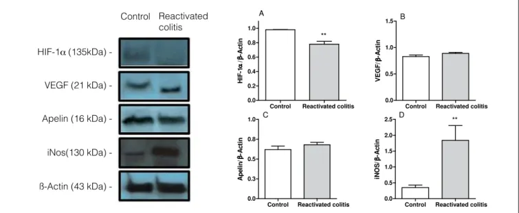
HIF-1a protein expression was similar in the colons of rats in the control and acute colitis groups, but VEGF and apelin expression were higher in the colons of rats in the acute colitis group compared to the control group (Figures 3A, B and C). In contrast, HIF-1a expression was lower in the colons of reactivated colitis group compared to the controls
(Figure 4A). Apelin and VEGF expression in the colons of rats with reactivated colitis were not altered (Figures 4B and C). iNOS expression was induced by intestinal inlammation in both groups (Figures 3D and 4D), but the increase was more pronounced in the reactivated colitis group (a 1.44-fold increase was observed in the acute colitis group versus a 5.25-fold increase in the reactivated colitis group).
FIGURE 4. Levels of HIF-1a (A), vascular endothelial growth factor (VEGF) (B), apelin (C) and inducible nitric oxide synthase (iNOS) (D) in rat colon
from control and colitis after repeated trinitrobenzenosulfonic acid (TNBS) administration (Reactivated colitis). Western blot analysis was performed on colon protein extracts with antibodies against HIF-1a, VEGF, apelin, iNos and ß-actin. The blot images are representative of one experiment. Bars show the densitometry quantiication of HIF-1a, VEGF, apelin, and iNos levels, normalized by densitometry quantiication of ß-actin level for the same sample. Data are expressed as the mean ± SEM of three experiments. **P<0.01 when compared with control group.
FIGURE 3. Levels of HIF-1a (A), vascular endothelial growth factor (VEGF) (B), apelin (C) and inducible nitric oxide synthase (iNOS) (D) in rat
colon from control and colitis after one trinitrobenzenosulfonic acid (TNBS) administration (Acute colitis). Western blot analysis was performed on colon protein extracts with antibodies against HIF-1a, VEGF, apelin, iNos and ß-actin. The blot images are representative of one experiment. Bars show the densitometry quantiication of HIF-1a, VEGF, apelin, and iNos levels, normalized by densitometry quantiication of ß-actin level for the same sample. Data are expressed as the mean ± SEM of three experiments. *P<0.05 and **P<0.01 when compared with control group.
Acute Control colitis
HIF-1a (135kDa)
VEGF (21 kDa)
Apelin (16 kDa)
iNos(130 kDa)
ßActin (43 kDa)
-Reactivated colitis
HIF-1a (135kDa)
VEGF (21 kDa)
Apelin (16 kDa)
iNos(130 kDa)
ßActin (43 kDa)
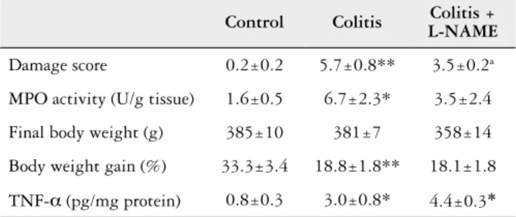
TABLE 1. Macroscopic evaluation, myeloperoxidase (MPO) acitivity, body weight and cytokine production by colon tissue in control treated with saline (Control), colitis with reactivation/treated with saline (Colitis) and colitis with reactivation treated with L-NAME (Colitis + L-NAME).
Control Colitis Colitis +
L-NAME
Damage score 0.2±0.2 5.7±0.8** 3.5±0.2a
MPO activity (U/g tissue) 1.6±0.5 6.7±2.3* 3.5±2.4
Final body weight (g) 385±10 381±7 358±14
Body weight gain (%) 33.3±3.4 18.8±1.8** 18.1±1.8
TNF-a (pg/mg protein) 0.8±0.3 3.0±0.8* 4.4±0.3*
*P<0.05 and **P<0.01 when compared control versus colitis group (n = 4-5). aP = 0.0571
when compared colitis/L-NAME with colitis group.
FIGURE 5. Pimonidazole-stained colonic sections from the control (A), acute colitis (B) and reactivated colitis (C) groups. Note the enhanced mucosal
epithelial hypoxia that occurred during colitis. 5.0 mm; magniication: 100x.
Hypoxia during TNBS-induced colitis
To determine the presence of hypoxia during TNBS-in-duced colitis, we evaluated the binding of pimonidazole to macromolecules, which occurs in the absence of adequate oxy-gen levels. Consistent with earlier reports, physiologic hypoxia on the colonic surface epithelium was observed in healthy animals (control group, Figure 5A). Following the induction of colitis by the single or repeated administration of TNBS (Figure 5), increased colonic epithelial pimonidazole uptake, extending to additional parts of the crypts and the submucosal layer, was observed in the model of reactivated colitis.
Effects of L-NAME treatment on reactivated colitis
L-NAME treatment during the inal 7 days of the experi-ment (after the inal TNBS administration in the reactivation protocol) reduced the number of macroscopically observed lesions (P = 0.0571). However, MPO activity was not
signii-cantly reduced (Table 1) in colitic rats that received only saline i.p. Body weight gain and TNF-a were not improved after L-NAME treatment compared with the colitic group (Table 1).
After L-NAME treatment, colitic animals displayed high-er levels of HIF-1a, apelin and VEGF expression (Figures 6A, B and C). iNOS expression was not reduced by L-NAME treatment (data not shown).
FIGURE 6. Levels of HIF-1a (A), vascular endothelial growth factor (VEGF) (B) and apelin (C) in the colon of rats from the control treated with
saline (Control), repeated trinitrobenzenosulfonic acid (TNBS) administration colitis treated with saline (Colitis) and repeated TNBS administration colitis treated with L-NAME (Colitis + L-NAME) groups. Western blot analysis were performed on the colon protein extracts using antibodies that had been raised against HIF-1a, VEGF, apelin and ß-actin. The blot images are representative of one experiment (the ß-actin igure is not shown). The bars show the densitometric quantiications of HIF-1a, VEGF and apelin levels after normalization by the densitometric quantiication of the ß-actin level for the same samples. The data are expressed as the mean ± SEM of three experiments.
* P<0.05 compared to the control group. #P<0.05 and ##P<0.01 when compared to the colitis group. Colitis +
L-NAME Control Coliis
Colitis + L-NAME Control Coliis
DISCUSSION
Active mucosal inlammation is characterized by al-terations in tissue metabolism and perfusion, resulting in hypoxia. Local hypoxia triggers an adaptive response
that is focused on HIF-1a expression. Karhausen and
colleagues demonstrated the presence of hypoxia, both in the superficial mucosa and the deeper submucosal regions, using an imidazole dye after 7 days of TNBS administration in mice(15). The expression of HIF-1a and HIF-1a–regulated gene products was increased in these mice(15). In rats, hypoxia was observed 7 days after the administration of a single dose of TNBS, but increases in HIF-1a were not observed. However, proteins that are regulated by HIF-1a, such as VEGF and apelin(10, 11) were increased in the colons of these animals. Interestingly, we observed that after repeated TNBS administration, a model that provides periods of relapse and remission(8), HIF-1a protein expression was lower in the colons of colitic rats than in healthy animals, despite the presence of hypoxia, as indicated by pimonidazole staining. Analysis of apelin and VEGF protein expression (HIF-1a-regulated factors) in colonic tissue revealed that they were not altered during reactivated colitis. Both of the TNBS-induced colitis proto-cols that were applied in this study induced inlammation and hypoxia, although in the colitis with reactivation model, hypoxia seemed to be more intense, affecting areas of the submucosa, but was not accompanied by hypoxia signaling. In the repeated administration protocol, iNOS was expressed at high levels, suggesting that higher NO production oc-curred in this experimental model. NO is considered to be an inlammatory mediator during IBD(24). NO can interfere with cell signaling and affect HIF-1a activation via different mechanisms, but much of the current knowledge of NO was acquired using NO-donor compounds in vitro(1, 3). Under normoxic conditions, low concentrations of NO facilitate HIF-1a degradation and impair HIF-1a signaling, while high concentrations of NO stabilize HIF-1a and mimic a hypoxic response(18). However, NO inhibits HIF-a activation during hypoxia by inducing PHD transcription and increasing the bioavailability of free cellular iron, which is a co-factor for PHD activity(2, 16). In addition, NO can interact with cytochrome c oxidase (CcO) and modulate its activity when O2 concentrations are low and the enzyme is in a reduced state(25). In this state, O
2 utilization by the mitochondrial chain is reduced and free O2 can induce the PHD-mediated
de gradation of HIF-1a(25), resulting in inadequate cellular signaling during hypoxia. L-NAME administration to rats with TNBS-induced colitis with reactivation at a dose previ-ously described to block NO synthesis in vivo(4, 5), improved HIF-1a expression and increased apelin and VEGF protein expression. These results suggest that iNOS inhibition re-stored HIF-1a signaling during intestinal inlammation and that this restoration was not dependent on an anti-inlam-matory response induced by L-NAME. In addition, HIF-1a
expression did not induce a robust anti-inlammatory re-sponse because a reduction in macroscopic damage, but not in TNF-a or MPO levels, was observed. The role of HIF-1a
signaling in experimental models of colitis is controversial. Chronic increases in HIF signaling by the disruption of von Hippel-Lindau tumor suppressor protein (VHL) in mice trigger colonic inlammation and potentiate dextran sodium sulfate (DSS)-induced colitis(23). However, the use of a PHD inhibitor (FG-4497), which stabilizes HIF-1a, had a benei-cial inluence on weight loss, histological modiications and
TNF-a production in a murine model of TNBS-induced
colitis(21). T-cells that iniltrated the inlamed mucosa in IBD patients expressed more HIF-1a, and HIF-1a KO mice dis-played more severe DSS-induced colonic inlammation(12). Based on these positive reports, PHD inhibitors have been suggested for use as a potential therapeutic approach to treat IBD patients(7). However, inliximab (an anti-TNF-a therapy) has demonstrated positive results by inhibiting an-giogenic processes through VEGF-A inhibition in IBD pa-tients(22). The results of the present study demonstrated that HIF-1a expression is variable in different experimental models of TNBS-induced colitis, despite the presence of hypoxia, and that iNOS inhibition restored HIF-1a expres-sion in TNBS-induced colitis with reactivations, suggesting that NO potentially interferes with hypoxia signaling during intestinal inlammation. iNOS expression and rectal NO levels were upregulated during active IBD(17) and were able to modulate the expression of several genes involved in perpetuating the inlammatory response(9) indicating that NO is an important mediator of intestinal inlammation.
CONCLUSIONS
REFERENCES
1. Ball KA, Nelson AW, Foster DG, Poyton RO. Nitric oxide produced by cytochrome C oxidase helps stabilize hif-1alpha in hypoxic mammalian cells. Biochem Biophys Res Commun. 2012;420:727-32.
2. Berchner-Pfannschmidt U, Tug S, Trinidad B, Oehme F, Yamac H, Wotzlaw C, et al. Nuclear oxygen sensing: induction of endogenous prolyl-hydroxylase 2 activity by hypoxia and nitric oxide. J Biological Chem. 2008;283:31745-53.
3. Brüne B, Zhou J. The role of nitric oxide (NO) in stability regulation of hypoxia inducible factor-1alpha (HIF-1alpha). Curr Medicinal Chemistry. 2003;10:845-55. 4. Chaves HV, Ribeiro R de A, De Souza AM, Rodrigues e Silva AA, Gomes AS,
Vale ML, et al. Experimental model of zymosan-induced arthritis in the rat tem-poromandibular joint: role of nitric oxide and neutrophils. J Biomed Biotechnol. 2011;2011:707985.
5. Cheng S, Yan Wm, Yang B, Shi JD, Song MM, Zhao Y. A Crucial role of nitric oxide in acute lung injury secondary to the acute necrotizing pancreatitis. Hum Exp Toxicol. 2010;29:329-37.
6. Colgan SP, Taylor CT. Hypoxia: an alarm signal during intestinal inlammation. Nat Rev Gastroenterol Hepatol. 2010;7:281-7.
7. Cummins EP, Doherty GA, Taylor CT. Hydroxylases as therapeutic targets in inlammatory bowel disease. Lab Invest. 2013;93:378-83.
8. Gambero A, Marostica M, Abdalla Saad MJ, Pedrazzoli J Jr. Mesenteric adipose tissue alterations resulting from experimental reactivated colitis. Inlamm Bowel Dis. 2007;13:1357-64.
9. Gillberg L, Varsanyi M, Sjöström M, Lördal M, Lindholm J, Hellström PM. Nitric oxide pathway-related gene alterations in inlammatory bowel disease. Scand J Gastroenterol. 2012;47:1283-97.
10. Grenz A, Clambey E, Eltzschig HK. Hypoxia signaling during intestinal ischemia and inlammation. Curr Opin Crit Care. 2012;18:178-85.
11. Han S, Wang G, Qi X, Lee HM, Englander EW, Greeley GH Jr. A possible role for hypoxia-induced apelin expression in enteric cell proliferation. Am J Physiol Regul Integr Comp Physiol. 2008;294:R1832-9.
12. Higashiyama M, Hokari R, Hozumi H, Kurihara C, Ueda T, Watanabe C, et al. Hif-1 in T cells ameliorated dextran sodium sulfate-induced murine colitis. J Leukoc Biol. 2012;91:901-9.
13. Hindryckx P, De Vos M, Jacques P, Ferdinande L, Peeters H, Olievier K, et al. Hydroxylase inhibition abrogates TNF-alpha-induced intestinal epithelial dam-age by hypoxia-inducible factor-1-dependent repression of FADD. J Immunol. 2010;185:6306-16.
Caria CRP, Moscato CH, Tomé RBG, Pedrazzoli Jr. J, Ribeiro ML, Gambero A. A interferência do óxido nítrico na sinalização da hipóxia durante a inlamação colônica. Arq Gastroenterol. 2014,51(4):302-8.
ABSTRACT - Contexto – A inlamação intestinal pode induzir uma redução local nos níveis de oxigênio e ativar uma resposta adaptativa relacionada à expressão de fatores induzíveis por hipóxia (HIFs). O óxido nítrico, um mediador inlamatório bem descrito, pode interferir com a sinalização de hipóxia. Objetivos – O objetivo foi avaliar o papel do óxido nítrico na sinalização de hipóxia durante a inlamação colônica. Método – A colite foi
induzida em ratos pela administração única (aguda) ou repetida (com reativações) de ácido trinitrobenzenosulfônico. Adicionalmente, um grupo de ratos de colite com reativações foi também tratado com Nw-Nitro-L-arginina metil éster para inibir a óxido nítrico sintase. A colite foi avaliada através do escore macroscópico e da atividade de mieloperoxidase em amostras de cólon. A hipóxia foi determinada usando uma sonda dependente de oxigênio, o pimonidazol. A expressão de HIF-1a e de fatores induzidos pelo HIF (factor de crescimento endotelial vascular - VEGF e apelina) foi avaliada pela técnica de Western blotting. Resultados – A administração única ou repetida de ácido trinitrobenzenosulfônico a ratos induziu colite
que foi caracterizada por um alto escore macroscópico e alta atividade de mieloperoxidase. Hipóxia foi observada em ambos os protocolos. Durante a colite aguda, a expressão de HIF-1a não aumentou, enquanto a de VEGF e apelina aumentou. A expressão de HIF-1a esteve inibida durante a colite com reativações e, a expressão de VEGF e apelina não se modiicou. O bloqueio com Nw-Nitro-L-arginina metil éster durante a colite com reativações restabeleceu a expressão de HIF-1a, VEGF e apelina. Conclusões – O óxido nítrico interfere com a sinalização da hipóxia durante a colite
com reativações modiicando a expressão de proteínas que são reguladas pelo HIF-1a.
DESCRITORES – Colite. Fator 1 induzível por hipóxia. Óxido nítrico. NG-Nitroarginina metil éster. Fatores de crescimento endotelial.
14. Kaelin WG, Jr., Ratcliffe PJ. Oxygen sensing by metazoans: the central role of the hif hydroxylase pathway. Mol Cell. 2008;30:393-402.
15. Karhausen J, Furuta GT, Tomaszewski JE, Johnson RS, Colgan SP, Haase VH. Epithelial hypoxia-inducible factor-1 is protective in murine experimental colitis. J Clin Invest. 2004;114:1098-106.
16. Kozhukhar AV, Yasinska IM, Sumbayev VV. Nitric Oxide Inhibits HIF-1alpha protein accumulation under hypoxic conditions: implication of 2-oxoglutarate and iron. Biochimie. 2006;88:411-8.
17. Ljung T, Lundberg S, Varsanyi M, Johansson C, Schmidt PT, Herulf M, et al. Rectal nitric oxide as biomarker in the treatment of inlammatory bowel disease: responders versus nonresponders. World J Gastroenterol. 2006;12:3386-92. 18. Mateo J, Garcia-Lecea M, Cadenas S, Hernandez C, Moncada S. Regulation of
hypoxia-inducible factor-1alpha by nitric oxide through mitochondria-dependent and -independent pathways. Biochem J. 2003;376:537-44.
19. Olson N, Van Der Vliet A. Interactions between nitric oxide and hypoxia-inducible factor signaling pathways in inlammatory disease. Nitric Oxide. 2011;25:125-37. 20. Rafa H, Saoula H, Belkhelfa M, Medjeber O, Souli I, Toumi R, et al. IL-23/ IL-17A axis correlates with the nitric oxide pathway in inlammatory bowel disease: immunomodulatory effect of retinoic acid. J Interferon Cytokine Res. 2013;33:355-68.
21. Robinson A, Keely S, Karhausen J, Gerich ME, Furuta GT, Colgan SP. Mucosal protection by hypoxia-inducible factor prolyl hydroxylase inhibition. Gastroen-terology. 2008;134:145-55.
22. Rutella S, Fiorino G, Vetrano S, Correale C, Spinelli A, Pagano N, et al. Inliximab therapy inhibits inlammation-induced angiogenesis in the mucosa of patients with Crohn’s disease. Am J Gastroenterol. 2011;106:762-70.
23. Shah Ym, Ito S, Morimura K, Chen C, Yim SH, Haase VH, Gonzalez FJ. Hy-poxia-inducible factor augments experimental colitis through an mif-dependent inlammatory signaling cascade. Gastroenterology. 2008;134:2036-48, 2048.e1-3. 24. Sohn JJ, Schetter AJ, Yfantis HG, Ridnour LA, Horikawa I, Khan MA, et al.
Macrophages, nitric oxide and microRNAs are associated with DNA damage response pathway and senescence in inlammatory bowel disease. PLoS One. 2012;7:E44156.
25. Taylor Ct, Moncada S. Nitric Oxide, Cytochrome C Oxidase, and the cellular response to hypoxia. Arterioscler Thromb Vasc Biol. 2010;30:643-7.