ARQGA/1750
AR
TIGO ORIGINAL / ORIGINAL AR
TICLE
v. 51 no. 4 - out./dez. 2014 Arq Gastroenterol 337
INTRODUCTION
Detection of incidental renal masses is increasing because of the widespread use of imaging studies. Over two thirds of renal masses noted incidentally on abdominal CT-scans for non-urological indications are most likely to be renal cell carcinoma (RCC)(1, 4, 8, 10, 15).
Needle biopsy has the potential to decrease the number of unnecessary treatments for benign patho-logical indings(15). Traditionally, tissue sampling of renal lesions has been performed via the percutaneous approach or laparoscopicaly, in order to characte-rize radiographically indeterminate lesions, conirm malignancy in not surgical candidates or to guide preoperative planning(1, 4, 10, 15).
Few studies addressed this issue and it remains unclear if endoscopic ultrasound (EUS) may have a role in the diagnostic work-up evaluation for RCC(4). It has been previously demonstrated the utility of EUS to biopsy the prostate and adrenal gland and to evaluate metastatic renal tumors(1). There are few data that describe the safety and feasibility of EUS for biopsy of the kidney.
INITIAL EXPERIENCE WITH ENDOSCOPIC
ULTRASOUND-GUIDED FINE NEEDLE
ASPIRATION OF RENAL MASSES:
indications, applications and limitations
Renata Nobre
MOURA
1, Roberto Iglesias
LOPES
2, Miguel
SROUGI
2,
Marcos Francisco
DALL’OGLIO
2, Paulo
SAKAI
1and Everson L A
ARTIFON
1ABSTRACT – Context - Tissue sampling of renal masses is traditionally performed via the percutaneous approach or laparoscopicaly.
The utility of endoscopic ultrasound to biopsy renal lesions it remains unclear and few cases have been reported. Objectives - To evaluate the feasibility and outcome of endoscopic ultrasound ine needle aspiration of renal tumors. Method - Consecutive subjects undergoing attempted endoscopic ultrasound ine needle aspiration of a kidney mass after evaluation with computerized tomography or magnetic resonance. Results - Ten procedures were performed in nine male patients (median age 54.7 years) on the right (n = 4) and left kidney (n = 4) and bilaterally (n = 1). Kidney masses (median diameter 55 mm ; range 13-160 mm) were located in the upper pole (n = 3), the lower pole (n = 2) and the mesorenal region (n = 3). In two cases, the mass involved more than one kidney region. Surgical resection conirmed renal cell carcinoma in six patients in whom pre-operative endoscopic ultrasound ine needle aspiration demonstrated renal cell carcinoma. No complications were reported. Conclusions - Endoscopic ultrasound ine needle aspiration appears as a safe and feasible procedure with good results and minimal morbidity.
HEADINGS – Kidney neoplasms, diagnosis. Ultrasonography. Endoscopic ultrasound-guided ine needle aspiration.
Declared conflict of interest of all authors: none No competing financial interests exist.
1 Departamento de Gastroenterologia; 2 Divisão de Urologia. Universidade de São Paulo, São Paulo, SP, Brasil.
Correspondence: Renata Nobre Moura. Divisão de Endoscopia Gastrointestinal. Faculdade de Medicina da Universidade de São Paulo. Rua Dr. Enéas de Carvalho Aguiar, 455, 6º andar. São Paulo, SP, Brasil. E-mail: [email protected]
The aim of this study is to describe the largest single-center case series in the literature and review our initial experience regarding feasibility and out-come of echoendoscopic ine needle aspiration of kidney tumors.
METHODS
This retrospective study protocol evaluate the usefulness of echoendoscopic ultrasonography with ine needle aspiration (EUS FNA) of renal masses ac-cording to recommended guidelines. Written informed consent approved by the Institutional Review Board at Sao Paulo Medical School was obtained before each procedure. EUS guided renal biopsies were performed by a single endosonographer with over 10 years of EUS experience.
All patients had abdominal evaluation with com-puterized tomography with endovenous contrast or magnetic resonance with gadolinium before the procedure.
Moura RN, Lopes RI, Srougi MF, Dall’Oglio MF, Sakai P, Artifon ELA.
Initial experience with endoscopic ultrasound-guided ine needle aspiration of renal masses: indications, applications and limitations
338 Arq Gastroenterol v. 51 no. 4 - out./dez. 2014
platelet count, international normalized ratio and partial thrombloplastin time within 1 month of biopsy. Patients were advised to discontinue aspirin and nonsteroidal antiin-lammatory drugs 7 to 10 days before the procedure and to stop warfarin in time to establish an acceptable international normalized ratio, which usually requires 5 days. Continuous heparin was stopped 4 hours before the procedure.
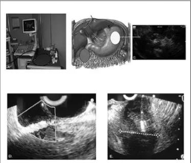
Sectorial EUS (echoendoscope GF-UCT 140, Olympus, America Corp., Melville, NY) sectorial array probe with 0.5 MHz, reaching 12.5 cm was used in this study .The anatomic location of both kidneys allows endosonographic imaging and direct needle access for tissue acquisition(1). The echo-endoscope used reaches 12.5 cm or 7.5 MHz and with the movement of the probe within the duodenum or stomach, this range is suficient to visualize both kidneys. The right kidney may be approached from the second portion of the duodenum with the EUS transducer rotated laterally. The left kidney may be approached from within the body of the stomach with the EUS transducer facing posterolaterally. The proximity of the EUS tip to the kidney from within the gastrointestinal lumen allows precise location and accurate access for tissue acquisition. Schematization of the echoen-doscope, EUS areas of interest for kidney approach (green duodenal area for the right kidney and yellow stomach area for the left kidney), appropriate visualization of the kidney (cortex and medulla), EUS visualization of tumor with needle insertion and aspiration and inally, cytologic aspirate is represented in Figure I. In all cases, three pass-es with a 22G needle (Cook Medical) were performed for echoendoscopic ine needle aspiration of renal tumors for adequate cytologic sampling. EUS FNA was performed on an outpatient basis. Only in one case, the procedure
was done during hospitalization (EUS FNA of bilateral renal masses).
Data collected included patient age and sex, clinical indication of renal biopsy, location and size of renal tumor, EUS FNA cytology, inal pathological indings, surgery re-sults, postoperative hospital stay, complications, and clinical follow-up. The criterion standard for diagnosis of any renal mass was histopathological indings from surgical resection. Nephron-sparing procedures were performed depending on tumor site, location and intraoperative evaluation. Cytolo-gical analysis was compared with inal patholoCytolo-gical results. Histopathological evaluation of the FNA was performed after hematoxylin-eosin staining. Histochemical techniques for surgical specimens included Hale and PAS staining. Immunohistochemistry for the antibodies used for renal cell tumors included pancytokeratin, CK7, CK20, vimentin, EMA, CD10, CD117 (c-kit), E-cadherin, WT1 and HMB-45, desmin and SMA.
RESULTS
Ten EUS FNA of renal masses were performed in nine male patients (mean age 56.5 years, median age 54.7 years). The procedure was on the right kidney (n = 4), on the left kidney (n = 4) and bilaterally in one. Tumors involved the upper pole (n = 3), the lower pole (n = 2), the mesorenal region (n = 3) and was considered a large mass (more than one kidney region involved) in two cases. Median tumor diameter was 55 mm (ranging 13 mm to160 mm).
Clinical indication for CT or MRI abdominal evaluation was macroscopic hematuria (n = 3), lank pain (n = 1) and abdominal mass (n = 2). In the three remaining cases, renal tumors were incidentally discovered. Indication for renal mass biopsy according to clinical guidelines were small renal mass (n = 4), suspicion of lymphoma (n = 1), suspicion of metastasis (n = 1), suspicion of oncocytoma (n = 1) in the case of bilateral EUS renal FNA, to distinguish between RCC and nephroblastoma in a young adult patient with renal mass and metastatic disease in one case and inally for histologic subtype analysis to predict response to immuno-therapy in a RCC case with brain metastasis. The clinical features are summarized in Table 1.
Final EUS FNA cytology was available in nine of the 10 attempted biopsies. One biopsy failure occurred in a case of a small posterior kidney tumor (biopsy number 4, Table 1). The renal hilum was visualized across the tract of the EUS FNA and although one pass with 22G needle was attempted, no more attempts were performed and tissue was insuficient. In all other cases, an accurate biopsy was performed which revealed clear cell RCC (n = 5), papillary RCC in two aspi-rates (bilateral tumors in the same patient), nephroblastoma (n = 1) and pulmonary carcinoma (n = 1).
Contact was done via telephone to determine any com-plication after the biopsy. No comcom-plications were reported. Patients with RCC were followed according to the guidelines of European Association of Urology (available on http:// www.uroweb.org/guidelines/online-guidelines).
Moura RN, Lopes RI, Srougi MF, Dall’Oglio MF, Sakai P, Artifon ELA.
Initial experience with endoscopic ultrasound-guided ine needle aspiration of renal masses: indications, applications and limitations
v. 51 no. 4 - out./dez. 2014 Arq Gastroenterol 339
DISCUSSION
More than 200.000 new cases of kidney cancer are diag-nosed annually, with more than 100.000 related deaths per year worldwide. RCC accounts for 3% of all adult malignan-cies and is increasing at a rate of 2.5% per year(1).
An enhancing renal neoplasm on CT or MRI has been considered by most urologists to be a suficient indication for surgery because about 80% of such lesions prove to be RCC(9). Currently, if local experience is suficient and the biopsy result has the potential to impact treatment decisions, urologists should consider increasing the use of core biopsy and FNA to better characterize suspicious renal masses preoperatively(2, 12, 14). The advantages of a biopsy in these cases are the potential to decrease unnecessary treatment of small renal masses and better selection of tumors for active surveillance and minimally invasive ablative therapies(7, 14).
The role of needle core biopsy and FNA of renal masses is primarily to rule out non renal cell primary tumors (metas-tasis and lymphoma) or benign conditions (abscess), which
may not require surgery(4). Biopsy has also been used to
conirm the diagnosis and the histological subtype of a renal primary lesion in patients with disseminated metastasis or unresectable retroperitoneal mass. In metastatic RCC, there is evidence that patients with clear cell subtype histology are more likely to beneit from adjuvant immunotherapy following cytoreductive nephrectomy(3). A role for biopsy in the new target therapies demonstrate different response rates with different RCC subtypes(6).
FNA with immunocytochemistry analysis can help dis-tinguish between RCC and oncocytomas. Even though RCC may be present in as many as 18% of oncocytomas, a EUS FNA showing oncocytoma, might allow surveillance for a renal lesion, especially if the patient prefers conservative management(13).
Percutaneous renal mass biopsy must not be performed routinely for renal lesions less than 40 mm but it should be indicated for incompletely accurate renal imaging diagnosis after a full imaging evaluation. Almost in 30% of the selected patients, a surgical procedure became no mandatory after renal biopsy results were obtained(9, 11).
The risk of complications associated with EUS FNA ranges from less than 1-6%. Tracheal suction (5%), vomiting (0.3%), aspiration (0.3%), esophageal perforation and death (less than 0.06%) are reported complications of EUS. Tumor seeding is a potential unlikely complication of EUS FNA
with few cases reported(5). The incidence of hemorrhage
after biopsy was low (1%). In the current study, there were no complications reported.
Preoperative biopsy of renal masses should be indicated only in selected cases. Good results for EUS FNA of selected renal tumors were observed in this study. Our overall techni-cal success rate of EUS-guided FNA was 90%, which is with-in the range previously reported(1, 2, 4, 14). The cause of failed procedure was due to the posterior aspect of this tumor and the smaller needle-tumor distance. In this patient, it appears to us that a computerized tomography guided posterior renal biopsy should be more appropriate. Some renal masses may
TABLE 1. Echoendoscopic ultrasonography with ine needle aspiration (EUS-FNA) renal biopsy Biopsy
number Age/
Sex Clinical Picture Indication
Kidney
(side) Kidney pole
Diameter
(cm) Final citology Surgery Histology Follow-up
1 61/M Macroscopic
hematuria
Suspicion of lymphoma
R Large mass 16 Clear cell RCC No - DOD,
2 months
2 73/M Incidental, family
history RCC
Oncocytoma vs RCC
R Inferior 7 Papillary RCC Yes (partial
nephrectomy)
RCC NED,
2 years
3 73/M Incidental, family
history RCC
Oncocytoma vs RCC
L Inferior 10 Papillary RCC Yes (partial
nephrectomy)
RCC NED,
2 years
4 53/M Incidental Small mass L Mesorenal 1.3 Unavailable
(biopsy not performed)
Yes (Laparoscopic
crioablation)
RCC NED,
1.5 years
5 51/M Convulsion, brain
metastasis + renal mass
Histologic diagnosis
R Mesorenal 5 Clear cell RCC No - DOD,
11 months
6 50/M Macroscopic
hematuria
Small mass L Mesorenal 1.3 Clear cell RCC Yes (radical
nephrectomy)
RCC NED,
2.5 years
7 49/M Macroscopic
hematuria
Small mass L Superior 3.5 Clear cell RCC Yes (partial
nephrectomy)
RCC NED,
3 years
8 67/M Incidental Small mass L Superior 2.6 Clear cell RCC Yes (partial
nephrectomy)
RCC NED,
2 years
9 27/M Abdominal mass Nephroblastoma
vs RCC
L Large mass 8
Nephroblas-toma
Yes (nephrectomy)
Nephro-blastoma
DOD, 6 months
10 78/M Abdominal pain Suspicion of
pulmonary cancer metastasis to
kidney
R Superior 6 Pulmonary
carcinoma
No - DOD,
3 months
Moura RN, Lopes RI, Srougi MF, Dall’Oglio MF, Sakai P, Artifon ELA.
Initial experience with endoscopic ultrasound-guided ine needle aspiration of renal masses: indications, applications and limitations
340 Arq Gastroenterol v. 51 no. 4 - out./dez. 2014
be unsuitable for EUS-guided biopsy because of anatomical limitations. Among other reasons, these limitations are likely to restrict widespread application of this method. EUS-FNA will be best applied to central anterior renal masses. For lesions on the posterior kidney aspect close to abdominal wall, percutaneous approach is probably the best choice.
EUS FNA appears as a safe and feasible procedure with good results, minimal morbidity and a short hospital stay(4). Although this paper is the second largest case series
REFERENCES
1. Artifon ELA, Lopes RI, Kumar A, Lucon AM, Dall’oglio M, Hawan B, et al.
Endoscopic ultrasound facilitates histological diagnosis of renal cell cancer. Journal of Endourology. 2008;22:2447-50.
2. Bardales RH, Stelow EB, Maallery S, Lai R, Stanley MW. Review of endoscopic
ultrasound-guided ine-needle aspiration cytology. Diagnostic Cytopathology. 2006;34:140-175.
3. Bex A, Horenblas S, Meinhardt W, Verra N, de Gast GC. The role of initial
immunotherapy as selection for nephrectomy in patients with metastatic renal cell carcinoma and the primary tumor in situ. Eur Urol. 2002;42:570-6.
4. DeWitt J, Gress FG, Levy MJ, Hernandez LV, Eloubeidi MA, Mishra G, et al.
EUS-guided FNA aspiration of kidney masses: a multicenter U.S experience. Gastrointest Endosc. 2009;70:573-8.
5. Doi S, Yasuda I, Iwashita T, Ibuka T, Fukushima H, Araki H, et al. Neddle
tract implantation on the esophageal wall after EUS-guided FNA of metastatic mediastinal lymphadenopathy. Gastrointest Endosc. 2008;67:988-90.
6. Farrell JJ, Brugge WR. EUS-guided ine-needle aspiration of a renal mass: an
alternative method for diagnosis of malignancy. Gastrointest Endosc. 2002;56:450-2.
7. Jhala NC, Jhala DN, Chhieng DC, Eloubeidi MA, Eltoum IA. Endoscopic
ultrasound-guided ine-needle aspiration - A cytopathologist´s perspective. Am J Clin Pathol. 2003;120:351-67.
8. Kutikov A, Fossett LK, Ramchandani P, Tomaszewski JE, Siegelman ES, Banner
MP, et al. Incidence of bening pathologic indings at partial nephrectomy for solitary renal mass presumed to be renal cell carcinoma on preoperative imaging. Urology. 2006;68:737-40.
of EUS FNA of renal masses in the literature and the irst on a single-center, our results should be interpreted carefully, especially due to the small number of cases submitted to FNA. The most important questions pertain to the role of EUS-FNA of renal tumors and the patients most likely to beneit from the procedure.
Further research should evaluate the beneits of preopera-tive renal biopsy use and randomization of percutaneous, lap-aroscopic and echoendoscopic approach should be compared.
Moura RN, Lopes RI, Srougi MF, Dall’Oglio MF, Sakai P, Artifon ELA. Ecoendoscopia com punção aspirativa de massas renais: indicações, aplicações e limitações. Arq Gastroenterol. 2014,51(4):337-40.
RESUMO - Contexto - A amostragem de tecido de massas renais é tradicionalmente realizada através da abordagem percutânea ou pelo método lapa-roscópico. A utilidade do ultrassom endoscópico para biópsia de lesões renais ainda não está clara e poucos casos foram relatados na literatura.
Objetivos - Avaliar a factibilidade e os resultados da biópsia de tumores renais guiada por ultrassom endoscópico. Método - Trata-se de uma série retrospectiva de casos de biópsias ecoguiadas de massa renal após avaliação com tomograia computadorizada ou ressonância magnética. Resultados
- Dez procedimentos foram realizados em nove pacientes do sexo masculino (idade média de 54,7 anos), no rim direito (n = 4), esquerdo (n = 4) e bilateralmente (n = 1). Massas renais (diâmetro médio 55 mm; variando de 13 a 160 mm) localizadas no pólo superior (n = 3), inferior (n = 2) e na região mesorenal (n = 3). Em dois casos, a massa envolvida mais de uma região renal. A ressecção cirúrgica conirmou carcinoma de células renais em seis pacientes nos quais a biópsia ecoguiada pré-operatória demonstrava carcinoma de células renais. Não foram relatadas complicações. Con-clusão - A biópsia de massas renais guiadas por ultrassom endoscópico é um procedimento seguro e viável, com bons resultados e mínima morbidade. DESCRITORES - Neoplasias renais, diagnósticos. Ultrassonograia. Aspiração por agulha ina guiada por ultrassom endoscópico
9. Lane BR, Samplaski MK, Herts BR, Zhou M, Novick AC, Campbell SC. Renal
mass biopsy – a renaissance?. J Urol. 2008;179:20-7.
10. Lebret T, Poulain JE, Molinie V, Herve JM, Denoux Y, Guth A, et al. Percu-taneous core biopsy for renal masses: indications, accuracy and results. J Urol. 2007;178:1184-8.
11. Lechevalier E, Andre M, Barriol D, Daniel L, Eghazarian C, De Fromont M, et al. Fine-needle percutaneous biopsy of renal masses with helical Ct guidance. Radiology. 2000;216:506-10.
12. Neuzillet Y, Lechevallier E, Andre M, Daniel L, Coulange C. Accuracy and clinical role of ine needle percutaneous biopsy with computerized tomography guidance of small (less than 4.0cm) renal masses. J Urol. 2004;171:1802-5.
13. Neuzillet Y, Lechevallier E, Andre M, Daniel L, Nahon O, Coulange C. Fol-low-up of renal oncocytoma diagnosed by percutaneous tumor biopsy. Urology.
2005;66:1181-5.
14. Volpe A, Kachura JR, Geddie WR, Evans AJ, Gharajeh A, Saravanan A, Jewett MA. Techiniques, safety and accuracy of sampling of renal tumors by ine needle aspiration and core biopsy. J Urol. 2007;178:379-86.
15. Wood BJ, Khan MA, McGovern F, Harisinghani M, Hahn PF, Mueller PR. Imaging guided biopsy of renal masses: indications, accuracy and impact on clinical management. J Urol. 1999;161:1470-4.