Involvement of DNA-PKcs in the Type I IFN
Response to CpG-ODNs in Conventional
Dendritic Cells in TLR9-Dependent or
-Independent Manners
Chi Ma, Narrissa P. Spies, Ting Gong, Can Xin Jones, Wen-Ming Chu*
Department of Cancer Biology, University of Hawaii Cancer Center, Honolulu, Hawaii 96813, United States of America
Abstract
CpG-ODNs activate dendritic cells (DCs) to produce interferon alpha (IFNα) and beta (IFNβ). Previous studies demonstrated that Toll-like receptor 9 (TLR9) deficient DCs exhib-ited a residual IFNαresponse to CpG-A, indicating that yet-unidentified molecules are also involved in induction of IFNαby CpG-A. Here, we report that the catalytic subunit of DNA-dependent protein kinase (DNA-PKcs) but not Ku70 deficient BMDCs showed defective IFNαand IFNβresponses to CpG-A or CpG-B. Loss of both DNA-PKcs and TLR9 further re-duced the IFNαresponse to CpG-A. These DNA-PKcs and TLR9 effects were mediated by their downstream Akt/mTORC1 pathway and downstream events IRAK1 and IKKα. Loss of DNA-PKcs, TLR9, MyD88 or IRAK4 impaired phosphorylation of Akt(S473), S6K, S6, IRAK1, or IKKαin BMDCs in response to CpG-ODNs. The residual IFNαand IFNβin DNA-PKcs-deficient BMDCs were partially responsible for the induction of IL-6 and IL-12 by CpG-ODNs and their stimulatory effect was blocked by IFNAR1 neutralizing antibodies. Further analysis indicated that CpG-ODN associated with DNA-PKcs and Ku70, and in-duced DNA-PKcs’s interaction with TRAF3. Intriguingly, DNA-PKcs but not Ku70 expres-sion level was reduced in TLR9-deficient BMDCs. Taken together, our data suggest that DNA-PKcs is an important mediator in the type I IFN response to CpG-ODNs in TLR9-dependent or -inTLR9-dependent fashions.
Introduction
Bacterial and viral genomic DNA containing the CpG motif (CpG-DNA) and its analog oli-godeoxynucleotides containing CpG motif (CpG-ODNs) are powerful activators of the in-nate immune system. There are two major types of CpG-ODNs, CpG-A and CpG-B. CpG-A prefers activating plasmacytoid DCs (pDCs), whereas CpG-B efficiently activates B cells, con-ventional dendritic cells (cDCs) and macrophages[1]. CpG-B strongly activates DCs and macrophages to produce pro-inflammatory IL-6 and IL-12, which is critical for the Th1 a11111
OPEN ACCESS
Citation:Ma C, Spies NP, Gong T, Jones CX, Chu W-M (2015) Involvement of DNA-PKcs in the Type I IFN Response to CpG-ODNs in Conventional Dendritic Cells in TLR9-Dependent or -Independent Manners. PLoS ONE 10(3): e0121371. doi:10.1371/ journal.pone.0121371
Academic Editor:Lucia Gabriele, Istituto Superiore di Sanità, ITALY
Received:March 23, 2014
Accepted:February 12, 2015
Published:March 26, 2015
Copyright:© 2015 Ma et al. This is an open access article distributed under the terms of theCreative Commons Attribution License, which permits unrestricted use, distribution, and reproduction in any medium, provided the original author and source are credited.
Data Availability Statement:All relevant data are within the paper.
Funding:This work is supported by the NIH (R01AI54128 to WMC). The funder had no role in study design, data collection and analysis, decision to publish, or preparation of the manuscript.
response and subsequent anti-infectious and anti-tumor activities. Also, CpG-A together with DOTAP (a lipid) triggers DCs to produce the type I IFN (IFNαand IFNβ), and CpG-B is able to induce IFNβexpression. It is known that CpG-ODNs activate TLR9, which in turn recruits the adaptor protein myeloid differentiation factor 88 (MyD88) and IL-1 receptor as-sociated protein kinase 4 (IRAK4) leading to activation of IRAK1 and IKKα, which then acti-vate the IFN regulatory factor 7, resulting in expression of the type I IFN[2,3]. However, loss of TLR9 does not abolish the IFNαresponse to CpG-A, but abolishes the IFNβresponse to CpG-B suggesting that an unidentified factor also mediates the IFNαresponse to CpG-A.
TNF receptor-associated factor 3 (TRAF3) has been found to be important for expression of type I IFNs but not pro-inflammatory cytokines in response to CpG-ODNs[3]. CpG-ODNs in-duce the association of TRAF3 with MyD88[3,4]. Yet, how TRAF3 mediates the type I IFN re-sponse to CpG-ODNs is still not well understood.
The Akt/mammalian target of rapamycin complex 1 (mTORC1) pathway is also suggested to be required for the type I IFN response to CpG-A in pDCs[5]. mTORC1 is an important downstream event of Akt, and can phosphorylate the ribosome 6 (S6) kinase (S6K), which in turn phosphorylates S6[6]. The mTORC1 inhibitor rapamycin was able to inhibit both the type I IFN and pro-inflammatory cytokine responses to CpG-ODNs[5]. Knockdown of S6K dimin-ished CpG-ODN-induced association of TLR9 with MyD88[5], indicating that Akt and S6K might act upstream of TLR9 in CpG-ODN signaling. Interestingly, chloroquine, which abol-ishes the activation of TLR9-dependent pro-inflammatory signaling, had no apparent inhibito-ry effect on Akt activation by CpG-ODN in THP1 macrophages[7]. Our results and others suggested that TLR9 was involved in Akt(S473) phosphorylation in bone marrow-derived mac-rophages (BMDMs) in response to CpG-ODNs [8,9].
In addition to above relay molecules, other proteins are suggested to be transducers in CpG-ODN signaling. One of them is DNA-PKcs, which is in both the cytoplasm and nucleus of mouse cells[8]. DNA-PKcs is an essential component of double-stranded DNA break re-pair complex and is vital for B and T cell development[10]. A high level of anti-DNA-PKcs autoantibody is frequently detected in serum of patients with polymyositis, scleroderma, sys-temic lupus erythematosus (SLE), and mixed connective tissue disease[11]. It is suggested that the type I IFN plays a principal role in the development of SLE[12] indicating that DNA-PKcs might be involved in the type I IFN expression. Indeed, in a bioassay it was found that loss of DNA-PKcs impaired CpG-B-induced type I IFN response [13]. Because the bioas-say is unable to tell whether IFNαor IFNβtriggers the anti-viral activity, it was unclear whether DNA-PKcs was involved in the production of IFNαor IFNβin the study[13]. An-other study suggested that DNA-PKcs is a DNA sensor for interferon regulatory factor 3-dependent innate immunity[14].
We previously reported that DNA-PKcs regulates the IL-6 and IL-12 responses to CpG-B in BMDCs in a time- and dose- dependent manner[15]. In this study, we investigated whether and how DNA-PKcs is involved in the IFNαand IFNβresponses to CpG-ODNs. Also, we de-termined whether Ku70, a regulatory subunit of DNA-PK, regulates the type I IFN response to CpG-ODNs, and whether inhibition of the type I IFN pathway further impairs IL-6 and IL-12 response to CpG-ODN in DNA-PKcs-deficient BMDCs. Additionally, we examined the impact of individual deficiency of DNA-PKcs or TLR9, or double deficiency of DNA-PKcs and TLR9 on the activation of the Akt/mTORC1 pathway, IKKαand IRAK1, and the type I IFN response to CpG-ODNs. We determined whether IRAK4 and MyD88 are involved in the activation of Akt and mTORC1 by CpG-ODNs. Finally, we determined the relationship between
Materials and Methods
Mice, regents and antibodies
Wild type(WT),DNA-PKcs-/-[6,8,15],Ku70-/-,TLR9-/-,IRAK4-/-and MyD88-/-[6,8,15–17] mice were on the C57/B6/129 genetic background, andDNA-PKcs-/-/Ku70-/-andDNA-PKcs-/-/ TLR9-/-[15] mice were generated by crossingDNA-PKcs-/-withKu70-/-orTLR9-/-mice. Ani-mals were bred at University of Hawaii, Honolulu, Hawaii, USA. All animal work was per-formed based on the protocols approved by the IACUC at University of Hawaii.
CpG-ODN 1018 (CpG-B) (50-TGACTGTGAACGTTCGAGATGA-30) and CpG-ODN
D19 (CpG-A) (50-GpsGpsTGCATCGATGCAGGpsGpsGpsGpsG-30)[6] were synthesized by
Trilinker Biotechnology (CA, USA). CpG-ODN 1018-biotin (CpG-biotin) was purchased from Trilinker Biotechnology. DOTAP Liposomal Transfection Reagent was from Roche (Mann-heim, Germany). Rapamycin was from LC Laboratories (MA, USA). Protein A/G Sepharoses were purchased from Amersham Biosciences (Uppsala, Sweden) and streptavidin-agarose beads were from Life Technologies (NY, USA).
Anti-phospho (p) antibodies (Abs) against Akt(S473), S6K(T389), S6(S235/236), IKKα/β
(S176/180), IRAK1(T209), ERK1/2(T202/Y204), and Stat1(Y701) were from Cell Signaling (MA, USA). Anti-DNA-PKcs mouse monoclonal antibody (mAb) was from Neomarker (CA, USA); anti-Ku70 mAb, anti-ERK1/2 polyclonal (pAb), and anti-TRAF3 pAb were from Santa Cruz Biotech (CA, USA); anti-IFNAR1 and isotype Abs were from Biolegend (CA, USA); and Alexa fluorescence-conjugated secondary Abs were from Life Technologies (NY, USA).
Bone marrow-derived dendritic cell culture
As described previously[15], bone marrow cells were isolated from femurs and tibias of mice, seeded at 2–3×10⁵cells/ml, and then cultured with 10% fetal bovine serum (FBS, Life Technol-ogies, NY, USA) RPMI1640 medium supplemented with granulocyte/macrophage-colony stimulatory factor [GM-CSF, Biolegend (CA, USA), 20 ng/ml] for 7 days. On day 2 and day 4, 5 ml of fresh 10% FBS RPMI1640 medium containing GM-CSF (20 ng/ml) were added to the cultures. On day 7, BMDCs in culture medium were harvested, and cells remained in dishes were collected by gently rinsing with 1× phosphate buffered saline (PBS) for three times. BMDCs in both medium and rinsed PBS were combined and used for the
following experiments.
ELISA assay
BMDCs harvested on day 7 were seeded in 96-well plates at 1 or 2x10⁵/well in triplicate with 10% FBS RPMI1640 medium. DOTAP was pre-incubated with CpG-A for 30 minutes at room temperature at 10μl perμg of DNA. The cells were treated with CpG-ODNs or left untreated
for 12 or 24 hours. The levels of IL-6, IL-12p40 or IFNβin the supernatants were determined by enzyme-linked immunosorbent assay (ELISA) via respective cytokine ELISA kits (Biole-gend, CA, USA; PBL Assay Science, NJ, USA) according to the manufacturer’s manual. The level of IFNαwas detected by ELISA using kits from PBL Assay Science (NJ, USA) according to manufacturer’s instruction.
Immunostainning for flow cytometry
with 1% paraformaldehyde in PBS. The samples were analyzed on a FACSCalibur (BD, CA, USA) and processed with Flowjo software.
For intracellular staining, 1×10⁷BMDCs were seeded in petri dishes and treated with CpG-B (1μM), CpG-A (3μM) with DOTAP, or left untreated for 24 hours. After the cells were
stained with PE-conjugated CD11c, APC-conjugated MHC-II-I-A/I-E and PE-Cy7-conjugated CD11b (Biolegend, CA, USA), cells were fixed with 1% paraformaldehyde in PBS in the dark for 20 minutes at room temperature. Fixed cells were resuspended in Permeabilization Wash Buffer (Biolegend, CA, USA), centrifuged at 350x g for 5 minutes, and then washed with Per-meabilization Wash Buffer twice. BMDCs were stained with either FITC-conjugated IFN-αor FITC-conjugated IFN-β(PBL Assay Science, NJ, USA) for 30 minutes at room temperature, and fixed with 1% paraformaldehyde in PBS. The samples were analyzed on a BD LSRFortessa (BD, CA, USA) and analyzed with Flowjo software. Only CD11c+CD11b+MHC-II+cells were analyzed for the IFNs expression.
Immunoblotting and immunoprecipitation assays
BMDCs were harvested, washed with 1x PBS twice and starved for 2 hours in pre-warmed serum-free RPMI-1640 medium (SFM). The cells were treated with CpG-B (1μM) or CpG-A
(3μM) in pre-warmed SFM for the indicated durations or left untreated. Whole cell lysates
(WCLs) were prepared in the lysis buffer (1% Triton-X100)[15] and 60μg proteins were
sepa-rated on 10% SDS-PAGE and transferred onto PVDF membranes. The membranes were blocked with 5% non-fat milk in PBS containing 0.1% Tween 20 or SuperBlock1T20 blocking buffer (Thermo-Pierce, IL, USA) for 1 hour and probed with correspondent primary Abs and secondary Abs-conjugated with horseradish peroxidase (HRP), and detected by enhanced chemiluminescence (ECL, Thermo-Piers, IL, USA).
For immunoprecipitation (IP) assays[6,8,15], after starvation, BMDCs were treated with CpG-B (1μM) in pre-warmed SFM for the indicated durations or left untreated, and then lysed with the
lysis buffer (1% NP-40). WCLs (400μg) were pre-cleared with 40μl of protein A/G Sepharose
(beads) (1:1) for 30 minutes. Pre-cleared WCLs were incubated with anti-DNA-PKcs Ab (0.8μg) or
control mouse IgG (0.8μg) together with 50μl of protein A/G beads at 4°C overnight with rotation.
The beads were washed four times with the lysis buffer (0.1% NP-40) containing 1 mM phenyl-methylsulfonylfluoride (PMSF, Sigma, USA). Proteins with beads were boiled, loaded on 10% SDS-PAGE and transferred onto PVDF membranes followed by immunoblotting (IB) analysis.
Pull-down assay
For pull-down assays, starved BMDCs were treated with CpG-biotin (1μM) for 10, 30 minutes
or left untreated and then washed twice with cold 1× PBS[15]. Cells were lysed with the lysis buffer [1% digitonin (Sigma, USA)][18]. WCLs (400μg) were pre-cleared with protein A/G
beads, incubated with 22μg CpG-biotin for 2 hours, and then with 50μl streptavidin-agarose
beads at 4°C overnight with rotation. The beads were washed four times with the lysis buffer (0.1% digitonin, 1 mM PMSF), boiled, separated on 10% SDS-PAGE, and transferred onto PVDF membranes followed by IB analysis.
Immunofluorescence staining
The immunofluorescence staining was described previously[6,8,15]. Briefly, harvested BMDCs were seeded in 8-well chamber slides at 2×10⁵cells/chamber and cultured with 10% FBS RPMI-1640 medium overnight. Cells were starved for 2 hours in pre-warmed SFM and then treated with CpG-B (1μM) for 30 minute or left untreated. Cells were fixed with 4% paraformaldehyde,
anti-DNA-PKcs and anti-TRAF3 Abs in PBS with 2% BSA at 4°C overnight. Alexa fluorescence-conjugated secondary Abs (Alexa 488 or 594) (Lifetechnologies, NY, USA) was added and incu-bated for 2 hours. Slides were mounted with anti-fade mounting solution (Electron Microscopy Sciences, PA, USA) and observed under an IX81 Olympus microscope with 60X oil immersion objective powered by 1.6x magnification. The images were captured by an ORCA R2 CCD mono camera and processed by the Metamorph Image Analysis Software (Olympus, Japan).
Results
DNA-PKcs but not Ku70 is involved in the type I IFN response to
CpG-ODNs
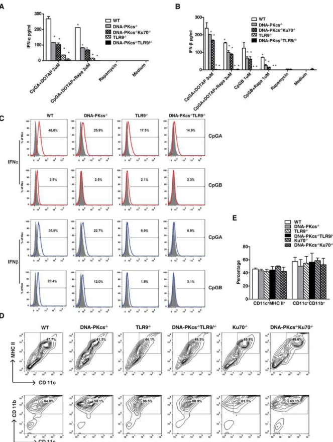
We previously reported that DNA-PKcs regulated the IL-6 and IL-12 responses to CpG-ODNs in BMDCs in a time- and dose-dependent fashion[15]. It was also suggested that DNA-PKcs regulated the type I IFN response to CpG-B in a bioassay[13]. Because the bioassay cannot dis-tinguish the anti-viral effect triggered by IFNαor IFNβ, it is unclear whether DNA-PKcs regu-lates the IFNαor IFNβresponse, or both IFNαand IFNβresponses to CpG-B. Thus, we examined secretion of IFNαor IFNβfrom WT and DNA-PKcs-deficient BMDCs in response to CpG-A together with DOTAP or CpG-B. As shown, CpG-A induced the secretion of both IFNαand IFNβ, whereas CpG-B only induced the secretion of IFNβ(Fig. 1A and 1B). Loss of DNA-PKcs impaired the IFNαand IFNβresponses to CpG-A, and reduced the IFNβresponse to CpG-B (Fig. 1A and 1B). In contrast, loss of Ku70 had no significant effects on the IFNαand IFNβresponses to CpG-A or CpG-B (Fig. 1C to 1D). Loss of Ku70 significantly enhanced the IL-6 and IL-12 responses to CpG-A, but had no significant effects on the IL-6 and IL-12 re-sponses to CpG-B (Fig. 1E to 1H). As a control, we also examined the IFNαand IFNβ re-sponses to CpG-ODNs in TLR9-deficient BMDCs. As expected, loss of TLR9 severely abrogated the IFNβresponse to CpG-A and abolished the IFNβresponse to CpG-B, but re-duced the IFNαresponse to CpG-A to 1/3 in BMDCs (Fig. 1A and 1B).
Taken together, our data suggest that DNA-PKcs but not Ku70 is important for the type I IFN response to CpG-ODNs.
Both DNA-PKcs and TLR9 contribute to the IFN
α
response to CpG-A
Our previous study suggested that DNA-PKcs might be parallel to TLR9 in CpG-DNA signal-ing[8,15]. Thus, we determined the type I IFN response to CpG-ODNs in DNA-PKcs/TLR9 double knockout (DKO) BMDCs. Consistent with the results inFig. 1A, loss of DNA-PKcs or TLR9 reduced the IFNαresponse to CpG-A (Fig. 2A). However, loss of both DNA-PKcs and TLR9 further reduced this response (Fig. 2A) indicating that DNA-PKcs may be parallel to TLR9 in CpG-A signaling. We also generated DNA-PKcs/Ku70 DKO mice and found that the IFNαand IFNβresponses to CpG-A were not further reduced in DNA-PKcs/Ku70 DKO BMDCs as compared to DNA-PKcs-deficient BMDCs (Fig. 2A, 2B and 2C). Consistent with our previous report[15], loss of DNA-PKcs, Ku70 or TLR9 had no apparent effect on BMDC development or maturation as indicated by cell surface expression of CD11c/MHC-II and CD11b/CD11c (Fig. 2D and 2E). Collectively, our data suggest that both DNA-PKcs and TLR9 are important for the IFNαand IFNβresponses to CpG-ODNs.
mTORC1 inhibition further diminishes the type I IFN response to
CpG-ODNs in DNA-PKcs- and TLR9- deficient BMDCs
Fig 1. DNA-PKcs but not Ku70 is involved in the type I IFN response to CpG-ODNs.Bone marrow was isolated from WT, DNA-PKcs-/-, TLR9-/-and
Ku70-/-mice and then cultured with GM-CSF for 7 days. (A-D) Harvested BMDCs were seeded in 96-well plates at 2x10⁵/well in triplicate, treated with CpG-ODNs (CpG-A 0.3μM or 3μM with DOTAP; CpG-B 0.1μM or 1μM), or left untreated for 24 hours. The levels of IFNα(A, C) and IFNβ(B, D) were determined by ELISA. Similar results were obtained in at least three independent experiments. (E-H) BMDCs from WT and Ku70-/-mice were seeded at
cDCs (e.g. GM-CSF-driven BMDCs in this study), we treated BMDCs with CpG-ODNs in the presence or absence of rapamycin. Similar to pDCs, rapamycin inhibited the IFNαand IFNβ
responses to CpG-A and the IFNβresponse to CpG-B in WT BMDCs. Rapamycin further re-duced the IFNαresponse to CpG-A in DNA-PKcs-, DNA-PKcs/Ku70- or TLR9-deficient BMDCs, and almost abolished this response in DNA-PKcs/TLR9 DKO BMDCs (Fig. 2A). Rapamycin additionally diminished the IFNβresponse to CpG-A and CpG-B in DNA-PKcs-or DNA-PKcs/Ku70-deficient BMDCs (Fig. 2B). Overall, our data suggest that the residual type I IFN response to CpG-ODN in DNA-PKcs or TLR9-deficient BMDCs is, at least in part, due to the residual activation of mTORC1 by CpG-ODNs.
The type I IFN is involved in the IL-6 and IL-12 responses to CpG-ODNs
in DNA-PKcs-deficient BMDCs
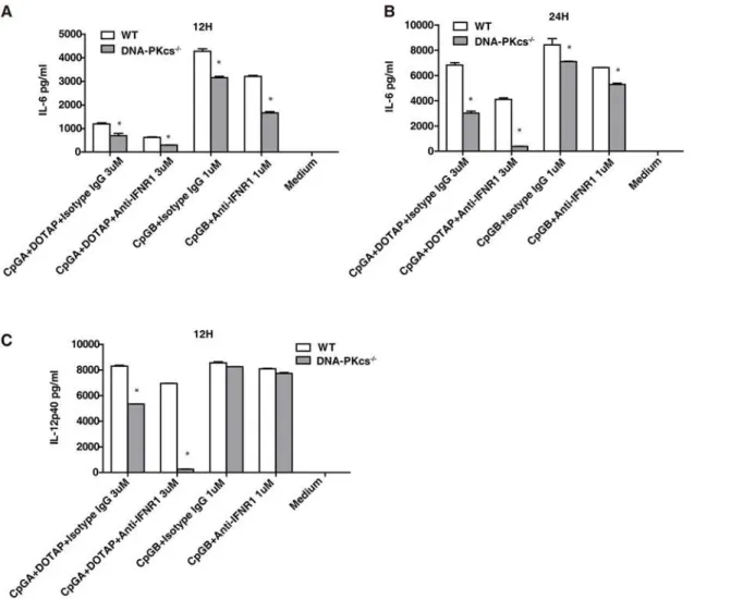
It was previously reported that the type I IFN contributes to the inflammatory cytokine re-sponses to TLR ligands[19]. Because the type I IFN response (Figs.1and2) and the IL-6 and IL-12 responses to CpG-ODNs were reduced[15], but not abolished in DNA-PKcs-deficient BMDCs, we reasoned that inhibition of type I IFN signaling would further reduce the IL-6 or IL-12 responses to CpG-ODNs in DNA-PKcs-deficient BMDCs. To test this scenario, we treat-ed both WT and DNA-PKcs-deficient BMDCs with CpG-ODNs in the presence or absence of anti-IFNα/βreceptor 1 (IFNAR1) neutralizing antibodies. Anti-IFNAR1 antibodies further sig-nificantly reduced the IL-6 response to CpG-A and CpG-B (Fig. 3A and 3B) and the IL-12 response to CpG-A in DNA-PKcs-deficient BMDCs (Fig. 3C). In contrast, IFNAR1 anti-bodies failed to decrease the IL-12 response to CpG-B in either WT or DNA-PKcs-deficient BMDCs (Fig. 3C). Taken together, our data suggest that IFNAR1 signaling contributes to resid-ual pro-inflammatory cytokine response to CpG-ODNs in DNA-PKcs-deficient BMDCs.
DNA-PKcs and TLR9 are important for the activation of the Akt/
mTORC1 pathway by CpG-ODN
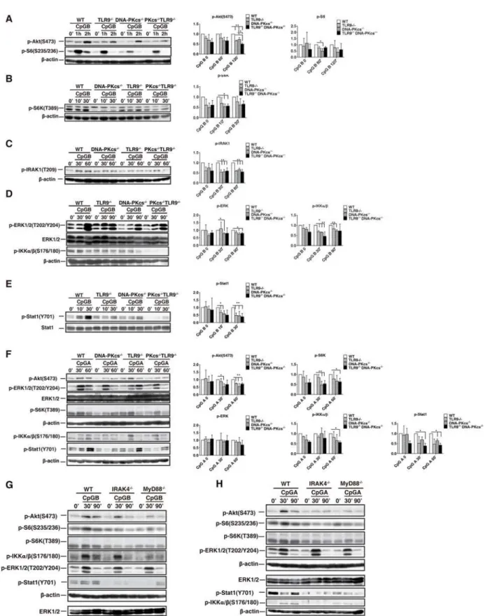
We previously reported that DNA-PKcs was required, and TLR9 was involved in activation of Akt by CpG-ODN in BMDMs [8]. Also, a study suggested that TLR9 was involved in this acti-vation in BMDMs in response to CpG-B[9]. Because rapamycin inhibits the type I IFN re-sponse to CpG-ODNs[5], we determined whether DNA-PKcs and TLR9 are required for activation of the Akt/mTORC1 pathway by CpG-ODNs in BMDCs. As shown, loss of either DNA-PKcs or TLR9 impaired phosphorylation of Akt(S473), S6(S235/236), or S6K(T389) in response to CpG-B (Fig. 4A and 4B); loss of both DNA-PKcs and TLR9 further reduced Akt (S473) phosphorylation (Fig. 4A). Statistically, for Akt(S473) phosphorylation, the significant difference between WT and KO cells occurred at 120 minutes after CpG-ODN stimulation; for S6(S235/236) phosphorylation, the significant difference occurred at 60 minutes; for S6K phos-phorylation, the significant difference occurred at 10 minutes.
A similar defect in the above phosphorylation was observed in DNA-PKcs-, TLR9- or DNA-PKcs/TLR9- deficient BMDCs in response to CpG-A (Fig. 4F) except that DNA-PKcs/ TLR9 double deficiency did not further significantly impair Akt(S473) phosphorylation. Taken together, our results suggest that both DNA-PKcs and TLR9 are important for the activation of the Akt/mTORC1 pathway in BMDCs by CpG-ODNs.
IL-6 (E, F) and IL-12 (G, H) was determined by ELISA using IL-6 or IL-12 ELISA kits. Similar results were obtained in three independent experiments. Note: bars represent the average of triplicates±SD.*P<0.01 KO groups versus WT groups.
DNA-PKcs is important for phosphorylation of IRAK1, IKK
α
, and Stat1 in
response to CpG-ODNs
To further understand how DNA-PKcs is involved in the type I IFN response to CpG-ODNs, first, we determined IKKαand IRAK1 activation, which is required for the type I IFN response to CpG-ODNs[20,21], in DNA-PKcs-, TLR9- or DNA-PKcs/TLR9- deficient BMDCs. As shown inFig. 4C, 4D and 4F, loss of either DNA-PKcs or TLR9 reduced IKKα/β(S176/180) and IRAK1(T209) phosphorylation. This reduction became more severe in DNA-PKcs/TLR9 average of triplicates±SD.*P<0.01 versus WT groups. (C) After 7 days culture, BMDCs from WT, DNA-PKcs-/-, TLR9-/-and DNA-PKcs-/-/TLR9-/-mice were
seeded in 3.5 cm petri-dishes at 1x10⁷/dish, and then treated with CpG-A (3μM) plus DOTAP or CpG-B (1μM) for 24 hours. The cells were harvested and
stained with anti-CD11b, CD11c and MHC-II Abs followed by internal staining for IFNαor IFNβ. The samples were analyzed by flow cytometry and CD11c+CD11b+MHC-II+cells were gated and used for the IFN expression comparison. (D) BMDCs at day 7 were stained for CD11b, CD11c and MHC-II,
and analyzed by flow cytometry. The live cells were gated. (E) The percentage of CD11c+MHC-II+and CD11c+CD11b+BMDCs was analyzed by one-way ANOVA and Tukey’s multiple comparisons test. Note: bars represent the average of three independent experiments±SD.
doi:10.1371/journal.pone.0121371.g002
Fig 3. The type I IFNs are involved in the IL-6 and IL-12 responses to CpG-ODNs in DNA-PKcs-deficient DCs.(A-C) BMDCs from WT and DNA-PKcs -/-mice were harvested on day 7, and then seeded in 96-well plates at 2x10⁵/well in triplicate. The cells were pre-incubated with 5μg/ml of anti-IFNAR1 or
isotype control Abs for 30 minutes, and then treated with CpG-A (3μM) plus DOTAP, or CpG-B (1μM), or left untreated for 12 or 24 hours. The supernatants were collected and the levels of IL-6 (A, B) and IL-12p40(C) were detected by ELISA. Similar results were obtained in three independent experiments. Note: bars represent the average of triplicates±SD.*P<0.01 versus WT groups.
p-DKO BMDCs in response to CpG-ODNs but was not significant as compared to individual de-ficient cells. Consistent with a previous report[22], loss of DNA-PKcs reduced ERK1/2(T202/ Y204) phosphorylation in response to CpG-ODNs (Fig. 4D). However, clear residual activation of ERK1/2 by CpG-ODNs was observed in DNA-PKcs KO, TLR9 KO, or DNA-PKcs/TLR9 DKO BMDCs (Fig. 4D and 4F), indicating that other protein factors are involved in the ERK1/ 2 activation by CpG-ODNs.
Finally, we examined phosphorylation of signal transducer activator of transcription 1 at the tyrosine (Y) 701 residue [Stat1(Y701)], which is a key transcription factor in type I IFN sig-naling[3], in above deficient BMDCs. Loss of either DNA-PKcs or TLR9 reduced Stat1(Y701) phosphorylation (Fig. 4E and 4F), and loss of both DNA-PKcs and TLR9 further diminished Stat1(Y701) phosphorylation in response to CpG-B (Fig. 4E). The most significant difference for the Stat1 phosphorylation between WT, single KO and DKO cells occurred at 30 minutes. Taken together, our data suggest that DNA-PKcs and TLR9 are important for activation of IKKα, IRAK1 and Stat1 by CpG-ODNs.
MyD88 and IRAK4 are involved in activation of the Akt/mTOR1 pathway
by CpG-ODNs
To elucidate the relationship between the TLR9 pathway and the Akt/mTORC1 pathway, we examined phosphorylation of Akt, S6K and S6 in BMDCs generated from mice with deletion of MyD88 or IRAK4, which are key downstream events of TLR9 in CpG-DNA signaling. Our results showed that loss of either IRAK4 or MyD88 reduced but not abolished phosphorylation of Akt(S473), S6K and S6 in BMDCs in response to CpG-ODNs (Fig. 4G and 4H). Also, the levels of phosphorylation of IKKα/β, ERK1/2 or Stat1 were determined. Interestingly, while phosphorylation of IKKα/βand Stat1 was severely impaired in both IRAK4- and MyD88-deficient BMDCs, phosphorylation of ERK1/2 was not markedly reduced (Fig. 4G and 4H), in-dicating that other molecules but not components in the TLR9 pathway are required for ERK1/ 2 activation by CpG-ODNs. Taken together, our results suggest that, in addition to their roles in activation of signaling evens in the IFN signaling pathway, IRAK4 and MyD88 are also in-volved in activation of the Akt/mTOCRs pathway by CpG-ODNs.
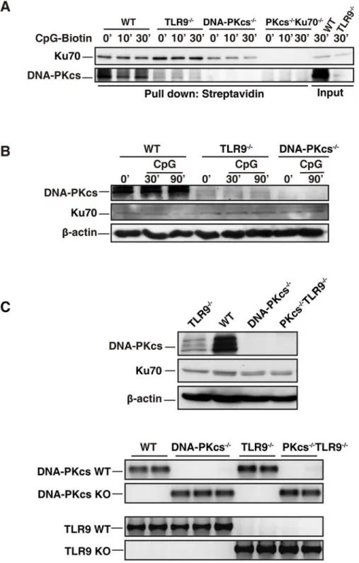
The level of DNA-PKcs is reduced in TLR9-deficient BMDCs
We previously reported that CpG-ODN bound to DNA-PKcs and TLR9[15]. Here, we con-firmed that CpG-ODN associated with DNA-PKcs (Fig. 5A). However, less DNA-PKcs was precipitated by CpG-ODN-biotin from TLR9-deficient BMDCs than WT controls (Fig. 5A). Because Ku70 is a regulatory subunit of DNA-PK, we examined whether Ku70 was pulled down by CpG-ODN-biotin and whether less Ku70 was pulled down from TLR9-deficient BMDCs. As shown, CpG-ODN was able to pull down Ku70 (Fig. 5A). Interestingly, the associ-ation of CpG-ODN-biotin with Ku70 was slightly enhanced in TLR9-deficient BMDCs (Fig. 5A) indicating that Ku70 may compete with TLR9 for binding to CpG-ODNs. This may explain why loss of Ku70 enhanced the IL-6 and IL-12 responses to CpG-A, which was deliv-ered to the cytoplasm by DOTAP (lipid).
The above observation led us to hypothesize that TLR9 is important for maintaining the protein level of DNA-PKcs. Indeed, immunoblotting analysis showed that loss of TLR9 clearly ERK1/2(T202/Y204), ERK1/2, p-IKKα/β(S176/180), p-Stat1(Y701) and Stat1 were detected by IB analysis. (A-D, F-H)β-actin was used as an equal loading control. Similar results were obtained from three (A-F) or two (G, H) independent experiments.**p<0.01,*p<0.05;Student’s t test. Note: bars represent the
average of three independent experiments±SD.
Fig 5. DNA-PKcs level is reduced in TLR9-deficient BMDCs.(A) CpG-ODN associates with Ku70 and DNA-PKcs in BMDCs. WT, TLR9-/-, DNA-PKcs-/-(PKcs-/-), and PKcs-/-/Ku70-/-BMDCs were treated with
CpG-ODN-biotin (CpG-B-biotin, 1μM) for the indicated time durations. WCLs were prepared with the lysis buffer (1% digitonin). After pre-cleared with protein A/G beads, 400μg proteins of WCLs were incubated with 15μg of CpG-B-biotin for 2 hours, and then incubated with 50μl of streptavidin-agarose beads at 4°C overnight with rotation. The agarose beads were washed four times with the lysis buffer (0.1% digitonin). The samples were separated on 10% SDS-PAGE and transferred onto a PVDF membrane followed by IB with anti-Ku70 or anti-DNA-PKcs Abs. Similar results were obtained from three independent experiments. (B) The level of DNA-PKcs is reduced in TLR9-dificient BMDCs. BMDCs from WT, DNA-PKcs-/-(PKcs-/-) and TLR9
decreased DNA-PKcs at the protein level, while Ku70 protein level was not affected by loss of DNA-PKcs, TLR9 or both DNA-PKc and TLR9 (Fig. 5B and 5C).
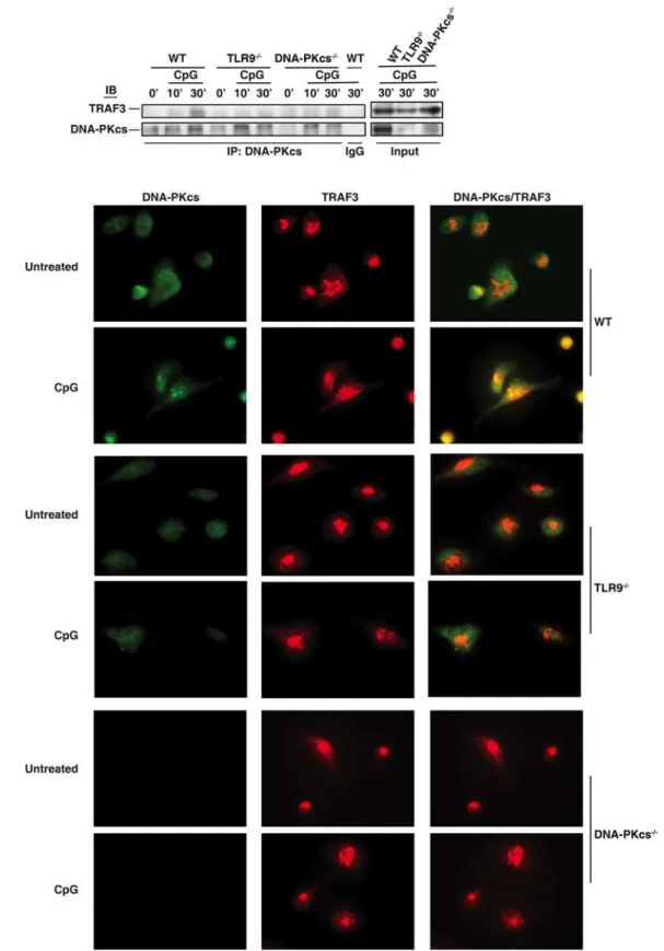
CpG-ODN induces the association of DNA-PKcs with TRAF3
TRAF3 has been suggested to be required for the type I IFN response to CpG-ODN[3,4]. Be-cause DNA-PKcs is also involved in this response, we hypothesized that CpG-ODN induces the interaction of DNA-PKcs with TRAF3. To test this hypothesis, we treated WT, DNA-PKcs-or TLR9-deficient BMDCs with CpG-ODN and then immunoprecipitated DNA-PKcs from WT and deficient BMDCs. As shown, CpG-ODN induced the association of DNA-PKcs with TRAF3 (Fig. 6A). Loss of TLR9 largely impaired CpG-ODN-induced association of DNA-PKcs with TRAF3 (Fig. 6A). To confirm the above observation, we performed an immunofluores-cence assay. More DNA-PKcs was observed in WT BMDCs compared to TLR9-deficient BMDCs. Much less co-localization of DNA-PKcs with TRAF3 was observed in TLR9-deficient BMDCs in response to CpG-ODN (Fig. 6B). Taken together, our results suggest that
CpG-ODN induced the association of DNA-PKcs with TRAF3 in a TLR9- dependent manner.
Discussion
TLR9 was thought to be the sole receptor for CpG-ODNs until it was found that a significant amount of CpG-A-induced IFNαwas still left in TLR9-deficinet pDCs[23]. Yet, loss of MyD88 and IRAK4, which are key downstream events of TLR9, abolished the IFNαresponse to CpG-A[23,24], indicating that other cytoplasmic proteins exist and transduce signal from CpG-A to MyD88 and IRAK4. However, additional molecules involved in the IFNαresponse to CpG-A remain to be identified. Here, we demonstrate that DNA-PKcs but not Ku70 is also involved in the type I IFN response to CpG-ODNs. Loss of DNA-PKcs reduced the type I IFN response to CpG-A and the IFNβresponse to CpG-B. Loss of both DNA-PKcs and TLR9 fur-ther diminished the IFNαresponse to CpG-A, suggesting that both DNA-PKcs and TLR9 are important for the IFNαresponse to CpG-A (Fig. 7A and 7B).
A previous study suggested that mTORC1 inhibitor rapamycin inhibited the type I IFN re-sponse to CpG-A in pDCs and that S6K was required for the interaction of TLR9 with MyD88 and the subsequent IFN response to CpG-ODNs[5]. In this study, we found rapamycin not only inhibited the IFNαresponse to CpG-A in WT BMDCs, but it also further reduced the IFNαresponse in either DNA-PKcs- or TLR9-deficient BMDCs (Fig. 2A and 2B), suggesting that mTORC1 activity, at least in part, remains in either deficient cells. We previously reported that loss of DNA-PKcs impaired Akt activation by CpG-ODNs in BMDMs[8]. In BMDCs, loss of either DNA-PKcs or TLR9 reduced Akt(S473) phosphorylation in response to CpG-ODNs suggesting that both DNA-PKcs and TLR9 contribute to the activation of Akt by CpG-ODNs (Figs.4Aand7A). Similarly, loss of DNA-PKcs or TLR9 reduced phosphorylation of S6K and S6 in response to CpG-ODNs (Fig. 4A and 4B). Our results explain why rapamycin further in-hibited the IFNαresponse to CpG-A in either DNA-PKcs- or TLR9-deficient BMDCs. To fur-ther understand how TLR9 is involved in activation of Akt and mTORC1 by CpG-ODN, we examined this activation in IRAK4- or MyD88-deficient BMDCs. Loss of either IRAK4 or durations or left untreated. Cell lysates were prepared with lysis buffer (1% Triton-X 100) and the expression levels of DNA-PKcs, Ku70 andβ-actin were determined by IB. Similar results were obtained from four independent experiments. (C) After genotyping, the splenocytes were isolated form WT, TLR9-/-,
DNA-PKcs-/-(PKcs-/-), and PKcs-/-/TLR9-/-mice. The red blood cells were lysed and the rest cells were
washed twice with PBS. Cell lysates were prepared, and DNA-PKcs, Ku70 andβ-actin were detected by IB analysis (left panel). Genotyping results for WT, TLR9-/-, DNA-PKcs-/-and DNA-PKcs-/-/TLR9-/-mice (right
panel).
Fig 6. CpG-ODN induces the association of DNA-PKcs with TRAF3.(A) WT, TLR9-/-, and DNA-PKcs-/-BMDCs were treated with CpG-B (CpG, 1μM) for
MyD88 reduced but not abolished phosphorylation of Akt, S6K and S6, thus suggesting that the activation of the Akt/mTORC1 pathway by TLR9 is mediated by its downstream events MyD88 and IRAK4 (Figs.4G,4H, and7A).
IRAK1 and IKKαare required for CpG-ODN-induced type I IFN expression[3]. Our data showed that IRAK1 and IKKαactivation was more defective in DNA-PKcs/TLR9 DKO BMDCs than that in individual KO BMDCs (Fig. 4C and 4D). Our results further explain why the IFNαresponse to CpG-ODN was more severely impaired in DNA-PKcs/TLR9 DKO BMDCs than in either DNA-PKcs- or TLR9-deficient BMDCs. Additionally, CpG-ODN-induced Stat1(Y701) phosphorylation was more severely reduced in DNA-PKcs/TLR9 DKO BMDCs than in individual deficient BMDCs (Fig. 4E).
Rapamycin and the blockade of the type I IFN signaling inhibited the pro-inflammatory cy-tokine response to CpG-ODN[5]. We found that rapamycin and anti-IFNAR1 antibodies not only inhibited the IL-6 and IL-12 responses to CpG-ODN in WT BMDCs, but they also further decreased this response in DNA-PKcs-deficient BMDCs (Fig. 2A and 2B, and data not shown). and then treated with CpG-ODN (CpG-B, 1μM) for 30 minutes or left untreated. The cells were fixed, permeabilized and immunostained with anti-DNA-PKcs Ab (primary)/anti-mouse Alexa 488 (2ndAb) (green) and anti-TRAF3 Ab (primary)/anti-rabbit Alexa 594 (2ndAb) (red). Co-localization of DNA-PKcs and
TRAF3 were indicated in yellow color in merged images. The cells were observed under an IX81 Olympus microscope with 60X oil objective powered by 1.6x magnification. The images were recorded by an ORCA R2 CCD mono camera and analyzed by the Metamorph advanced for imaging software. Similar results were obtained from two independent experiments.
doi:10.1371/journal.pone.0121371.g006
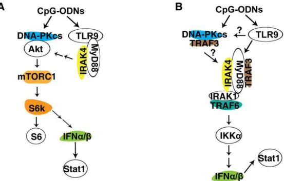
Fig 7. Models of the involvement of DNA-PKcs and TLR9 in the IFNα/βpathway. (A) DNA-PKcs and TLR9 are involved in activation of Akt and mTORC1 by CpG-ODN.In response to CpG-ODN, DNA-PKcs associates with Akt, leading to its phosphorylation and activation of downstream event mTORC1, which phosphorylates S6K, which in turn phosphorylates S6. S6K may involve expression of IFNα/βinduced by CpG-ODN. Also, CpG-ODN binds and activates TLR9, which in turn recruits MyD88 and IRAK4 leading to activation of Akt and mTORC1.(B) DNA-PKcs and TLR9 are important for the IFNα/βresponse to CpG-ODN. In response to CpG-ODN, DNA-PKcs associates with TRAF3 and activates IRAK1, leading to activation of IKKαand subsequent secretion of IFNα/β, which activate Stat1. Also, in response to CpG-ODN, TLR9 recruits MyD88, which in turn recruits and activates TRAF3 and IRAK4. Activated IRAK4
phosphorylates IRAK1, which then recruits TRAF6 and phosphorylates IKKα, leading to expression of IFNα/β
and phosphorylation of Stat1.
Because DNA-PKcs is also involved in activation of the Akt/mTORC1 pathway and the type I IFN response to CpG-ODN, our data suggest that the involvement of DNA-PKcs in the IL-6 and IL-12 responses to CpG-ODN is partially due to its contribution to the type I IFN response and the activation of the Akt/mTORC1 pathway.
We previously reported that CpG-ODN-biotin was able to precipitate both DNA-PKcs and TLR9[15]. Here, we showed that CpG-ODN-biotin was able to pull down DNA-PKcs and Ku70 (Fig. 5A). However, loss of TLR9 reduced the precipitated amount of DNA-PKcs by CpG-ODN, but this deficiency seemed to enhance Ku70 pull-down (Fig. 5A). Three possible situations may lead to this phenomenon. First, because DNA-PKcs can form a complex with Ku70[10], it is very possible that Ku70 competes with DNA-PKcs for binding to CpG-ODN in the absence of TLR9. The second possibility is that TLR9 assists in DNA-PKcs’s binding to CpG-ODN or its association with Ku70. The third possibility is that DNA-PKcs is less stable or less expressed in TLR9-deficient BMDCs. Because in WT BMDCs CpG-ODN efficiently pulled down both DNA-PKcs and Ku70, it is less likely that the absence of TLR9 will strikingly de-crease DNA-PKcs-CpG-ODN binding. Also, DNA-PKcs is a cytoplasmic protein in mouse cells[8,15] and its distribution is not limited to the endosome or lysosome compartments. It is less likely that TLR9 will stimulate DNA-PKcs’s binding to CpG-ODN or its interaction with Ku70. Thus, we favor the third possibility that TLR9 may control DNA-PKcs expression or sta-bility. Indeed, we found that loss of TLR9 clearly reduced DNA-PKcs protein level (Fig. 5B and 5C). The reduction of DNA-PKcs in TLR9-deficient BMDCs always occurred, but the degree of the reduction varied reflecting the nature of primary BMDC culture with growth factors or a plausible alteration of activities of protease inhibitors provided to cells when they were lysed each time. Nevertheless, the molecular mechanism of how TLR9 deficiency leads to a low level of DNA-PKcs in BMDCs remains elusive.
TRAF3 is critical for the type I IFN response to TLR ligands including CpG-ODN[3,4]. TRAF3 associates with MyD88 and TRIF, and loss of TRAF3 severely impairs production of type I IFN in response to CpG-ODN, poly(I:C), R848 (TLR7/8) and lipopolysaccharide [4]. Our data showed that the association of DNA-PKcs with TRAF3 was clearly induced by CpG-ODN in WT but not in TLR9-deficient BMDCs (Fig. 6A and 6B). The later could be due to reduced expression of DNA-PKcs in TLR-deficient cells. However, it is also possible that TLR9 is indeed required for the interaction of DNA-PKcs with TRAF3 in response to CpG-ODNs. Nonetheless, this finding further supports our conclusion that DNA-PKcs is in-volved in the type I IFN response to CpG-ODNs.
In summary, DNA-PKcs and TLR9 are important for the type I IFN response to
CpG-ODNs. Both DNA-PKcs and TLR9 contribute to the IFNαresponse to CpG-A, whereas TLR9 is essential for the IFNβresponse to CpG-ODNs. These IFN responses involve both the MyD88/IRAK4/IRAK1/IKKαand the Akt/mTORC1/S6K pathways (Fig. 7A and 7B). There-fore, DNA-PKcs regulates the type IFN response to CpG-ODNs in TLR9-dependent or independent manners.
Acknowledgments
We are grateful to Drs. G-C Li, S. Akira and W. Yeh for providing DNA-PKcs-/-and Ku70-/-, MyD88-/-, TLR9-/-, and IRAK4-/-mice, respectively.
Author Contributions
References
1. Wang H, Rayburn E, Zhang R. Synthetic oligodeoxynucleotides containing deoxycytidyl-deoxyguano-sine dinucleotides (CpG ODNs) and modified analogs as novel anticancer therapeutics. Current phar-maceutical design. 2005; 11(22):2889–907. PMID:16101444
2. Kumar H, Kawai T, Akira S. Pathogen recognition by the innate immune system. International reviews of immunology. 2011; 30(1):16–34. doi:10.3109/08830185.2010.529976PMID:21235323
3. Kawai T, Akira S. Toll-like receptor and RIG-I-like receptor signaling. Annals of the New York Academy of Sciences. 2008; 1143:1–20. doi:10.1196/annals.1443.020PMID:19076341
4. Hacker H, Redecke V, Blagoev B, Kratchmarova I, Hsu LC, Wang GG, et al. Specificity in Toll-like re-ceptor signalling through distinct effector functions of TRAF3 and TRAF6. Nature. 2006; 439 (7073):204–7. PMID:16306937
5. Cao W, Manicassamy S, Tang H, Kasturi SP, Pirani A, Murthy N, et al. Toll-like receptor-mediated in-duction of type I interferon in plasmacytoid dendritic cells requires the rapamycin-sensitive PI(3)K-mTOR-p70S6K pathway. Nature immunology. 2008; 9(10):1157–64. doi:10.1038/ni.1645PMID:
18758466
6. Wang Z, Dela Cruz R, Ji F, Guo S, Zhang J, Wang Y, et al. G(i)alpha proteins exhibit functional differ-ences in the activation of ERK1/2, Akt and mTORC1 by growth factors in normal and breast cancer cells. Cell communication and signaling: CCS. 2014; 12:10. doi:10.1186/1478-811X-12-10PMID:
24521094
7. Sanjuan MA, Rao N, Lai KT, Gu Y, Sun S, Fuchs A, et al. CpG-induced tyrosine phosphorylation occurs via a TLR9-independent mechanism and is required for cytokine secretion. The Journal of cell biology. 2006; 172(7):1057–68. PMID:16567503
8. Dragoi AM, Fu X, Ivanov S, Zhang P, Sheng L, Wu D, et al. DNA-PKcs, but not TLR9, is required for ac-tivation of Akt by CpG-DNA. The EMBO journal. 2005; 24(4):779–89. PMID:15678105
9. Sester DP, Brion K, Trieu A, Goodridge HS, Roberts TL, Dunn J, et al. CpG DNA activates survival in murine macrophages through TLR9 and the phosphatidylinositol 3-kinase-Akt pathway. J Immunol. 2006; 177(7):4473–80. PMID:16982883
10. Smith GC, Divecha N, Lakin ND, Jackson SP. DNA-dependent protein kinase and related proteins. Bio-chemical Society symposium. 1999; 64:91–104. PMID:10207623
11. Jafri F, Hardin JA, Dynan WS. A method to detect particle-specific antibodies against Ku and the DNA-dependent protein kinase catalytic subunit in autoimmune sera. Journal of immunological methods. 2001; 251(1–2):53–61. PMID:11292491
12. Liu Z, Davidson A. IFNalpha Inducible Models of Murine SLE. Frontiers in immunology. 2013; 4:306. doi:10.3389/fimmu.2013.00306PMID:24106491
13. Katakura K, Lee J, Rachmilewitz D, Li G, Eckmann L, Raz E. Toll-like receptor 9-induced type I IFN pro-tects mice from experimental colitis. The Journal of clinical investigation. 2005; 115(3):695–702. PMID:
15765149
14. Ferguson BJ, Mansur DS, Peters NE, Ren H, Smith GL. DNA-PK is a DNA sensor for IRF-3-dependent innate immunity. eLife. 2012; 1:e00047. doi:10.7554/eLife.00047PMID:23251783
15. Ma C, Muranyi M, Chu CH, Zhang J, Chu WM. Involvement of DNA-PKcs in the IL-6 and IL-12 response to CpG-ODN is mediated by its interaction with TRAF6 in dendritic cells. PloS one. 2013; 8(3):e58072. doi:10.1371/journal.pone.0058072PMID:23533581
16. Lye E, Mirtsos C, Suzuki N, Suzuki S, Yeh WC. The role of interleukin 1 receptor-associated kinase-4 (IRAK-4) kinase activity in IRAK-4-mediated signaling. The Journal of biological chemistry. 2004; 279 (39):40653–8. PMID:15292196
17. Adachi O, Kawai T, Takeda K, Matsumoto M, Tsutsui H, Sakagami M, et al. Targeted disruption of the MyD88 gene results in loss of IL-1- and IL-18-mediated function. Immunity. 1998; 9(1):143–50. PMID:
9697844
18. Park B, Brinkmann MM, Spooner E, Lee CC, Kim YM, Ploegh HL. Proteolytic cleavage in an endolyso-somal compartment is required for activation of Toll-like receptor 9. Nature immunology. 2008; 9 (12):1407–14. doi:10.1038/ni.1669PMID:18931679
19. Yasuda K, Richez C, Maciaszek JW, Agrawal N, Akira S, Marshak-Rothstein A, et al. Murine dendritic cell type I IFN production induced by human IgG-RNA immune complexes is IFN regulatory factor (IRF) 5 and IRF7 dependent and is required for IL-6 production. J Immunol. 2007; 178(11):6876–85. PMID:
17513736
21. Saitoh T, Satoh T, Yamamoto N, Uematsu S, Takeuchi O, Kawai T, et al. Antiviral protein Viperin pro-motes Toll-like receptor 7- and Toll-like receptor 9-mediated type I interferon production in plasmacytoid dendritic cells. Immunity. 2011; 34(3):352–63. doi:10.1016/j.immuni.2011.03.010PMID:21435586
22. Yotsumoto S, Saegusa K, Aramaki Y. Endosomal translocation of CpG-oligodeoxynucleotides inhibits DNA-PKcs-dependent IL-10 production in macrophages. J Immunol. 2008; 180(2):809–16. PMID:
18178819
23. Honda K, Ohba Y, Yanai H, Negishi H, Mizutani T, Takaoka A, et al. Spatiotemporal regulation of MyD88-IRF-7 signalling for robust type-I interferon induction. Nature. 2005; 434(7036):1035–40. PMID:
15815647