121 121 121 121 121 Mem Inst Oswaldo Cruz, Rio de Janeiro, Vol. 99(Suppl. I): 121-126, 2004
Clinical and Immunological Consequences of H uman T Cell
Leukemia Virus Type-I and
Schistosoma mansoni
Co-infection
Silvane Braga Santos/* , Aurélia Fonseca Porto, André Luiz M uniz, Amélia Ribeiro de Jesus, Edgar M Carvalho/* * /+
Serviço de Imunologia, Hospital Universitário Professor Edgard Santos, Universidade Federal da Bahia, Rua João das Botas s/n, 40110-160 Salvador, BA, Brasil *Departamento de Ciências Biológicas, Universidade Estadual de Feira de Santana, Feira de
Santana, BA, Brasil **Instituto de Investigação em Imunologia, São Paulo, SP, Brasil
Human T cell leukemia virus type-I (HTLV-I) infection is associated with spontaneous T cell activation and uncontrolled lymphocyte proliferation. An exacerbated type-1 immune response with production of pro-inflamma-tory cytokines (interferon-γ and tumor necrosis factor-α) is significantly higher in patients with myelopathy asso-ciated to HTLV-I than in HTLV-I asymptomatic carriers. In contrast with HTLV-I, a chronic Schistosoma mansoni
infection is associated with a type-2 immune response with high levels of interleukin (IL-4, IL-5, and IL-10) and low levels of IFN-γ. In this study, clinical and immunological consequences of the HTLV-I and S. mansoni infection were evaluated. The immune response in patients with schistosomiasis co-infected with HTLV-I showed low levels of IL-5 (p < 0.05) in peripheral blood mononuclear cells cultures stimulated with S. mansoni antigen (SWAP) and de-creased SWAP-specific IgE levels when compared with patients with only schistosomiasis (p < 0.05). Liver fibrosis was mild in all HTLV-I co-infected patients. Immunological response was also compared in individuals who had only HTLV-I infection with those who were co-infected with HTLV-I and helminths (S. mansoni and Strongyloides stercoralis). In patients HTLV-I positive co-infected with helminths the IFN-γ levels were lower than in individuals who had only HTLV-I. Moreover, there were fewer cells expressing IFN-γ and more cells expressing IL-10 in individu-als co-infected with HTLV-I and helminths. These dates indicate that HTLV-I infection decrease type 2-response and IgE synthesis and are inversely associated with the development of liver fibrosis. Moreover, helminths may protect HTLV-I infected patients to produce large quantities of pro-inflammatory cytokines such as IFN-γ.
Key words: human T cell leukemia virus type-1 - Schistoma mansoni - co-infection
The human T cell leukemia virus type-I (HTLV-I) is an oncogenic exogenous retrovirus that infects between 10 and 20 million people worldwide (Edlich et al. 2000). HTLV-I is the recognized cause of adult T-cell leukemia (ATL) as well as HTLV-I-associated myelopathy/tropical spastic paraparesis (HAM/TSP) (Osame et al. 1986, Uchiyama 1997), but other disorders have been associated with HTLV-I infection.The immunological response in HTLV-I infection is characterized by a spontaneous lymphopro-liferation and an exaggerated T cell response with high production of important inflammatory mediators of tissue damage as interferon-γ (IFN-γ), tumor necrosis factor-α (TNF-α) and interleukin (IL-6) (Nishimoto et al. 1990, Kubota et al. 1998, Carvalho et al. 2001). Although the pathogenesis of neurological disease associated to HTLV-I is not completely understood, there are various evidences that immunological response participate and is respon-sible by inducing tissue damage (Hanon et al. 2000, Nagai & Jacobson 2001, Osame 2002). By the other hand, helminthes infections such as strongyloidiasis and in
Financial support: Brazilian Research Council, Fundação de Amparo à Pesquisa do Estado da Bahia
+Corresponding author and CNPq Senior Investigator. Fax:
+55-71-245.7110. E-mail: [email protected]. Received 28 May 2004
Accepted 26 July 2004
particular a chronicdisease caused by infection with Schis-tosoma mansoni are associated with a predominant anti-inflammatory type-2 immune response with increased lev-els of IL-4, IL-5 and IL-10 and low levlev-els of IFN-γ (Araujo et al. 1996, Finkelman et al. 1997). The high degree of in-fection and the host’s immune reaction to parasite eggs contribute to granuloma formation. Liver fibrosis is the most important pathological finding in schistosomiasis, being registered in about 5% of chronically S. mansoni
infected patients (Bina & Prata 2003). Although initial ex-perimental studies suggested that type-1 cytokines were associated with granulomatous reaction to S. mansoni
im-122 122 122 122
122 H TLV-1 and Schistosom iasis • Silvane Braga Santos et al.
mune response observed in patients with schistosomia-sis and the impact of HTLV-I on the development of liver fibrosis. Moreover, considering that pathogenesis of dis-eases associated to HTLV-I is dependent of high produc-tion of pro-inflammatory cytokines it was evaluated if co-infection with S. mansoni and HTLV-I decrease the type-1 immune response
MATERIALS AND METHODS
Patients’ selection - Patients were selected from the HTLV-I clinic of the Hospital Universitário Professor Edgard Santos, Federal University of Bahia, Brazil.The clinic follows more than 500 HTLV-I infected individuals, most of then referred from two blood banks in Salvador, the capital of the state of Bahia.The diagnosis of HTLV-I infection was confirmed by Western blot (HTLV blot 2.4, Genelabs, Singapore). All patients admitted in the HTLV-I clinic are asked to perform three stool examinations. From 500 HTLV-I infected individuals, 309 had stool examina-tion. Frequency of S. mansoni infection was assayed in 309 HTLV-I positive individuals and 331 seronegative blood donors were screened as negative controls also by stool examination;14 HTLV-I positive patients had S. stercoralis larvae in their stool examination and all of them participated of the study. S. mansoni eggs were found in 26 HTLV-I infected individuals, but only 22 of them ac-cepted to participate in the study. The diagnosis of schis-tosomiasis was made by a positive fecal examination for eggs by Hoffman technique and the criterion for a diag-nosis of strongyloidiasis was a positive fecal examination for larvae by the Baermann concentration technique. Forty-four (n = 44) patients with S.mansoni without HTLV-I infection were selected from an existing cohort of patients from an area endemic for S. mansoni infection (Caatinga do Moura, Bahia). These patients were used to select a ratio of 2 to 1 by matching age and sex with the group of patients with co-infection. Clinical history and a complete physical examination and abdominal ultrasound were per-formed in both groups with S. mansoni infection (HTLV- I positive and negative).
Neurological exam - Motor dysfunction was deter-mined by Osame’s Motor Disability Score (OMDS) (Izumo et al. 1996) and Expanded Disability Status Scale (EDSS) (Kurtzke 1983). Patients with HAM/TSP had a marked neurological impairment with EDSS ≥ 3 and OMDS ≥ 1 and all asymptomatic subjects had OMDS and EDSS of zero. Based on exclusion criteria that included the use of antiviral drugs or immunomodulators in the previous 90 days, co-infection with HIV, HCV or hepatitis B and pres-ence of helminthes infection or other neurological dis-eases, 17 patients with HAM/TSP performed immunologi-cal evaluation. Thirty-six HTLV-I asymptomatic carriers, without clinical manifestations associated with HTLV-I were also selected. Healthy University Hospital employ-ees who were seronegative for HTLV-I and seronegative normal donors without helminthes infection were used as negative controls.
The Ethical Committee of the Hospital Universitário Professor Edgard Santos approved this study and in-formed consent was obtained from all prospectively en-rolled patients.
Cell preparation and cytokine determination - Pe-ripheral blood mononuclear cells (PBMC) were obtained by density gradient centrifugation using lymphocyte sepa-ration media (LSM; Organon Teknika Coorposepa-ration, Durham, NS, US). PBMC were cultivated in RPMI 1640 (Gibco, Grand Island, NY, US) plus 10% heat inactivated human AB Rh+ serum (Sigma Chemical Co., St. Louis, MO), antibiotics and glutamine (complete media) and adjusted to 3 x 10⁶ cells/ml in complete media. The cells were cul-tured unstimulated or stimulated with 2 mg/mlsoluble adult
S. mansoni worm antigen (SWAP) when patients were co-infected with S. mansoni.All cultures were incubated at 37°C in 5 % CO2 atmospherefor 72 h until supernatant fluids were collected. IFN-γ and IL-5 levels were mea-sured by sandwich ELISA technique (R&D system, Min-neapolis, MN) and the results were expressed as pg/ml using a standard curve generated using recombinant cytokines.
Flow cytometric analysis (FACS) - FACS was per-formed after 20 h of incubation. Briefly, the PBMC were immunophenotyped by double immunofluorescence us-ing a FACScalibor flow cytometer and a panel of phyco-erythrin (PE)-conjugated monoclonal antibodies. In all cases the cells were double stained for cytokine and for cell surface markers. Specifically the total percentage of cells producing IFN-γ and IL-10 was assayed. In all cases, 30,000 gates events were acquired for later analysis due to the low frequency of positive events being analyzed.
IgE specific to S. mansoni antigen - Analysis of IgE specific to SWAP was performed by ELISA as previously described (Souza-Atta et al. 1999). The ELISA was devel-oped with 100 µl of p-nitrophenyl phosphate and the ab-sorbance changes (optical density, OD) were measured by a spectrophotometer at 405 nm. The cut-offs of the immunoassay were determined using the mean plus 3 SD of the absorbance obtained with serum from 15 healthy individuals.
Ultrasonography - Ultrasonography examination was performed with the Quantum 2000 Siemens ultrasound with a convex transductor of 3.5 Mhz, according to a previ-ously published technique (Abdel-Wahab et al. 1992). Grading of hepatic fibrosis was determined according with WHO criteria established in 1993 and previously revali-dated (de Jesus et al. 2000). Patients were classified in four different degrees according to the mean thickness of four portal tracts after the first division from the right and left branches of portal vein.
Statistical analysis - A non-parametric Mann-Whitney U Test was used to evaluate differences among the groups. Fisher’s exact test was used to compare proportions. These statistical analyses were performed using the pro-gram Instat for Windows. An alpha (α) of 5% was consid-ered significant.
RESULTS
123 123 123 123 123 Mem Inst Oswaldo Cruz, Rio de Janeiro, Vol. 99(Suppl. I), 2004
levels in 17 myelopathy patients (4,246 ± 2,924pg/ml, range: 375 to 10,750), was higher than that observed in 36 asymptomatic carriers (1,362 ± 1,408 pg/ml range: 15 to 6,995)or in 15 negative controls (1 ± 4 pg/ml), p = 0.0001, Mann-Whitney U test. Evaluation of the frequency of helminthes infection in 309 HTLV-I infected subjects re-vealed that S. mansoni infection was 4.6 fold higher in HTLV-I infected individuals (26/309 – 8.4%) than a com-parable group of HTLV-I seronegative individuals (6/331 – 1.8%, p = 0.0003, Fisher’s exact test; data not shown). With the aim to evaluate whether HTLV-I infection modify the immune response in patients with S. mansoni, the lev-els of IL-5 in co-infected patients were measured and com-pared with IL-5 levels found in controls patients (schisto-somiasis without HTLV-I infection) from an endemic area of S. mansoni infection. The mean ± SD of IL-5 levels in patients co-infected with S. mansoni and HTLV-1 was 258 ± 692 pg/ml with variation of 0 – 2943 pg/ml. This value was lower than (p < 0.05, Mann-Whitney U test) that ob-served in patients only infected with S. mansoni (907 ± 1289 pg/ml with ranging of 0 – 4747 pg/ml (Fig. 2).
To evaluate the role of HTLV-I infection on antigen-specific IgE levels in patients with schistosomiasis, the distribution of the IgE, expressed in OD, in patients with schistosomiasis without HTLV-I infection and in those co-infected with HTLV-I was assayed (Fig. 3). The mean IgE in 40 patients without HTLV-I infection was 0.195 + 0.169 compared to 0.123 + 0.04 in 22 patients with schisto-somiasis associated with HTLV-I infection (p < 0.01, Mann Whitney U test).
Fig. 1: interferon-γ (IFN-γ) levels (pg/ml) in HTLV-I-associated myelopathy/tropical spastic paraparesis (HAM/TSP) (n = 17) pa-tients compared with asymptomatic carries (n = 36) and negative controls (n=15). The bars represent the median of IFN-γ concen-trations and the difference were considered significant when p < 0.05 (Mann-Whitney U Test).
Fig. 2: HTLV-I down regulates IL-5 production to Schistosoma mansoni antigen. IL-5 levels in peripheral blood mononuclear cells stimulated with SWAP in S. mansoni and HTLV-1 co-infected pa-tients (n = 22) and in chronic schistosomiasis papa-tients (n = 44) from an endemic area.
Fig. 3: SWAP-specific IgE from patients with Schistosoma mansoni infection (n = 40) and S. mansoni co-infected with HTLV-I (n = 22).
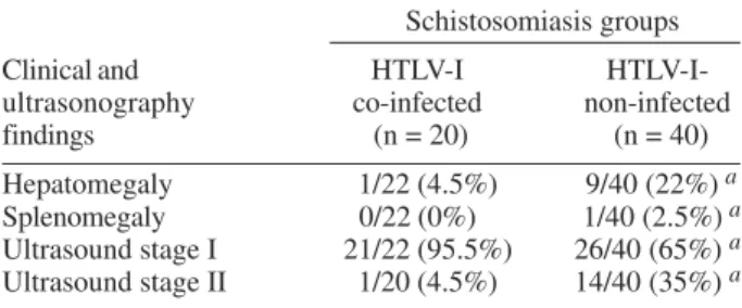
Ultrasonography studies were used to further quan-tify the observed clinical findings. The ultrasound find-ings of 22 schistosomiasis patients co-infected with HTLV-I and 40 schistosomiasis HTLV-HTLV-I-seronegative controls are shown in the Table. None of the 22 co-infected pa-tients had splenomegaly (0/22), an index of severe hepatic fibrosis. Only one case had mild hepatomegaly (1/22). In contrast, age and sex-matched control schistosomiasis HTLV-1-seronegative cases showed a significantly higher frequency of clinical parameters for hepatic fibrosis. Sple-nomegaly was observed in 2.5% (1/40) and hepatomegaly was registered in 22% (9/40) of the HTLV-I-seronegative schistosomiasis controls from Caatinga do Moura. An absence of or a mild degree of fibrosis was noted in 21 schistosomiasis patients co-infected with HTLV-I stud-ied by ultrasound. In contrast, 35% of the HTLV-I-serone-gative schistosomiasis control group had degree II, evi-dence of liver fibrosis that was significant different (p < 0.05, Fisher’s exact test) between the two groups.
124 124 124 124
124 H TLV-1 and Schistosom iasis • Silvane Braga Santos et al.
helminthic infection (893 ± 1,174 pg/ml, p < 0.05, Mann-Whitney U test). Moreover there were less cells express-ing IFN-γ and more cells expressing IL-10 in HTLV-I pa-tients co-infected with helminths than in individuals only infected with HTLV-I. Fig. 5 shows the frequency of CD8+ T cells secreting IFN-γ or IL-10 and the total frequency of cells secreting IL-10 in unstimulated cultures of 4 HTLV-I carriers co-infected with helminths and 7 HTLV-I carriers without co-infection. Co-infection of HTLV-I with helminthes significantly decreases the frequency of CD8 T cells secreting IFN-γ (p < 0.05). In contrast, the total frequency of cells secreting IL-10 and the frequency of CD8+ T cells secreting IL-10 was higher in HTLV-I indi-viduals co-infected with helminthes (0.58%) in compari-son with these only infected with HTLV-I (0.21%), p < 0.05.
stercoralis and/or S. mansoni) can down modulate the exaggerated inflammatory response observed in HTLV-I infected patients by reducing spontaneous IFN-γ syn-thesis.
The pathogenesis of HAM/TSP is not completely understood. Increased proviral load and the expansion of HTLV-I tax-specific CD8+ cytotoxic T lymphocytes, both in cerebrospinal fluid and in peripheral blood, have been associated with the central nervous system involvement in patients with HAM/TSP (Hanon et al. 2000, Kubota et al. 2000, Nagai & Jacobson 2001). Moreover, many stud-ies have demonstrated that pro-inflammatory cytokines as IFN-γ, TNF-α, and IL-15 contribute to tissue damage of the central nervous system of HAM/TSP patients (Umehara et al. 1994, Biddison et al. 1997, Azimi et al. 2000). Additionally, occurrence of fibrosis in neurological tis-sue was also associated with the immunopathogenesis of the neurological disease associated to HTLV-I (Nagai & Jacobson 2001). Although the lymphocyte response was quite variable in some HTLV-I asymptomatic carriers, our date indicates a higher and significant IFN-γ production in HAM/TSP patients as compared to asymptomatic car-riers. Although both type-1 and type-2 cytokines are found to increase in unstimulated lymphocyte cultures of HTLV-I infected individual when compared with controls (Carvalho et al. 2001), this finding confirm an exacerbated type-1 immune response in these HTLV-I infected sub-jects
Schistosomiasis is one the most important helminthic disease found in Northeast region of Brazil. Although the majority of patients infected with S. mansoni have an in-testinal or hepatoinin-testinal form, liver fibrosis is observed in 5% of patients with long-standing chronic S. mansoni
infection. Schistosomiasis is a well-characterized Th2 re-sponse-dominated disease (Grzych et al. 1991). Immuno-logical response in chronic schistosomiasis patients is characterized by decreased IFN-γ production and en-hancement in IL-4, IL-5, and IL-10 levels. This predomi-nant type-2 immune response is independent of the de-gree of infection measured by egg/stool gram and occurs in all clinical form of schistosomiasis (de Jesus et al. 1993, Araujo et al. 1996).
TABLE
Inverse association between human T cell leukemia virus-I (HTLV-I) infection and liver fibrosis in patients with
schistosomiasis
Schistosomiasis groups
Clinical and HTLV-I
HTLV-I-ultrasonography co-infected non-infected findings (n = 20) (n = 40)
Hepatomegaly 1/22 (4.5%) 9/40 (22%) a
Splenomegaly 0/22 (0%) 1/40 (2.5%) a
Ultrasound stage I 21/22 (95.5%) 26/40 (65%) a
Ultrasound stage II 1/20 (4.5%) 14/40 (35%) a
a: p < 0.05 (Fisher’s exact test)
Fig. 4: IFN-γ levels of HTLV-I infected patients (n = 35) co-in-fected or not (n = 35) by helminthes (Strongyloides stercoralis and Schistosoma mansoni).
Fig. 5: frequency of CD8+T cell secreting IFN-γ or IL-10 in HTLV-I carriers co-infected with helmints (n = 4) and HTLV-HTLV-I carriers without co-infection with helmints (n = 7).
DISCUSSION
125 125 125 125 125 Mem Inst Oswaldo Cruz, Rio de Janeiro, Vol. 99(Suppl. I), 2004
High frequency of strongyloidiasis is registered in ar-eas where both HTLV-I and S. stercoralis infection are endemic (Nakada et al. 1984). Additionally, our finding showed that the frequency of S. mansoni was increased in HTLV-I infected patients when compared with HTLV-I seronegative controls. Considering that no previous study evaluated the impact of HTLV-I infection on S. mansoni
infection, we evaluated the changes in the immune re-sponse to S. mansoni antigen in co-infected patients. We observed that IL-5, a typical type-2 cytokine secreted in schistosomiasis, was down regulated in S. mansoni pa-tients co-infected with HTLV-I. In addition, we showed that HTLV-I co-infection decrease the levels of SWAP-specific IgE in S. mansoni infected patients. We have pre-viously shown that HTLV-I decreases antigen specific type-2 immune response in patients with S. stercoralis
infection (Neva et al. 1998, Porto et al. 2001a). Since there is a tendency for an inverse correlation between IFN-γ and IL-5 levels, it is possible that the down regulation of IL-5 and specific-IgE levels is related to the enhancement of type-1 pro-inflammatory cytokines synthesized during HTLV-I infection.
The most severe form of S. mansoni infection is the hepatosplenic form. Patients who develop this chronic form show extensive liver fibrosis and hepatosplenom-egaly. It is the parasite eggs that, by accumulating in the liver, leading the granuloma formation. Several factors have been associated to liver fibrosis including the ge-netic background, the degree of infestation and host im-munological response. Initially, experimental studies sug-gested that type-1 cytokines were associated with granu-loma formation (Leptak & McKerrow 1997, Rezende et al. 1997). However, more recent date point to the importance of type-2 cytokines such as IL-4 and IL-13 in inducing fibrosis and the ability of IL-12 and IFN-γ to decrease it (Wynn et al. 1994, Chiaramonte et al. 1999b, Jankovic et al. 1999).
HTLV-I infection modifies the immune response to S. mansoni antigen. It is possible that the HTLV-I co-infec-tion also modify the clinical manifestaco-infec-tion of schistoso-miasis in these patients. When ultrasound of S. mansoni
and HTLV-I co-infected patients were compared with a group of patients having only schistosomiasis but with similar degree of infection than those dually co-infected, the co-infected patients had significant lower liver fibro-sis. In such case it is possible that the high type-1 im-mune response observed by spontaneous IFN-γ produc-tion in HTLV-I infecproduc-tion or the decrease of type-2 cytokines are able to preventing the development of fibrosis in co-infected patients.
By the other hand, the evaluation of the role of helminthes infections (S. stercoralis and/or S. mansoni)
on the immune response of HTLV-I infected subjects with-out symptoms showed that co-infection HTLV-I and helminthes had immunological implications. The documen-tation that IFN-γ levels decreased in HTLV-I carriers co-infected with helminthes indicates that helmintic infec-tion may down regulate IFN-γ production. It is probably that helminthes down regulate IFN-γ production by in-ducing secretion of IL-10. This date is consistent with the date that show that exogenous IL-10 can decrease IFN-γ
production in lymphocytes cultures of HTLV-I carriers (Carvalho et al. 2001). Together, these observations sug-gest that HTLV-I alter the clinical and immunological find-ings of HTLV-I and S. mansoni co-infection and those HTLV-I individuals co-infected with S. stercoralis and/or
S. mansoni may decrease IFN-γ production and protect I carriers to develop diseases associated to HTLV-I as HAM/TSP.
ACKNOWLEDGMENT
To Serviço de Transfusão de Sangue, Salvador, BA and to Hemocentro da Bahia by guiding the blood donors with HTLV positive serology. To Elbe Myrtes Souza Silva for her technical assistance in preparing this manuscript.
REFERENCES
Abdel-Wahab M F, Esmat G, Farrag A, el-Boraey Y A, Strickland G T 1992. Grading of hepatic schistosomiasis by the use of ultrasonography. Am J Trop Med Hyg46: 403-408. Araujo MI, de Jesus AR, Bacellar O, Sabin E, Pearce E, Carvalho
EM 1996. Evidence of a T helper type 2 activation in hu-man schistosomiasis. Eur J Immunol26: 1399-1403. Azimi N, Mariner J, Jacobson S, Waldmann TA 2000. How
does interleukin 15 contribute to the pathogenesis of HTLV type 1-associated myelopathy/tropical spastic parapare-sis? AIDS Res Hum Retroviruses16: 1717-1722.
Biddison WE, Kubota R, Kawanishi T, Taub DD, Cruikshank WW, Center DM, Connor EW, Utz U, Jacobson S 1997. Human T cell leukemia virus type I (HTLV-I)-specific CD8+ CTL clones from patients with HTLV-I-associated neuro-logic disease secrete proinflammatory cytokines, chemokines, and matrix metalloproteinase. J Immunol159: 2018-2025.
Bina JC, Prata A 2003. Shistosomiasis in hyperendemic area of Taquarendi: I - Schistosoma mansoni infection and severe clinical forms. Rev Soc Bras Med Trop36: 211-216. Carvalho EM, Bacellar O, Porto AF, Braga S, Galvão-Castro B,
Neva F 2001. Cytokine profile and immunomodulation in asymptomatic human T-lymphotropic virus type 1-infected blood donors. J Acquir Immune Defic Syndr27: 1-6. Chiaramonte MG, Dnaldson DD, Cheever AW, Wynn TA 1999a.
An IL-13 inhibitor blocks the development of hepatic fi-brosis during a T-helper type 2-dominated inflammatory response. J Clin Invest104: 777-785.
Chiaramonte MG, Schopf LR, Neben TY, Cheever AW, Donaldson DD, Wynn TA 1999b. IL-13 is a key regulatory cytokine for Th2 cell-mediated pulmonary granuloma for-mation and IgE responses induced by Schistosoma mansoni eggs. J Immunol 162: 920-930.
de Jesus AM, Almeida RP, Bacellar O, Araujo MI, Demeure C, Bina JC, Dessein AJ, Carvalho EM 1993. Correlation be-tween cell-mediated immunity and degree of infection in subjects living in an endemic area of schistosomiasis. Eur J Immunol23: 152-158.
de Jesus AR, Miranda DG, Miranda RG, Araujo I, Magalhaes A, Bacellar M, Carvalho EM 2000. Morbidity associated with Schistosoma mansoni infection determined by ultra-sound in an endemic area of Brazil, Caatinga do Moura. Am J Trop Med Hyg63: 1-4.
Edlich RF, Arnette JA,Williams FM 2000. Global epidemic of human T-cell lymphotropic virus type-I (HTLV-I). J Emerg Med18: 109-119.
126 126 126 126
126 H TLV-1 and Schistosom iasis • Silvane Braga Santos et al.
Rev Immunol15: 505-533.
Grzych JM, Pearce E, Cheever A, Caulada ZA, Caspar P, Heiny S, Lewis F, Sher A 1991. Egg deposition is the major stimu-lus for the production of Th2 cytokines in murine schisto-somiasis mansoni. J Immunol 146: 1322-1327.
Hanon E, Hall S, Taylor GP, Saito M, Davis R, Tanaka Y, Usuku K, Osame M, Weber JN, Bangham CR 2000. Abundant tax protein expression in CD4+ T cells infected with human T-cell lymphotropic virus type I (HTLV-I) is prevented by cytotoxic T lymphocytes. Blood95: 1386-1392.
Hayashi J, Kishihara Y, Yoshimura E, Furusyo N, Yamaji K, Kawakami Y, Murakami H, Kashiwagi S 1997. Correlation between human T cell lymphotropic virus type-1 and Strongyloides stercoralis infections and serum immunoglo-bulin E responses in residents of Okinawa, Japan. Am J Trop Med Hyg56: 71-75.
Izumo S, Goto I, Itoyama Y, Okajima T, Watanabe S, Kuroda Y, Araki S, Mori M, Nagataki S, Matsukura S, Akamine T, Nakagawa M, Yamamoto I, Osame M 1996. Interferon-alpha is effective in HTLV-I-associated myelopathy: a multicenter, randomized, double-blind, controlled trial. Neurology46: 1016-1021.
Jankovic D, Kullberg MC, Noben-Trauth N, Caspar P, Ward J M, Cheever AW, Paul WE, Sher A 1999. Schistosome-in-fected 4 receptor knockout (KO) mice, in contrast to IL-4 KO mice, fail to develop granulomatous pathology while maintaining the same lymphokine expression profile. J Immunol163: 337-342.
Kubota R, Kawanishi T, Matsubara H, Manns A, Jacobson S 1998. Demonstration of human T lymphotropic virus type I (HTLV-I) tax-specific CD8+ lymphocytes directly in pe-ripheral blood of HTLV-I-associated myelopathy/tropical spastic paraparesis patients by intracellular cytokine de-tection. J Immunol161: 482-488.
Kubota R, Kawanishi T, Matsubara H, Manns A, Jacobson S 2000. HTLV-I specific IFN-gamma+ CD8+ lymphocytes correlate with the proviral load in peripheral blood of in-fected individuals. J Neuroimmunol102: 208-215. Kurtzke JF 1983. Rating neurologic impairment in multiple
sclerosis: an expanded disability status scale (EDSS). Neu-rology33: 1444-1452.
Leptak CL, McKerrow JH 1997. Schistosome egg granulomas and hepatic expression of TNF-alpha are dependent on immune priming during parasite maturation. J Immunol 158: 301-307.
Nagai M, Jacobson S 2001. Immunopathogenesis of human T cell lymphotropic virus type I-associated myelopathy. Curr Opin Neurol14: 381-386.
Nakada K, Kohakura M, Komoda H, Hinuma Y 1984. High incidence of HTLV antibody in carriers of Strongyloides stercoralis. Lancet1: 633.
Neva FA, Filho JO, Gam AA, Thompson R, Freitas V, Melo A, Carvalho E M 1998. Interferon-gamma and interleukin-4 responses in relation to serum IgE levels in persons
in-fected with human T lymphotropic virus type I and Strongy-loides stercoralis. J Infect Dis178: 1856-1859.
Newton RC, Limpuangthip P, Greenberg S, Gam A, Neva FA 1992. Strongyloides stercoralis hyperinfection in a carrier of HTLV-I virus with evidence of selective immunosup-pression. Am J Med92: 202-208.
Nishimoto N, Yoshizaki K, Eiraku N, Machigashira K, Tagoh H, Ogata A, Kuritani T, Osame M, Kishimoto T 1990. Elevated levels of interleukin-6 in serum and cerebrospinal fluid of HTLV-I-associated myelopathy/tropical spastic paraparesis. J Neurol Sci97: 183-193.
Osame M 2002. Pathological mechanisms of human T-cell lymphotropic virus type I-associated myelopathy (HAM/ TSP). J Neurovirol8: 359-364.
Osame M, Usuku K, Izumo S, Ijichi N, Amitani H, Igata A, Matsumoto M, Tara M 1986. HTLV-I associated myel-opathy, a new clinical entity. Lancet1: 1031-1032. Phelps KR, Ginsberg SS, Cunningham AW, Tschachler E, Dosik
H 1991. Case report: adult T-cell leukemia/lymphoma as-sociated with recurrent strongyloides hyperinfection. Am J Med Sci302: 224-228.
Porto AF, Neva FA, Bittencourt H, Lisboa W, Thompson R, Alcantara L, Carvalho EM 2001a. HTLV-1 decreases Th2 type of immune response in patients with strongyloidiasis. Parasite Immunol 23: 503-507.
Porto AF, Oliveira Filho J, Neva FA, Orge G, Alcantara L, Gam A, Carvalho EM 2001b. Influence of human T-cell lymphocytotropic virus type 1 infection on serologic and skin tests for strongyloidiasis. Am J Trop Med Hyg65: 610-613.
Rezende SA, Lambertucci JR, Goes AM 1997. Role of immune complexes from patients with different clinical forms of schistosomiasis in the modulation of in vitro granuloma research. Mem Inst Oswaldo Cruz92: 683-687.
Robinson RD, Lindo JF, Neva FA, Gam AA, Vogel P, Terry SI, Cooper ES 1994. Immunoepidemiologic studies of Strongy-loides stercoralis and human T lymphotropic virus type I infections in Jamaica. J Infect Dis169: 692-696.
Souza-Atta ML, Araujo MI, D’Oliveira Junior A, Ribeiro-de-Jesus A, Almeida RP, Atta AM, Carvalho EM 1999. Detec-tion of specific IgE antibodies in parasite diseases. Braz J Med Biol Res32: 1101-1105.
Uchiyama T 1997. Human T cell leukemia virus type I (HTLV-I) and human diseases. Annu Rev Immunol 15, 15-37. Umehara F, Izumo S, Ronquillo AT, Matsumuro K, Sato E,
Osame M 1994. Cytokine expression in the spinal cord lesions in HTLV-I-associated myelopathy. J Neuropathol Exp Neurol53: 72-77.