Hypothalamic-Pituitary-Ovarian Axis Leads to Ovarian
Epithelial Tumorigenesis in Mice
Mary J. Laws1, Athilakshmi Kannan1, Sandeep Pawar2, Wanda M. Haschek3, Milan K. Bagchi2*, Indrani C. Bagchi1*
1Department of Comparative Biosciences, University of Illinois Urbana/Champaign, Urbana, Illinois, United States of America,2Department of Molecular and Integrative Physiology, University of Illinois Urbana/Champaign, Urbana, Illinois, United States of America,3Department of Pathobiology, University of Illinois Urbana/Champaign, Urbana, Illinois, United States of America
Abstract
The etiology of ovarian epithelial cancer is poorly understood, mainly due to the lack of an appropriate experimental model for studying the onset and progression of this disease. We have created a mutant mouse model in which aberrant estrogen receptor alpha (ERa) signaling in the hypothalamic-pituitary-ovarian axis leads to ovarian epithelial tumorigenesis. In these mice, termed ERad/d, the ERa gene was conditionally deleted in the anterior pituitary, but remained intact in the hypothalamus and the ovary. The loss of negative-feedback regulation by estrogen (E) at the level of the pituitary led to increased production of luteinizing hormone (LH) by this tissue. Hyperstimulation of the ovarian cells by LH resulted in elevated steroidogenesis, producing high circulating levels of steroid hormones, including E. The ERad/dmice exhibited formation of palpable ovarian epithelial tumors starting at 5 months of age with 100% penetrance. By 15 months of age, 80% of ERad/dmice die. Besides proliferating epithelial cells, these tumors also contained an expanded population of luteinized stromal cells, which acquire the ability to express P450 aromatase and synthesize E locally. In response to the elevated levels of E, the ERasignaling was accentuated in the ovarian epithelial cells of ERad/dmice, triggering increased ERa-dependent gene expression, abnormal cell proliferation, and tumorigenesis. Consistent with these findings, treatment of ERad/dmice with letrozole, an aromatase inhibitor, markedly reduced circulating E and ovarian tumor volume. We have, therefore, developed a unique animal model, which serves as a useful tool for exploring the involvement of E-dependent signaling pathways in ovarian epithelial tumorigenesis.
Citation:Laws MJ, Kannan A, Pawar S, Haschek WM, Bagchi MK, et al. (2014) Dysregulated Estrogen Receptor Signaling in the Hypothalamic-Pituitary-Ovarian Axis Leads to Ovarian Epithelial Tumorigenesis in Mice. PLoS Genet 10(3): e1004230. doi:10.1371/journal.pgen.1004230
Editor:Marshall S. Horwitz, University of Washington, United States of America ReceivedSeptember 18, 2013;AcceptedJanuary 27, 2014;PublishedMarch 6, 2014
Copyright:ß2014 Laws et al. This is an open-access article distributed under the terms of the Creative Commons Attribution License, which permits
unrestricted use, distribution, and reproduction in any medium, provided the original author and source are credited.
Funding:MJL was supported by NIH T32ES007326. The funders had no role in study design, data collection and analysis, decision to publish, or preparation of the manuscript.
Competing Interests:The authors have declared that no competing interests exist. * E-mail: [email protected] (MKB); [email protected] (ICB)
Introduction
Ovarian cancer is the most lethal malignancy of the female reproductive system and the fifth leading cause of cancer-related death among women [1]. Approximately 90% of malignant ovarian tumors are derived from either the ovarian surface epithelium (OSE) or fallopian tube epithelium (FTE) [2]. Due to the absence of specific symptoms and the lack of strategies for early detection of ovarian cancer, the majority (70%) of women with this disease are diagnosed at a late stage when the cancer has spread beyond the confines of the ovary [1]. Despite its clinical significance, the etiology of ovarian cancer is poorly understood, mainly due to the lack of an appropriate experimental model for studying the onset and progression of this disease.
Multiple theories regarding the etiology of ovarian cancer have been proposed, but the precise molecular defects underlying the development of this disease remain elusive [3]. The ‘‘gonadotropin hypothesis’’ proposes that high gonadotropin levels can have a stimulatory effect on OSE cells, promoting their neoplastic transformation [4,5]. It was reported that the addition of
gonadotropins to rodents in which ovarian cancer was induced upon treatment with the chemical carcinogen, 7,12-dimethylben-z(a)anthracene (DMBA) led to increased lesion severity, suggesting that gonadotropins play a role in tumor progression [6]. In humans, epidemiologic evidence, indirectly supporting this hypothesis, includes the well-documented protective effects of oral contraceptives and multiparity, which suppress gonadotropin secretion by the pituitary gland [5,7]. The majority of women with epithelial ovarian cancer present the disease at a postmen-opausal stage where circulating follicle stimulating hormone (FSH) and lutenizing hormone (LH) levels are elevated, indicating a causal relationship between chronically elevated gonadotropin levels and ovarian cancer development [5,8].
[9–13]. While most of these studies comprise a small number of subjects and fail to control for all of the factors that may influence cancer risk, in patients with ovarian cancers, intratumoral production of E via in situ aromatization has been suggested to promote growth of breast, endometrial and ovarian cancer cells [14]. However, only few animal models have been used to investigate the role of E in ovarian tumorigenesis. Baiet alreported the effects of prolonged E exposure on the morphology of rabbit ovaries and found an increase in both OSE cell proliferation and the number of papillae covering the ovarian surface, but no ovarian tumors [15]. In a recent study, Laviotteet alconditionally activated an oncogene, SV40 TAg, in OSE cells and treated the mice with exogenous E [16]. These investigators reported that E treatment resulted in an earlier onset of ovarian tumors and a significantly decreased survival time [16]. While the results from this animal model underscore the importance of E in the progression of ovarian cancer, it is clear that new animal models independent of specifically directed single oncogenic mutations are needed for assessment of the role of E signaling in ovarian epithelial tumorigenesis.
In this study, we present a novel transgenic mouse model of ovarian tumorigenesis. In this model, termed ERad/d, the estrogen receptor alpha (ERa) gene is dysregulated in the hypothalamic-pituitary-ovarian axis. Conditional deletion of this gene in the anterior pituitary, but not in the hypothalamus and the ovary, led to elevated circulating LH. Hyperstimulation by LH resulted in luteinization of the ovarian stromal cells, expression of P450 aromatase in these cells, and increased E synthesis in the ovarian microenvironment. Our study suggests that E critically controls ovarian tumor growth, presumably by stimulating the proliferation of OSE cells to drive epithelial tumorigenesis. The ERad/dmouse, therefore, provides a useful model to study the mechanisms by which dysregulated E signaling promotes the initiation and progression of ovarian epithelial tumors.
Results
ERa conditional knockout mice (ERad/d) were generated by crossing progesterone receptor cre recombinase (PR-Cre) knock-in mice with ERa floxed (ERaf/f) mice [17,18]. By five months of age, the ERad/d mice developed palpable ovarian tumors with 100% penetrance. In contrast, the ERaf/f and the global ERa knockout mice did not develop any tumor (Fig. 1A). The ovarian tumors of ERad/dmice grew progressively with age and became as large as 11 mm in size with an average weight of 300 mg by eight
months of age (Fig. S1A). Because of this large tumor burden, 80% of the ERad/dmice die by 68 weeks of age (Fig. S1C). Histological analyses of the ERad/dovaries showed cystic hemorrhagic follicles at 3 months of age. By 6 months, there was evidence of neoplastic epithelial cells migrating into the ovarian stroma, and by 11 months, extensive cellular proliferation occurred, resulting in the formation of a large tumor mass (Fig. S1B). Immunohistochemical analysis of ovaries of ERaf/fmice at 6 months of age, using cell proliferation markers, revealed that the follicular granulosa cells were proliferative but the OSE cells were quiescent (Fig. 1B, panel a,c). In sharp contrast, both OSE and the tumor cells within ERad/dovaries exhibited pronounced proliferative activity (panels b, d; proliferative cells indicated by arrow) (Fig. 1B).
We next assessed the expression of ERain the key tissues of the hypothalamic-pituitary-ovarian (HPO) axis. As shown in Fig. 2A, ERa expression was detected near the third ventricle of the hypothalamus in ERaf/fmice, and this expression remained intact in ERad/dmice. Widespread expression of ERawas also observed in the anterior pituitary of ERaf/f mice. However, the pituitary expression of ERa was absent in ERad/d mice. The ERa expression was evident in OSE of ERaf/f mice and remained intact in ERad/dOSE (panels e,f). In addition theca cell expression of ERaalso remained intact in the ERad/dovaries (panel h). Most notably, ERa was present in the tumor cells of ERad/d ovaries
Figure 1. The ERad/dmice form proliferative ovarian tumors.(A)
Gross morphology of ERad/d, ERaf/f, and ERaglobal KO mouse ovaries at
3, 5, and 8 months of age. (B) Immunohistochemistry of ERaf/fovary
(panels a, c) and ERad/dovaries (panels b, d) with tumors at 6 months of
age using anti-PCNA antibody. Red staining indicates proliferating PCNA positive cells. Arrows point to hyperproliferative OSE (b) and tumor cells (d) in ERad/dovarian tumors. F indicates follicle.
doi:10.1371/journal.pgen.1004230.g001
Author Summary
Ovarian cancer is currently the most lethal gynecological cancer in the United States. Multiple epidemiological studies indicate that women who take hormone replace-ment therapy, estrogen or estrogen with progesterone, peri- or postmenopause will have an increased chance of developing ovarian cancer. Unfortunately, the five-year survival rate after diagnosis is very low indicating that better tools are needed to diagnose and treat ovarian cancer. The models that would allow investigation of this disease are severely limited. In this article we introduce a mouse model that develops epithelial ovarian tumors, and by employing inhibitors of estrogen synthesis, we show that ovarian tumorigenesis in this model is dependent on estrogen production within the ovarian tumor. These studies suggest that estrogen may play a role in promoting ovarian tumor growth.
Estrogen Signaling and Ovarian Tumorigenesis
(inset j, Fig. 2A). The Cre-mediated excision of the floxed ERa gene in the anterior pituitary is consistent with earlier reports indicating high levels of progesterone receptor (PR) expression in this tissue. The lack of Cre-mediated excision of the ERagene in the hypothalamus and OSE, on the other hand, is presumably due to relatively low levels of PR expression in these tissues [18,19].
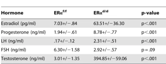
Due to selective ablation of pituitary ERa expression, the ERad/dmice are likely to experience a loss of negative-feedback regulation by E at the level of pituitary. Consistent with this prediction, the serum level of LH was significantly elevated in ERad/dmice (Table 1). Hyperstimulation of ovarian cells by LH resulted in increased steroidogenesis, leading to high circulating levels of progesterone, testosterone and E in ERad/d mice (Table 1). In contrast, the level of FSH was not statistically different between ERad/dand ERaf/fmice. According to previous reports, the levels of LH, progesterone, testosterone, and E are also elevated in ERaKO mice [19,20]. Consistent with the ERaKO mouse phenotype, ERad/d mice are infertile. Adult mice fail to ovulate due to chronic high levels of LH. Due to the lack of ERa expression in uterine epithelial and stromal cells, the ERad/duteri are unable to receive an implanting embryo. Furthermore, uterine tumors are not found in the ERad/dmice, presumably because the major uterine cell types do not express ERa. However, in contrast to the ERaKO mice, which lack ERa in all cells, including the ovarian cells, ERawas intact in OSE of ERad/dmice. This raised the possibility that elevated systemic E levels contribute to tumor initiation by stimulating ER signaling in OSE of ERad/dmice but fails to do so in OSE of ERaKO mice.
In agreement with this view, we observed marked up-regulation of a transcriptionally active form of ERa, phosphorylated at serine 118, in OSE of ERad/dmice (Fig. 2B, b). We also examined the status of the phosphoinositide 3-kinase (PI3K)/AKT pathway, which is reported to be activated in response to E treatment of ovarian cancer cell lines [21–23]. We noted that the level of AKT phosphorylated at Ser 473 (p-AKT) is elevated in the OSE and tumor cells of ERad/dovaries, while p-AKT level is maintained at a low level in ERaf/fovaries (Fig. 2B, c,d). It is likely that the increased level of phosphorylated AKT is linked to the elevated E signaling in ERad/dovaries.
To further characterize the nature of the ovarian tumor in ERad/dmice, we performed immunohistochemical analyses using epithelial and granulosa cell biomarkers. Anti-mullerian hormone (AMH) is a well-known marker for normal granulosa cells and granulosa cell tumors [24]. While both ERaf/fand ERad/dovaries expressed AMH exclusively in the granulosa cells of follicles, ERad/dovaries did not express AMH in the tumor cells, indicating that these tumors are not of granulosa cell origin (Fig. 3A). Analysis using anti-cytokeratin 8 (CK8) antibody revealed that ERaf/fmice express this epithelial marker exclusively in a single layer of OSE at 3, 6, and 11 months of age (Fig. 3B, panels a,c,e). In contrast, the OSE of ERad/dmice at 3 months of age exhibited multiple layers of cytokeratin-positive cells (panel b). At 6 months of age, we observed pronounced cytokeratin 8 expression within the ovaries of ERad/dmice, indicating the presence of epithelial cells within the tumor mass (panel d). By 11 months of age, widespread cytokeratin 8 immunostaining was observed within the ovarian tumor, highlighting its remarkable epithelial component (Fig. 3B, panel f).
Current literature suggests that the human ovarian epithelial tumors are derived from either OSE or FTE [2,25]. Although these epithelia are derived from a common embryologic precursor, OSE is thought to retain mesothelial characteristics, while FTE is terminally differentiated [25–27]. Recent studies on the serous subtype of ovarian cancer have suggested that either OSE
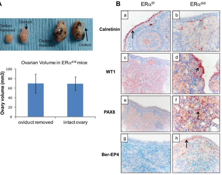
differentiates to resemble FTE or the cancer originates in the fallopian tube and spreads to the ovary [27]. To investigate further the origin of epithelial ovarian tumor cells in ERad/dmice, we removed the oviducts of these mice prior to tumor formation. Interestingly, removal of the oviducts from pre-pubertal ERad/d mice did not prevent the onset of ovarian tumor growth in these animals, indicating that the tumor cells originate from the OSE rather than the oviductal epithelium (Fig. 4A). We also examined the epithelia of ERaf/f and ERad/d ovaries by monitoring the expression of biomarkers specific for either OSE or FTE. As shown in Fig. 4B, we detected prominent expression of calretinin, a mesothelial marker [2,28], in OSE of ERaf/fovaries but not in OSE of ERad/dovaries. We also noted marked up regulation of tubal-specific makers, including PAX8, WT1, and Ber-EP4 in the ovaries of ERad/dmice, while the ovaries of ERaf/fmice lacked their expression. Since PAX8, WT1, and Ber-EP4 are normally expressed in FTE and are present in serous epithelial ovarian tumors [2,28–30], it is likely that the OSE cells of ERad/dovaries have undergone differentiation to resemble FTE. Furthermore, it has been reported previously that PAX8 is expressed in serous, endometrioid, and mucinous ovarian cancer while expression of WT1 is restricted to the serous subtype of ovarian cancer [29]. Currently there are no available biomarkers that can differentiate between high and low-grade serous ovarian carcinoma. It is clear that the ERad/dtumors do not grow aggressively.
To investigate the molecular pathways underlying ovarian tumorigenesis in ERad/dmice, we next performed gene expression profiling, using RNA isolated from the ovaries of ERaf/f and ERad/d mice. We identified more than 2500 genes that were differentially expressed in the tumor tissue compared to the normal ovaries. The GEO accession number for the microarray data is GSE39402. When we compared the differentially regulated genes to three different datasets of differential gene expression profiles of human serous adenocarcinoma versus control human ovaries that exist in the Oncomine database, we noted that a large number of genes, which are differentially expressed in human serous ovarian cancer specimens, are also present in ERad/d ovarian tumors (Fig. S2A). Remarkably, the identity of genes expressed in ERad/d ovaries and human serous ovarian cancer ranged from 25–40%. Prominent among these genes were those encoding platelet derived growth factor receptor alpha (PDGFRa), vascular cell adhesion molecule (VCAM), clusterin, intercellular adhesion molecule 1 (ICAM-1), and serine/threonine phosphatase 1 (Wip1), which are overexpressed in human serous ovarian cancer [31–36]. We observed that the levels of PDGFRa, VCAM, ICAM1, and clusterin were markedly elevated in the ovaries of ERad/d mice compared to those of ERaf/f mice (Fig. S2B). Collectively, the presence of these cancer biomarkers in ERad/d ovarian tumors underscored the importance of this model in deciphering the pathways involved in genesis and progression of epithelial ovarian tumorigenesis.
Estrogen Signaling and Ovarian Tumorigenesis
mRNA (Fig. S3A). To localize aromatase expression we digested ERad/d ovarian tumors into single-cell suspension, plated both fibroblast stromal and epithelial cells, and completed immunocy-tochemistry co-localizing both aromatase and a marker indicating the cell type. We observed that ovarian tumor cells isolated from ERad/d mice express high levels of P450 aromatase protein in luteinized stromal cells of the tumor, suggesting that these cells acquired the ability to synthesize E (Figs. S3B). Furthermore, the activated form of ERa, phosphorylated at Ser-118, is abundantly expressed in the OSC, while aromatase is expressed in ovarian stroma of ERad/d mice as early as 3 months (Fig. S3C). We postulated that the epithelial ERa signaling remains elevated in response to this locally produced E in ERad/d ovarian tumors, supporting increased ERa-dependent gene expression, abnormal cell proliferation, and tumorigenesis.
To examine whether E plays a critical role in ovarian tumor progression in ERad/dmice, we chronically treated these mice at 3 months of age with letrozole, a specific inhibitor of P450 aromatase, by implanting silastic capsules containing this drug. Following three months of letrozole treatment, ovarian tumors of 6-month old ERad/dmice displayed a remarkable reduction, up to 60%, in tumor volume when compared to sham-treated ERad/d mice (Fig. 5A). It is important to note that while ERad/d mice treated with letrozole exhibited significantly lower levels of serum E compared to sham-treated ERad/dmice, their serum LH levels were not altered in response to this treatment (Fig. 5A). Interestingly, ERad/d mice treated with the letrozole exhibited significantly lower levels of ovarian expression of PDGFRa and VCAM transcripts compared to sham-treated ERad/d mice (Fig. 5B). Similarly, the levels of Wip1 mRNA and protein were markedly decreased in ovarian tumors of ERad/d mice upon letrozole treatment (Fig. 5, B and C). Taken together, these results confirmed that elevated E signaling in the ovarian tumors of ERad/dmice leads to dysregulated expression of a subset of genes with known links to ovarian epithelial cancer.
Discussion
Genetically engineered mouse models are considered to be among the most powerful and promising tools presently available for studying the biology of various forms of cancer and for developing therapeutics. Although the creation of mouse models of
ovarian cancer has lagged behind models for many other neoplastic diseases, significant advances have been made in the last decade. Orsulicet al have shown that p53-deficient ovarian cells engineered to overexpress multiple oncogenes, c-myc, Kras, and Akt, develop ovarian tumors when injected in mice [42]. Similar mouse models for ovarian epithelial tumors were developed via inactivation of various tumor suppressors, such as
Pten, APC, p53 and Rb, through intrabursal administration of adenoviral vectors [43,44]. Conditional inactivation of multiple genes, such asPtenandKrasorPTENandDicer, by expression of Cre recombinase driven by theAmhr2promoter also led to ovarian cancer in mice [45,46]. These mouse models have provided compelling evidence that OSE or FTE can be transformed by altering the expression of a variety of oncogenic factors or tumor suppressors. Some of these models display tumor histotypes similar to ovarian cancer subtypes seen in women. However, it is clear that these models typically require multiple genetic changes and are limited by very rapid tumor onset, which limits their usefulness for studying early modulators of ovarian tumorigenesis. In the present study, we report the development of a unique animal model, in which initiation of ovarian tumorigenesis is independent of any oncogenic insult but dependent on elevated E signaling in the ovary. Since the onset and progression of tumorigenesis is relatively slow in ERad/dmice, this model is potentially useful in providing insights into the factors involved in the initiation and early phases of ovarian epithelial tumorigenesis.
In ERad/dmice, ERais conditionally ablated in the pituitary but retained in the hypothalamus and ovary. The loss of negative-feedback regulation by E in the HPO axis led to elevated production of LH by the pituitary. Interestingly, high levels of gonadotropins in women in early postmenopause have been postulated to play a role in the development of epithelial ovarian neoplasms [4,5]. Consistent with this notion, it has been found that women with polycystic ovary syndrome, which is accompanied by high LH levels, have a greater risk of developing ovarian cancer [47]. Further supporting a role of gonadotropins in ovarian cancer development, gonadotropin levels in cysts and peritoneal fluid from ovarian cancer patients have been shown to be elevated [48]. However, not all studies have led to similar findings and it is clear that elevated gonadotropin levels alone do not cause ovarian cancer. In fact, our studies using the ERad/d model suggested that hyperstimulation of ovarian cells by LH results in increased steroidogenesis, leading to high levels of circulating E as well as locally produced E in the ovarian tissue. High levels of testosterone coupled with increased expression of aromatase in the ovarian tissue would lead to increased synthesis of local E. We propose that this elevated E is an important factor in epithelial ovarian tumorigenesis as it stimulates signaling in the OSE, promoting its proliferation and phenotypic transformation. These results are supported by epidemiological and clinical studies, which indicate that postmenopausal women with elevated gonadotropin levels and receiving E replacement therapy exhibit an increased incidence of ovarian tumors [9–13]. Consistent with a role of E in the genesis of ovarian tumors, recent reports point to the clinical use of anti-estrogen drugs in stabilization of ovarian cancers [49,50].
Although many previous studies indicated that epithelial ovarian cancer arises from OSE, recent studies have revealed
Figure 2. ERalocalization in the tissues of HPO axis.(A) Histological sections of ERaf/f(a) and ERad/d(b) hypothalami, ERaf/f(c) and ERad/d(d)
pituitaries, ERaf/fovary (e, g) and ERad/dovarian tumor (f, h) from adult mice at 6 months of age stained with anti-ERa. Inserts i and j indicate higher
magnification depicting ERapositive cells (red staining) in the OSC and ovarian tumor cells. 3 V indicates the third ventricle. A indicates the anterior
lobe, I indicates the intermediate lobe, and P indicates the posterior lobe of the pituitaries. Arrows point to OSE cells or theca cells expressing ERa. (B)
Ovarian sections obtained from ERaf/f(left pictures) and ERad/d(right pictures) mice were subjected to immunohistochemistry using antibodies
against phospho-ERa(S118) (panels a, b) and phospho-Akt (S473) (panels c,d).
doi:10.1371/journal.pgen.1004230.g002
Table 1.Serum hormone measurements of ERaf/fand ERad/d mice at six months of age.
Hormone ERaf/f ERad/d p-value
Estradiol (pg/ml) 7.03+/2.84 63.51+/236.30 p,.001
Progesterone (ng/ml) 1.94+/2.61 8.78+/2.77 p,.001
LH (ng/ml) .17+/2.12 2.31+/2.51 p,.001
FSH (ng/ml) 6.30+/21.58 2.92+/2.57 p = .09
Testosterone (ng/ml) 3.01+/21.35 394.85+/259.06 p,.001
Estrogen Signaling and Ovarian Tumorigenesis
that the fimbriae of the fallopian tube is a possible site of origin of this malignancy, particularly high-grade serous carcinoma [51]. The common embryologic precursor of OSE and FTE is the coelomic epithelium, which gives rise to the epithelial linings of the fallopian tube and the ovary [27]. Unlike FTE, OSE retains mesothelial characteristics and is not terminally differentiated. It has been proposed that either OSE terminally differentiates to resemble FTE, or the cancer originates in fallopian tube and then spreads to the ovary. In support of the latter hypothesis, a recent study showed that conditional deletion ofPtenandDicer, using the Amhr2-Cre, led to tumor development in the fallopian tube, which subsequently metastasized to the ovary [46]. Our studies, on the other hand, appear to indicate that ovarian tumorigenesis in ERad/dmice is associated with differentiation of OSE to FTE. We
observed prominent expression of FTE marker proteins, such as PAX8, WT1, and Ber-EP4, which are not normally expressed in OSE, in the ovaries of ERad/d mice. Furthermore, removal of oviducts from ERad/dmice did not prevent the onset of ovarian tumorigenesis, indicating that FTE is not the precursor tissue for tumorigenesis in ERad/dmice.
Interestingly, we did not observe any intraperitoneal metastatic spread of the ovarian tumor in ERad/dmice. This could be partly due to the fact that the majority of the mutant mice died by 10 months of age due to the enlarged tumor, making it difficult to follow the progression of tumorigenesis beyond this point. The absence of overt malignancy in our model is not entirely surprising as several recent studies indicate that multiple genetic changes are necessary for metastatic transformation. It is conceivable that
Figure 3. Development of ovarian tumors in ERad/dmice.(A) Ovarian sections from ERaf/f(a, c) and ERad/d(b, d) mice at 11 months of age
were subjected to immunohistochemistry using an antibody against AMH. Red staining indicates AMH positive granulosa cells. (B) Ovarian sections from ERaf/fmice at 3, 6, and 11 months of age (left panels a, c, e respectively) and ERad/dmice at 3, 6, and 11 months of age (right panels b, d, f
respectively) were subjected to immunohistochemistry using anti-cytokeratin 8 antibody. Red staining indicates cytokeratin 8 positive epithelial cells. doi:10.1371/journal.pgen.1004230.g003
Figure 4. OSE is the site of origin for ovarian tumorigenesis in ERad/dmice.(A) Ovarian tumor growth in ERad/dmice after surgical removal
of oviducts. The right oviduct was surgically removed from ERaf/fand ERad/dmice at four weeks of age, leaving the left oviduct intact as an internal
control.Upper panel, Gross morphology of ERaf/fand ERad/dovaries at 5 months after oviduct removal surgery.Lower panel, The graph depicts
ovarian volume comparing ERad/dtumors with and without the oviduct. Two-tailed t-test was performed for statistical analysis: n = 5; p = 0.99. (B)
Immunohistochemistry of ERaf/f(left panel) and ERad/d(right panel) ovaries from mice at 6 months of age using calretinin (a, b); WT1 (c, d); PAX8 (e, f);
Estrogen Signaling and Ovarian Tumorigenesis
additional mutation(s) in tumor suppressor genes, such as p53, is required to drive the tumorigenic pathways in ERad/dovaries to rapidly progressing ovarian carcinoma, which will culminate in metastasis. Indeed, recent studies, utilizing genomic sequencing data from human high-grade serous ovarian cancer specimens, have shown that these cancers exhibit genomic instability and harbor genetic mutations in p53, Rb, BRCA1, and/or BRCA2 loci [52–55].
The ovarian tumors in ERad/dmice are composed of cells of both epithelial and stromal origins. These tumors appear to be distinct from the tubular or tubulostromal adenomas, which are reported to occur spontaneously in a number of mutant mouse strains, including the WXWX mice [56,57]. The adenomas, composed of numerous tube-like structures plus abundant large luteinized stromal cells, arise due to a defect in primordial germ cell proliferation and rapid loss of oocytes at birth, resulting in destruction of graafian follicles [58,59]. They also arise when mice are irradiated and there is a rapid loss of oocytes after radiation exposure [60]. However, these adenomas are not lethal and administration of E prevents rather than promotes their develop-ment [61]. Furthermore, in contrast to these mutant mouse strains, the ERad/dmice exhibit normal number of oocytes at 3 months of age, which then start to decline when ovarian epithelial and stromal cells expand and form the tumor mass at 6 months. Therefore, the initial stages of tumorigenesis in ERad/dmice are independent of the oocyte loss. Most importantly, the growth of the ovarian tumors exhibited by the ERad/d mice is inhibited by letrozole, indicating that these tumors, unlike adenomas, are E-dependent. The ovarian tumors in the ERad/d mice are presumably dependent on pituitary LH production, which help luteinize the stromal cells. However, the local production of E by these tumors and the resulting estrogenic effects on ovarian surface epithelial expansion and transformation appear to be the two key features that distinguish these tumors from the endocrinologically inactive tubular adenomas or tubulostromal adenomas.
Although the ovarian neoplasm in ERad/dmice did not show signs of overt malignancy, there was nevertheless clear evidence of tumorigenic transformation. Particularly striking is the finding that a large number of genes, associated with human serous ovarian cancer, are also expressed in ERad/dovarian tumors. Specifically, these tumors exhibit dysregulated expression of PDGFRa, VCAM, and Wip1, which were previously reported to be involved in human ovarian cancer. PDGFRa, a cell surface tyrosine kinase receptor for members of the platelet-derived growth factor family, is over-expressed in human serous ovarian tumors and is targeted in clinical trials to treat ovarian cancers [32,33]. VCAM, a vascular cell adhesion molecule, is found in the blood circulation of cancer patients and has recently been proposed as a marker to detect early stages of ovarian cancer [34,35]. Wip1, a p53-inducible phosphatase and an oncogene, is of particular interest. Under normal conditions, it restores cellular homeostasis following DNA-damage by cooperating with p53 to induce G2/M cell cycle arrest, thereby allowing ample time for repair of the damaged DNA [62]. However, amplification of Wip1 leads to sustained inhibition of DNA damage response and tumor suppressors, and consequently, its overexpression has been implicated in a variety of
human malignancies, including ovarian carcinoma [37,63]. Recent studies have revealed that Wip1 is regulated by ERa [64]. Consistent with this finding, administration of letrozole to ERad/d mice, which decreased the ovarian tumor size, also markedly reduced the expression of Wip1 along with PDGFRa, and VCAM. These results are consistent with our hypothesis that accentuated E signaling in the ovarian tissue promotes aberrant expression of genes that participate in tumorigenesis.
In summary, we describe a unique mouse model that allows us to identify hormonal effectors, particularly elevated E signaling, which play an important role in the development of ovarian epithelial tumorigenesis. In the future, the ERad/d model will serve as a valuable tool for exploring the involvement of E-dependent signaling pathways in the onset and progression of this deadly disease.
Materials and Methods
Animals
Mice (C57BL/6; Jackson Laboratory) were maintained in the designated animal care facility at the University of Illinois College of Veterinary Medicine according to the institutional guidelines for the care and use of laboratory animals. We crossed mice harboring ‘floxed’ ERa gene (Esr1tm1.2Mma), termed ERaf/f, with PR-Cre mice expressing Cre recombinase under the control of progester-one receptor promoter (Pgrtm2(cre)Lyd) to develop mice of genotype Esr1tm1.2Mma/Esr1tm1.2MmaPgrtm2(cre)Lyd/Pgr+, which we termed ERad/d. The PR-Cre knock-in mice expression of cre recombinase in pituitary, uterus, oviduct, mammary gland, and corpora lutea of the ovary have been described previously [18]. It has been used extensively to ablate ‘‘floxed’’ genes in tissues expressing PR [17,18].
Immunohistochemistry (IHC)
Paraffin-embedded ovarian tissue sectioned at 4mm, mounted on slides and subjected to immunohistochemistry as described previously [65]. Sections were incubated at 4uC with polyclonal antibodies against PCNA (Santa Cruz sc-56), cytokeratin 8 (Developmental Studies Hybridoma Bank, TROMA I), ERa (Novacastra Laboratories), p-Akt1/2/3 serine 473 (Santa Cruz SC-33437), AMH (Santa Cruz Biotechnology SC-6886), WT1 (Santa Cruz Biotechnology), PAX8 (Proteintech group 10336-1-AP), calretinin (Invitrogen 18-0291), Ber-EP4 (Dako), aromatase (Abcam ab35604), vimentin (Sigma Aldrich V5255). Biotinylated secondary antibodies were used followed by incubation with horseradish peroxidase-conjugated streptavidin (Invitrogen). Sec-tions were stained in AEC Solution.
Real-time PCR analysis
Total RNA was isolated from ovaries by standard Trizol-based protocols and converted to cDNA. The cDNA was amplified by real-time PCR to quantify gene expression using gene-specific primers and SYBR Green (Applied Biosystems). As a loading control, the expression level ofRPLP0 (36B4), which encodes a ribosomal protein, was determined. For each treatment, the mean threshold cycle (CT) and standard deviation were calculated from
CT values obtained individually from 3 to 4 replicates of that
Figure 5. ERad/dovarian tumor growth is inhibited by P450 aromatase inhibitor.(A) Ovaries with tumors from ERad/dmice treated with
letrozole, a P450 aromatase inhibitor, filled silastic capsules or sham empty silastic capsules for 3 months are analyzed by gross morphology and tumor volume. Serum estradiol and LH is assayed by radioimmunoassay. (B) Real-time quantitative PCR was employed to measure mRNA levels of genes associated with ovarian carcinoma, PDGFRa, VCAM, and Wip1, in ovaries with tumors of ERad/dmice treated with letrozole-containing or sham
empty silastic capsules. (C) Wip1 protein localization in ovaries with tumors of ERad/dmice either treated with sham control (a) or treated with
sample. Each sample was subjected to three independent real-time PCR trials. The fold change was derived from the meanCTvalues. Primer sequences recognizing each gene are located in Table S1.
Measurement of serum hormones
Hormones were measured by radioimmunoassay (RIA) at the Ligand Core facility, University of Virginia, Charlottesville. Statistical significance was determined on SAS program using the Tukey procedure to control for comparison-wise error rate. Significance cutoff value of p,.05 was determined to be statistically significant.
Isolation of cells from ovaries with tumors
Ovarian tumors were removed from mice and digested with either 6 g/liter dispase (Invitrogen) and 25 g/liter pancreatin (Sigma Aldrich), or 0.5 g/liter collagenase (Sigma Aldrich) in Hank’s balanced salt solution (HBSS). After incubation for 1 h at 37uC, the tubes were vortexed for 10–12 s until the supernatant became turbid with dispersed cells. The contents were then passed through an 80-mm gauze filter (Millipore). Cells were re-suspended in Dulbecco’s modified Eagle’s F12 medium (DMEM-F12; with 100 unit/liter penicillin, 0.1 g/liter streptomycin, 1.25 mg/liter fungizone) containing 10% heat-inactivated fetal calf serum. Cell culture was continued for 48 h after addition of fresh medium.
Immunocytochemistry
Ovarian tumor cells were fixed with 10% formalin solution for 10 m. Cells were treated with 25% Triton X-100 (Sigma Aldrich) in PBS for 10 m and exposed to a blocking serum for 1 h. Cells were treated with primary antibodies and incubated at 4uC and exposed to cy3 or cy5-conjugated secondary antibodies.
Silastic capsule implant
Silastic capsules were made by filling silastic laboratory tubing with 0.8 mg of ground Novartis Femara tablets (letrozole) and sealing with medical adhesive silicone type A (Dow Corning). For surgery, mice were first treated with analgesic 1 h prior to surgery and then anesthetized. A small dorsal incision was made just below the neck, and the silastic capsule was inserted underneath the skin. The incision was held together with wound clips until healed. After 3 months of exposure to either empty silastic capsules (sham control) or silastic capsules containing letrozole, mice were euthanized and ovarian tumors were fixed or frozen for analysis.
Statistical analysis
Statistical analysis was performed by ANOVA or two-tailed student’s ttest. Values ofP,0.05 were considered significant.
Supporting Information
Figure S1 (A) Average size and weight of ERaf/f ovaries and ERad/d ovaries with tumors at 8–11 months of age (n = 62). * indicates p,.05 using two-tailed t-test to calculate statistical significance. (B) Histological analysis of ERaf/fovary and ERad/d ovarian tumors. Ovarian sections from ERaf/fmice at 3 months of age (a & b) and ovarian sections from ERad/dmice at 3 months of age (c & d), 6 months of age (e & f), 11 months of age (g & h) are stained with hemotoxylin and eosin. Panels b, d, f, and h are higher magnified images of a, c, e, and g, respectively. (C) Survival
curve of ERaf/fand ERad/dmice indicating probability of survival at different weeks of age. Data analyzed using GraphPad Prism. P,.0001 indicated that the survival curves between ERaf/fand ERad/dmice are significantly different.
(TIF)
Figure S2 (A) Similarity between the genetic profiles of aberrantly regulated genes in ERad/dovarian tumors compared to the genetic profiles of aberrantly regulated genes in human serous adenocarcinoma from three independent microarray studies. Venn diagrams indicate similarity of aberrantly regulated genes in ERad/dovarian tumors compared to aberrantly regulated genes of human serous adenocarcinoma as assessed by microarray analysis published by Adib et al. (A), Hendrix et al. (B) and Lu et al. (C). (D) Table indicates the percentage of aberrantly regulated genes similar between ERad/dovarian tumors and human serous adenocarcinoma. Microarray lists indicating aberrantly regulated genes between human serous adenocarcinoma and normal human ovaries were exported from the Oncomine public database. Lists of aberrantly regulated genes between human serous adenocarci-noma and ERad/dovarian tumors were compared using Ingenuity software. (B) Real-time quantitative PCR was employed to measure mRNA levels of genes associated with ovarian carcinoma, PDGFRa, VCAM, clusterin, and ICAM-1, in ERaf/fand ERad/d ovaries at 3, 6, and 11 months of age. * indicates significant difference of p,.05 using two-tailed t-test for comparison of gene expressions of ERaf/fand ERad/dovaries.
(TIF)
Figure S3 Expression of P450 aromatase in ovarian tumor stromal cells of ERad/dmice. (A) Real-time quantitative PCR was employed to measure mRNA levels of P450 aromatase in ERaf/f ovaries and whole ERad/dovarian tumors from mice at 11 months of age. (B) Localization of aromatase in cultured ERad/dovarian tumor cells is assessed by immunocytochemistry. Upper; Epithelial cells dually stained with cytokeratin 8 in red (a), P450 aromatase in green (b), and co-localized with blue dapi staining (c).
Lower; Stromal cells are dually stained with vimentin in red (d), P450 aromatase in green (e) and co-localized with blue dapi staining (f). Yellow indicates co-localization of P450 aromatase and vimentin (f). (C) Localization of aromatase (a,b) in ERad/dovarian tumors and in ERaf/f ovaries (c). Ovarian section treated with non-immune IgG (d). Localization of pERa in ERad/dovarian tumors (e,f) and in ERaf/fovaries (g,h). Tissues are from mice at 3 months of age. Red color indicates localization of each protein. (TIF)
Table S1 Sequences of quantitative PCR Primer Sets. Primer sequences were designed to recognize coding regions of specific mRNA.
(TIF)
Acknowledgments
We thank Dr. Guo-Xia Tong of Columbia University Medical Centre, New York, NY, for histopathlogical analyses of ovarian tumors.
Author Contributions
Conceived and designed the experiments: MJL MKB ICB. Performed the experiments: MJL AK SP. Analyzed the data: MJL WMH MKB ICB. Wrote the paper: MJL WMH MKB ICB.
References
1. Siegel R, Naishadham D, Jemal A. (2012) Cancer statistics. CA Cancer J Clin 62(1): 10–29.
2. Auersperg N. (2011) The origin of ovarian carcinomas: a unifying hypothesis. Int J Gynecol Pathol 30(1): 12–21.
Estrogen Signaling and Ovarian Tumorigenesis
3. Bast RC, Hennessy B, Mills GB (2009) The biology of ovarian cancer: new opportunities for translation. Nature Reviews Cancer 9: 415–428.
4. Stadel BV (1975) Letter: the etiology and prevention of ovarian cancer. Am J Obstet Gynecol 123: 772–774.
5. Mertens-Walker I, Baxter RC, Marsh DJ (2012) Gonadotropin signaling in epithelial ovarian cancer. Cancer Letters 324: 152–159.
6. Stewart SL, Querec TD, Ochman AR, Gruver BN, Bao R, et al. (2004) Characterization of a carcinogenesis rat model of ovarian preneoplasia and neoplasia. Cancer Res 64: 8177–8183.
7. Riman T, Persson S, Nilsson S (1998) Hormonal aspects of epithelial ovarian cancer: review of epidemiological evidence. Clin Endocrinol 49: 695–707. 8. Chakravarti S, Collins WP, Forecast JD, Newton JR, Oram DH, et al. (1976)
Hormonal profiles after the menopause. Br Med J 2: 784–787.
9. Lacey JV Jr, Mink PJ, Lubin JH, Sherman ME, Troisi R, et al. (2002) Menopausal hormone replacement therapy and risk of ovarian cancer. JAMA 288: 334–341.
10. Glud E, Kjaer SK, Thomsen BL, Høgdall C, Christensen L, et al. (2004) Hormone therapy and the impact of estrogen intake on the risk of ovarian cancer. Arch Intern Med 164: 2253–2259.
11. Beral V; Million Women Study Collaborators, Bull D, Green J, Reeves G. (2007) Ovarian cancer and hormone replacement therapy in the Million Women Study. Lancet 369: 1703–1710.
12. Rossing MA, Cushing-Haugen KL, Wicklund KG, Doherty JA, Weiss NS. (2007) Menopausal hormone therapy and risk of epithelial ovarian cancer. Cancer Epidemiol Biomarkers Prev 16: 2548–2556.
13. Mørch LS, Løkkegaard E, Andreasen AH, Kru¨ger-Kjaer S, Lidegaard O. (2009) Hormone therapy and ovarian cancer. JAMA 302: 298–305.
14. Sasano H, Harada N (1998) Intratumoral aromatase in human breast, endometrial, and ovarian malignancies. Endocr Rev 19: 593–607.
15. Bai W, Oliveros-Saunders B, Wang Q, Acevedo-Duncan ME, Nicosia SV. (2000) Estrogen stimulation of ovarian surface epithelial cell proliferation. In Vitro Cell Dev Biol Anim 36: 657–66.
16. Laviolette LA, Garson K, Macdonald EA, Senterman MK, Courville K, et al. (2010) 17beta-estradiol accelerates tumor onset and decreases survival in a transgenic mouse model of ovarian cancer. Endocrinology 151: 929–38. 17. Dupont S, Krust A, Gansmuller A, Dierich A, Chambon P, et al. (2000) Effect of
single and compound knockouts of estrogen receptors alpha (ERalpha) and beta (ERbeta) on mouse reproductive phenotypes. Development 127: 4277–4291. 18. Soyal SM, Mukherjee A, Lee KY, Li J, Li H, et al. (2005) Cre-mediated
recombination in cell lineages that express the progesterone receptor. Genesis 41: 58–66.
19. Hewitt SC, Korach KS (2000) Progesterone action and responses in the alphaERKO mouse. Steroids 65: 551–7.
20. Couse JF, Yates MM, Walker VR, Korach KS (2003) Characterization of the hypothalamic-pituitary-gonadal axis in estrogen receptor (ER) Null mice reveals hypergonadism and endocrine sex reversal in females lacking ERalpha but not ERbeta. Mol Endocrinol 17: 1039–53.
21. Altomare DA, Wang HQ, Skele KL, De Rienzo A, Klein-Szanto AJ, et al. (2004) AKT and mTOR phosphorylation is frequently detected in ovarian cancer and can be targeted to disrupt ovarian tumor cell growth. Oncogene 23: 5853–5857.
22. Vivanco I, Sawyers CL (2002) The phosphatidylinositol 3-kinase AKT pathway in human cancer. Nat Rev Cancer 2: 489–501.
23. Kimura A, Ohmichi M, Kawagoe J, Kyo S, Mabuchi S, et al. (2004) Induction of hTERT expression and phosphorylation by estrogen via Akt cascade in human ovarian cancer cell lines. Oncogene 23: 4505–15.
24. Rey R, Sabourin JC, Venara M, Long WQ, Jaubert F, et al. (2000) Anti-Mullerian hormone is a specific marker of sertoli- and granulosa-cell origin in gonadal tumors. Hum Pathol 31: 1202–1208.
25. Auersperg N, Wong AS, Choi KC, Kang SK, Leung PC (2001) Ovarian surface epithelium: biology, endocrinology and pathology. Endocr Rev 22: 255–288. 26. Auersperg N, Woo MM, Gilks CB (2008) The origin of ovarian carcinomas: a
developmental view. Gynecol Oncol 110: 452–454.
27. King SJ, Burdette JE (2011) Evaluating the progenitor cells of ovarian cancer: analysis of current animal models. BMB reports 44: 435–445.
28. Kurman RJ, Shih LM (2010) The origin and pathogenesis of epithelial ovarian cancer-a proposed unifying theory. Am J Surg Pathol 34: 433–443. 29. Nonaka D, Chiriboga L, Soslow RA. (2008) Expression of pax8 as a useful
marker in distinguishing ovarian carcinomas from mammary carcinomas. Am J Surg Pathol 32: 1566–1571.
30. Tong GX, Devaraj K, Hamele-Bena D, Yu WM, Turk A, et al. (2011) Pax8: a marker for carcinoma of Mullerian origin in serous effusions. Diagn Cytopathol 39: 567–574.
31. Wilczynski SP, Chen YY, Chen W, Howell SB, Shively JE, et al. (2005) Expression and mutational analysis of tyrosine kinase receptors c-kit, PDGFRalpha, and PDGFRbeta in ovarian cancers. Hum Pathol 36: 242–9. 32. Giavazzi R, Nicoletti MI, Chirivi RG, Hemingway I, Bernasconi S, et al. (1994)
Soluble intercellular adhesion molecule 1 (ICAM-1) is released into the serum and ascites of human ovarian carcinoma patients and in nude mice bearing tumor xenogtafts. Eur J Cancer 30A: 1865–70.
33. Banks RE, Gearing AJ, Hemingway IK, Norfolk DR, Perren TJ, et al. (1993) Circulating intercellular adhesion molecule-1 (ICAM-1), E-selectin and vascular cell adhesion molecule-1 (VCAM-1) in human malignancies. Br J Cancer 68: 122–4.
34. Yurkovetsky Z, Skates S, Lomakin A, Nolen B, Pulsipher T, et al. (2010) Development of a multimarker assay for early detection of ovarian cancer. J Clin Oncol 28: 2159–66.
35. Gunawardana CG, Kuk C, Smith CR, Batruch I, Soosaipillai A, et al. (2009) Comprehensive analysis of conditioned media from ovarian cancer cell lines identifies novel candidate markers of epithelial ovarian cancer. J Proteome Res 8: 4705–13.
36. Tan DS, Lambros MB, Rayter S, Natrajan R, Vatcheva R, et al. (2009) PPM1D (Wip1) is a potential therapeutic target in ovarian clear cell carcinomas. Clin Cancer Res 15: 2269–2280.
37. MacLusky NJ, Voit R, Lazo JS, Schwartz PE, Merino MJ, et al. (1987) Aromatase activity in human ovarian cancer. Steroids 50: 423–433. 38. Thompson MA, Adelson MD, Kaufman LM, Marshall LD, Coble DA. (1988)
Aromatization of testosterone by epithelial tumor cells cultured from patients with ovarian carcinoma. Cancer Res 48: 6491–6497.
39. Zimniski SJ, Garola RE, Fendl K, Peterson CM. (1989) Endocrine character-ization of a human ovarian carcinoma (BG-1) established in nude mice. Steroids 54:593–606.
40. Cunata S, Hoffmannb P., Pujol P. (2004) Estrogens and epithelial ovarian cancer. Gynecologic Oncology 94(1): 25–32.
41. Goodman MT, Lurie G, Thompson PJ, McDuffie KE, Carney ME. (2008) Association of two common single-nucleotide polymorphisms in the CYP19A1 locus and ovarian cancer risk. Endocr Relat Cancer 15(4): 1055–60. 42. Orsulic S, Li Y, Soslow RA, Vitale-Cross LA, Gutkind JS, et al. (2002) Induction
of ovarian cancer by defined multiple genetic changes in a mouse model system. Cancer Cell 1: 53–62.
43. Flesken-Nikitin A, Choi KC, Eng JP, Shmidt EN, Nikitin AY (2003) Induction of carcinogenesis by concurrent inactivation of p53 and Rb1 in the mouse ovarian surface epithelium. Cancer Res 63: 3459–3463.
44. Wu R, Hendrix-Lucas N, Kuick R, Zhai Y, Schwartz DR, et al. (2007) Mouse model of human ovarian endometrioid adenocarcinoma based on somatic defects in the Wnt/beta-catenin and PI3K/Pten signaling pathways. Cancer Cell 11: 321–33.
45. Fan HY, Liu Z, Paquet M, Wang J, Lydon JP, et al. (2009) Cell type specific targeted mutation of Kras and Pten document proliferation arrest in granulosa cells versus oncogenic insult in ovarian surface epithelial cells. Cancer Res 69: 6463–6472.
46. Kim J, Coffey DM, Creighton CJ, Yu Z, Hawkins SM, et al. (2012) High-grade serous ovarian cancer arises from fallopian tube in a mouse model. Proc Natl Acad Sci U S A 109: 3921–6.
47. Schildkraut JM, Schwingl PJ, Bastos E, Evanoff A, Hughes C (1996) Epithelial ovarian cancer risk among women with polycystic ovary syndrome. Obstet Gynecol 88: 554–559.
48. Halperin R, Hadas E, Langer R, Bukovsky I, Schneider D (1999) Peritoneal fluid gonadotropins and ovarian hormones in patients with ovarian cancer. Int J Gynecol Cancer 9: 502–507.
49. Kothari R, Argenta P, Fowler J, Carter J, Shimp W (2010) Antiestrogen therapy in recurrent ovarian cancer resulting in 28 months of stable disease: a case report and review of the literature. Arch Oncol 18: 32–35.
50. Argenta PA, Thomas SG, Judson PL, Downs LS Jr, Geller MA, et al. (2009) A phase II study of fulvestrant in the treatment of multiply-recurrent epithelial ovarian cancer. Gynecol Oncol 113: 205–9.
51. Jarboe E, Folkins A, Nucci MR, Kindelberger D, Drapkin R, et al. (2008) Serous carcinogenesis in the fallopian tube: a descriptive classification. Int J Gynecol Pathol 27: 1–9
52. Cancer Genome Atlas Research Network (2011) Integrated genomic analyses of ovarian carcinoma. Nature 474: 609–615.
53. Mullany LK, Richards JS (2012) Minireview: Animal models and mechanisms of ovarian cancer development. Endocrinology 153: 1585–1592.
54. Hashiguchi Y, Tsuda H, Yamamoto K, Inoue T, Ishiko O, et al. (2001) Combined analysis of p53 and RB pathways in epithelial ovarian cancer. Hum Pathol 32: 988–96.
55. Risch HA, McLaughlin JR, Cole DE, Rosen B, Bradley L, et al. (2001) Prevalence and penetrance of germline BRCA1 and BRCA2 mutations in a population series of 649 women with ovarian cancer. Am J Hum Genet 68: 700– 710.
56. Rehm S., Dierksen D., Deerberg F. (1984). Spontaneous ovarian tumors in Han:NMRI mice: histological classification, incidence and influence of food restriction. J Natl Cancer Inst 72: 1383–96.
57. Allison, R. H., Morgan K. T. (1987) Ovarian neoplasms in F344 rats and B6C3F1 mice. Environ Helath Perspect 73: 91–106.
58. Capen, Charles C. (2004) Mechanisms of hormone-mediated carcinogenesis of the ovary. Toxicologic Pathology 32(Suppl.2): 1–5.
59. Murphy E.D. (1972) Hyperplastic and early neoplastic changes in the ovaries of mice after genic deletion of germ cells. J Natl Cancer Inst 48: 1283–95. 60. Gardner W. U. (1950). Ovarian and lymphoid tumors in female mice
subsequent to Roentgen-ray irradiation and hormone treatment. Proc Soc Exp Biol Med 75: 434–6.
61. Capen CC, Beamer WG, Tennent BJ, Stitzel KA. (1995). Mechanisms of hormone-mediated carcinogenesis of the ovary in mice. Mutation Research 333: 143–51.
63. Bulavin DV, Demidov ON, Saito S, Kauraniemi P, Phillips C, et al. (2002) Amplification of PPM1D in human tumors abrogates p53 tumor-suppressor activity. Nat Genet 31: 210–215.
64. Han HS, Yu E, Song JY, Park JY, Jang SJ, et al. (2009) The estrogen receptor alpha pathway induces oncogenic Wip1 phosphatase gene expression. Mol Cancer Res 7: 713–23.
65. Li, Q., Y. P. . Cheon, et al. (2004). ‘‘A novel pathway involving progesterone receptor, 12/15-lipoxygenase-derived eicosanoids, and peroxisome proliferator-activated receptor gamma regulates implantation in mice.’’ J Biol Chem 279(12): 11570–11581.
Estrogen Signaling and Ovarian Tumorigenesis