T
Social defeat protocol and relevant biomarkers,
implications for stress response physiology, drug abuse,
mood disorders and individual stress vulnerability: a
systematic review of the last decade
Protocolo de derrota social e biomarcadores relevantes, implicações para a isiologia
de resposta ao estresse, abuso de drogas, transtornos do humor e vulnerabilidade
individual ao estresse: revisão sistemática de estudos na última década
Mailton Vasconcelos,1 Dirson João Stein,2 Rosa Maria M. de Almeida1
Abstract
Introduction: Social defeat (SD) in rats, which results from
male intraspeciic confrontations, is ethologically relevant and useful to understand stress effects on physiology and behavior.
Methods: A systematic review of studies about biomarkers induced by the SD protocol and published from 2002 to 2013 was carried out in the electronic databases PubMed, Web of
Knowledge and ScienceDirect. The search terms were: social defeat, rat, neurotrophins, neuroinlammatory markers, and
transcriptional factors.
Results: Classical and recently discovered biomarkers were
found to be relevant in stress-induced states. Findings were summarized in accordance to the length of exposure to stress: single, repeated, intermittent and continuous SD. This review
found that the brain-derived neurotrophic factor (BDNF) is a
distinct marker of stress adaptation. Along with glucocorticoids
and catecholamines, BDNF seems to be important in
understanding stress physiology.
Conclusion: The SD model provides a relevant tool to study stress response features, development of addictive behaviors,
clinic depression and anxiety, as well as individual differences in
vulnerability and resilience to stress.
Keywords: Social stress, affective disorders, drug addiction, glucocorticoids, catecholamines
Resumo
Introdução: A derrota social (social defeat, SD) entre ratos,
resultado da confrontação intraespecíica entre machos, é etologicamente relevante e útil para o entendimento dos efeitos do estresse na isiologia e no comportamento.
Métodos: Foi realizada uma revisão sistemática de estudos sobre biomarcadores induzidos pelo protocolo de SD publicados entre 2002 e 2013, usando as bases de dados PubMed, Web of
Knowledge e ScienceDirect. Os termos usados na busca foram: derrota social, neurotroinas, marcadores neuroinlamatórios e
fatores de transcrição.
Resultados: Biomarcadores clássicos ou recentemente descobertos mostraram-se relevantes nos estados induzidos pelo
estresse. Os achados foram resumidos de acordo com o tempo de exposição ao estresse: SD única, repetida, intermitente ou contínua. O fator neurotróico derivado do cérebro se mostrou um marcador especíico de adaptação ao estresse. Assim como glicocorticóides e catecolaminas, o BDNF parece ser importante para o entendimento da isiologia do estresse.
Conclusão: O modelo de SD oferece uma ferramenta importante para
estudar características da resposta ao estresse, desenvolvimento de comportamentos aditivos, depressão clínica e ansiedade, bem como diferenças individuais de vulnerabilidade e resiliência ao estresse. Descritores: Estresse social, distúrbios afetivos, adição a
drogas, glicocorticóides, catecolaminas.
1 Instituto de Psicologia, Universidade Federal do Rio Grande do Sul (UFRGS), Porto Alegre, RS, Brazil. 2 Hospital de Clínicas de Porto Alegre (HCPA), UFRGS,
Porto Alegre, RS, Brazil.
This review was based on an academic dissertation presented at Instituto de Psicologia, Universidade Federal do Rio Grande do Sul (UFRGS), Porto Alegre, RS, Brazil, in 2014, as partial fulillment of the requirements for the degree of Master in Psychology. The original dissertation was entitled “Studies on social defeat: biomarkers of stress-induced states and inluences on individual reactivity to social stress.”
Financial support: none.
Submitted Jul 30 2014, accepted for publication Nov 04 2014. No conlicts of interest declared concerning the publication of this article.
Introduction
Mental disorders, increasingly characterized by their high worldwide prevalence,1,2 are neuropsychiatric
conditions that lead to signiicant loss of quality of life for
patients and relatives.3,4 In the World Health Organization
(WHO), studies about the global burden of diseases classify mortality and socioeconomic impact according to
disease causes.5 WHO reports aim to provide evidence
of the relative impact of health problems worldwide. Calculated projections based on these studies helped to raise awareness about the substantial effect of mental health around the world.2 According to the 2005 WHO
report, 31.7% of all years lived with disability may be
attributed to neuropsychiatric conditions. The ive major conditions contributing to this are unipolar depression (11.8%), alcohol abuse (3.3%), schizophrenia (2.8%),
bipolar depression (2.4%) and dementia (1.6%).5 The
analysis of mortality reveals that 1.2 million deaths every year are attributed to neuropsychiatric conditions, and that 40,000 are associated with mental disorders
and 182,000 with drug use and alcohol abuse.5 The WHO
report did not include suicide as a neuropsychiatric cause
of death, but almost 800,000 suicides are recorded
annually.2 These numbers seem to conirm that the
large prevalence of mental diseases worldwide is an
independent contributor, but interactions with other health problems, such as coronary disease, stroke,
diabetes, HIV/AIDS and medically unexplained somatic
symptoms, should also be taken into consideration.2 The
public health relevance of evidence-based arguments
should be evaluated in the study of mental health, and efforts should be directed to social and public policy
making throughout the preclinical stages of biomedical investigations. For this purpose, animal models of stress are important tools to construct knowledge about affective and drug-abuse disorders.
Animal models of stress are particularly useful,
because they focus on social life events that generalize across many mammal species, including humans.6
Social stress, a common stressor readily translated across species, is a recurrent factor in the life of all social species.7,8 Animal models of social stress have
different temporal and intensity characteristics: single, intermittent or continuous exposure of an individual to another or other conspeciics. Interactions are speciic to the animal species, sex, age, life history, and distinctive
environment.6 Within these models, the inluence of
social status may be determined by several factors:
the frequency, duration, and intensity of agonistic
interactions, their outcome, and the perception of controllability.9 A common protocol to generate social
defeat (SD) among rodents is the resident-intruder
paradigm.10,11 Under precise experimental conditions,
it is possible to measure the number of salient acts,
postures and displays, and to exert experimental control by determining whether the rat prevails as dominant, or
is defeated. The animal can perceive this confrontation
as extremely stressful, inducing characteristic
neuroendocrine and behavioral responses.12-14 This
pattern confers the model with ethological relevance and translational value for the elucidation of the physiologic
and behavioral adaptations to stress.
Animal models of social stress share many response characteristics with models that use other environmental stressors.15 After decades of studies about social stress,
the neuroendocrine responses to social challenges are largely understood as events that begin with the
activation of the sympathetic-adrenal-medullary (SAM)
axis, followed by stimulation of the hypothalamus-pituitary-adrenal (HPA) axis16 in response to stress. They
activate metabolism of energetic and immune responses that are crucial in coping with stressors. Glucocorticoids
and catecholamines are a well-studied set of molecules that act as effectors of those mechanisms.17
Corticosterone (CORT) secretion in animals under stress is mostly regulated by the activity of the HPA-axis and the negative feedback exerted by the levels of circulating glucocorticoids acting upon glucocorticoid receptors (GR).18 This mechanism is triggered by a set
of stress hormones, such as corticotropin releasing
hormone (CRH), adrenocorticotropic hormone (ACTH)
and arginine vasopressin,19,20 and is also determined by
stressor modality and its characteristic time course, as in the case of its effect on social status.21 The involvement
of catecholamines is more pronounced and well studied because of their rapid peripheral activation, as a result
of sympathetic discharges.22,23 Norepinephrine and
epinephrine are synthesized and released by cells in the
adrenal medullary region; also, nerve ibers in contact with target tissues release norepinephrine. As a result of acute activation of this catecholaminergic system, these
amines provide the necessary boost to master immediate stress. This response is characterized by rapid increases
in heart rate and increased blood low to skeletal muscles and other regions of the body.24-27 Individuals
need these endocrine responses to survive challenging situations, but inadequate or excessive adrenocortical
and autonomic functions are harmful to the body and
brain. A characteristic of intermittent exposure to social
stress is the continuous activation of these SAM and HPA
axes, which indicates lack of habituation to the stressful
situation.28
correspond to those seen in patients with depression.6,40
Several of these behavioral and neuroendocrine effects caused by psychosocial stress may be reversed with
antidepressants and anxiolytics.41,42 Not all individuals
experiencing social stress develop cardinal symptoms of depression or anxiety, which suggests that the response to stress is, to a signiicant extent, determined by
individual vulnerability or resilience, in both humans43
and several animal species.44,45 In animal models,
individual differences in the endophenotypes have been studied to act as substrates for stress vulnerability.46-48
One such example is attributing differences in hedonic
temperament between individuals as a candidate mechanism.49 Rats exposed to the mild stress of a novel
environment that respond with increased exploratory behaviors are termed high responders (HR), and those responding with decreased exploration are termed low
responders (LR).48,49 HR animals are more vulnerable to
depressive-like symptoms, such as decreased behavioral responses to SD stress, sweet solution preference, forced swim test and social avoidance.50,51
The molecular changes in the central nervous system (CNS) that trigger and sustain behavioral and physiologic changes in socially defeated animals are not
completely known. This systematic literature review will
discuss information about the extensive brain and CNS molecular changes induced by the rat SD protocol. Its
main focus is to provide an overview of this protocol by
using systematic and explicit methods to search, critically
review and synthesize selected information. The current review focuses on CNS biomarkers induced by the use of this SD protocol in the last 10 years.
Methods
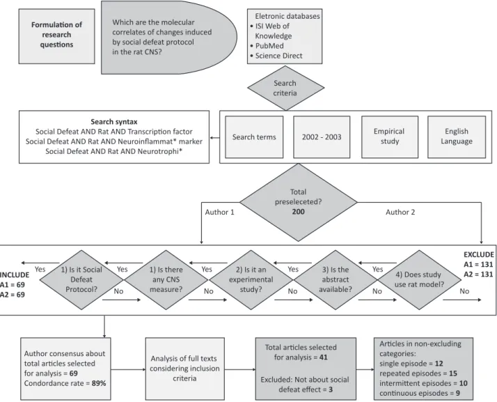
A systematic literature review was conducted in the second half of 2013 by means of an electronic search
of articles indexed in the databases Web of Knowledge,
PubMed, and ScienceDirect. Articles published in the
last ten years (2002-2013) were selected using the following terms: social defeat, rat, neurotrophins, neuroinlammatory markers, and transcriptional factors. Only empirical studies written in English were included.
The preselected studies were assessed independently by
two authors according to inclusion criteria: experimental
studies; abstract available; rats as animal model; SD protocol used in the methods; and analysis of CNS
biomarkers. After excluding duplicates, concordance
between authors was analyzed, as recommended by Lopes, Viacava & Bizarro (personal communication), to
deine the inal selection of studies. Figure 1 shows the lowchart of steps of this systematic search.
cortical and brainstem areas are implicated in the neural sites that are critical for adaptations to social
stress. Dopaminergic and glutamatergic connections in the ventral tegmental area (VTA), nucleus accumbens (NAc) and prefrontal cortex (PFC) are essential for coping with stressful environmental situations, as well for drug addiction.29,30 The association between social
stress, with its consequent physiological response,
and neural substrates involved in neuroadaptations
is of particular interest. Behavioral changes induced
by social stress seem to be relevant in increased
drug taking and affective disturbances in rodents.28
For instance, socially stressed animals respond more robustly to low doses of psychostimulants and increase
self-administration of psychomotor stimulants.31,32
This behavioral sensitization may contribute to drug-related behaviors, such as craving and relapse,33 and
acquisition of drug self-administration.34 An augmented
behavioral response to a drug challenge produced by an agent other than the challenge drug is termed
cross-sensitization. The establishment of this behavior occurs
initially due to intermittent exposure to social stress and, in the case of intense and chronic exposure, behavioral
sensitization deteriorates and behavioral impairments
emerge. The association of stress exposure and addictive behaviors follows an inverted U-shaped curve,
in accordance with the Yerkes-Dodson Law,35 and has a
powerful example in alcohol consumption. This biphasic effect of stress, which may also be found in cognitive
processes, such as memory and emotion, is relevant in
affective psychopathology.36
Brain-derived neurotrophic factor (BDNF), an important neurotrophin for synaptic plasticity, is one of
the molecular candidates underlying the development
of persistent neuroplastic adaptation to social and other types of stress. It is also a candidate molecule that
may trigger cross-sensitization induced by SD stress.
Mesocorticolimbic elevated BDNF in the VTA is a risk
factor for drug sensitivity.37,38 BDNF mediates synaptic
plasticity and cell responses to stress and drugs of abuse. Stress-induced lasting changes of BDNF signaling in mesocorticolimbic regions may regulate the reward
circuit.39 This neurotrophin has some opposite role
differences: episodically defeated and continuously subordinated rats may show, respectively, increased
and suppressed BDNF responses. These divergent
neuroadaptations to social stress may be representative
of the substrates for the intensiication of cocaine bingeing due to the anhedonia-like deterioration of reward processes during subordination stress.37
Psychiatric disorders are directly linked to social stress
etiologically. For example, defeated animals have signs
effect. Therefore, the inal sample had 38 studies, which were analyzed and classiied according to the length of SD episodes during the protocol. Some studies had more than one type of occurrence: single episode (12),
repeated episodes (15), intermittent episodes (10) and
continuous exposure (9). The analysis of the methods,
summarized in Table 1, revealed studies that used the rat SD protocol to obtain behavioral and molecular
changes associated with speciic brain areas or with the CNS. Excluded studies adopted a combination of distinct stress protocols together with SD, and did not,
therefore, clearly identify a SD effect on the results.52-54
The analysis of results was conducted separately for
each length of SD protocol.
Results
The searches in the three databases using the
search terms resulted in 200 articles. After the
inclusion of studies that met the deined criteria and the exclusion of duplicates, 69 were selected by author 1 (MV) and 69 by author 2 (DJS). The authors
discussed and reached the consensus that 41 studies met the previously established criteria. This consensus
resulted in a concordance rate of 89%, calculated using the following equation: total compatible studies / total
compatible studies + total incompatible studies =
compatibility index. However, during analysis, 3 studies were excluded because they did not discuss the SD
Figure 1 - Flow diagram of articles selected for inclusion
Formulaion of
research
quesions
Which are the molecular correlates of changes induced by social defeat protocol in the rat CNS?
Eletronic databases
• ISI Web of
Knowledge
• PubMed • Science Direct
Search syntax
Social Defeat AND Rat AND Transcripion factor Social Defeat AND Rat AND Neuroinflammat* marker
Social Defeat AND Rat AND Neurotrophi*
Search terms 2002 - 2003 Empirical study
English Language
Author 1 Author 2
Total
preseleceted? 200
INCLUDE
A1 = 69 A2 = 69
Yes Yes Yes Yes Yes
No No No No No
1) Is it Social Defeat Protocol?
1) Is there
any CNS measure?
2) Is it an
experimental study?
3) Is the
abstract available?
4) Does study
use rat model?
EXCLUDE
A1 = 131 A2 = 131
Author consensus about total aricles selected
for analysis = 69 Condordance rate = 89%
Analysis of full texts
considering inclusion criteria
Total aricles selected
for analysis = 41
Excluded: Not about social
defeat effect = 3
Aricles in non-excluding
categories: single episode = 12 repeated episodes = 15 intermitent episodes = 10 coninuous episodes = 9 Search
Behavioral impact and molecular effects Brain areas References Adult single SD
22 kHz USV
IGF-I protein levels Frontal and parietal cortices Burgdorf et al.55
BDNF, TrkB.T1 mRNA levels, BDNF epigenetic factors Hippocampus Duclot & Kabbaj46
Fos protein and CRH2 Arcuate nucleus, ventromedial
hypothalamus, posterior medial
amygdala
Fekete et al.56
c-Fos mRNA, CRH mRNA mPFC, LC and VTA/vBNST Funk et al.57
Increased passive coping behavior in maternally separated animals
c-Fos, TrpOH Dorsal raphe nucleus Gardner et al.58
Tph2 mRNA Dorsal raphe nucleus Gardner et al.59
slc6a4 mRNA Dorsal raphe nucleus Gardner et al.60
NOP receptor mRNA Amygdala, PVN Green & Devine61
Higher immobility in FST and lower latency to immobility, decreased total line crossing in OF
CAM-L1 protein, increased CORT concentrations,
ratio pCREB/CREB, GAP-43 expression Amygdala and hippocampus Kavushansky et al.
62
Increased 22 kHz USV, reduced number of line crossing in OF
Increased CORT serum levels, increased CRH and GR mRNA levels Hippocampus Marini et al.15
Increased ACTH, CORT and leptin plasma levels HPA-axis Razzoli et al.51
Short-term locomotor sensitization under amphetamine challenge
BDNF and ΔFosB VTA, NAc and PFC Wang et al.38
Adult repeated SD
Passive coping behaviors
c-Fos/TrpOH Dorsal raphe nucleus Paul et al.63
Increased anxiety-like behaviors
Proteins, including protein folding, signal transduction, synaptic
plasticity, cytoskeleton regulation and energy metabolism Hippocampus Carboni et al.
64
TrkB.FC-induced social avoidance in LR rats and 7,8-DHF-induced social
approach in HR rats
TrkB.FC and TrkB.T1 mRNA levels, PKB and pCREB, BNDF promoter 6 Hippocampus Duclot & Kabbaj46
Grooming frequency and time in OF
Decreased T3 and T4 serum levels Olivares et al.65
Reduced saccharin intake
5-HT1b mRNA expression NAcSh, dorsolateral striatum Furay et al. 66
NOP receptor mRNA PVN Green & Devine61
Increased self-grooming, shorter rearing duration, presence of risk assessment, lower sucrose preference, lower climbing in FST, lower general activity and sociability in social avoidance test
HPA-axis Razzoli et al.51
Increased rearing, decreased partition exploration, higher risk
assessment
Increased ACTH and CORT plasma levels in long-term condition HPA-axis Razzoli et al.23 Reduced sucrose consumption of HR rats
Histone 3, 4 and 2B Hippocampus Hollis et al.48
Lower FGF2 and FGFR1 mRNA levels Hippocampus Turner et al.67
IL-1β PVN Hueston et al.68
CAM-KIIb gene Hippocampus Kabbaj et al.69
Freezing during exposure, 22 kHz USV
Increased expression of CHRNB2 and ACHE genes PAG Kroes et al.70
Reduced mounting in copulatory behavior
Reduced testosterone plasma levels, c-Fos mRNA Medial preoptic area Niikura et al.71
Table 1 - Behavioral and molecular impact of distinct lengths of SD exposure and related brain areas
Adolescent repeated SD
Adult increased locomotion behavior, CPP for amphetamine
D2 receptor NAc Burke et al.72
Adult intermittent SD
Sensitized locomotor response to amphetamine
BDNF and ΔFosB Mesocorticolimbic structures Nikulina et al.39 Increased play initiation, increased submission in adolescence and less
and later submission in adulthood
Buwalda et al.73
Sensitized locomotor response to amphetamine
Long-term Fos-like immunoreactivity VTA and AMY Nikulina et al.32
Anhedonic sexual disinterest
Reduced orexin and dynorphin VTA, mPFC and hypothalamus Nocjar et al.74
Decreased horizontal and vertical locomotion, increased inactivity in OF,
decreased sucrose preference
Decreased T3, T4 and CORT serum levels HPA-axis Olivares et al.65
Increased cocaine intake in 24 h binge
zif268 mRNA expression Amygdala and frontal cortex Covington III et al.75
BDNF and/or mRNA expression mPFC, AMY, substantia nigra,
VTA
Fanous et al.76
Reduced social behavior
BDNF VTA Fanous et al.77
Reduced saccharin intake Furay et al.66
Increased cocaine self-administration
Increased dopamine levels and BDNF NAc and VTA Miczek et al.37
Adult continuous SD
Anhedonia
Increased CORT, mRNA and protein NET, decreased PKA, PKC and
pCREB
Locus coeruleus, and its terminal
regions Chen et al.
18
Increased defeat-related behaviors
Increased BrdU-positive labeling Hippocampus Buwalda et al.73
Increased CORT, decreased serotonin, increased MAO A mRNA
expression, increased KLF11 mRNA and protein expression Cortex and thalamus Grunewald et al.
78
TNF-α BNST Hueston et al.68
Increased immobility in FS test
Phosphorylated MEK1/2, phosphorylated ERK1/2 and MKP-1 Hippocampus Iio et al.79
Reduced sucrose consumption and increased struggling of HR rats Decreased expression of Gsk3b and Taf2, increased expression of HMox3 and Eno3
Frontal cortex and hippocampus Kanarik et al.80
Reduced sucrose consumption
Lower BMP7 gene expression LC Ordway et al.81
Increased CORT level, 5-HT1a receptor mRNA PFC Kieran et al.82
Suppressed cocaine intake, decreased preference and intake of sugar and decreased exploratory behavior
Suppression of DA and BDNF responses NAc and VTA Miczek et al.37
5-HT1a = 5-hydroxytryptamine receptor 1a or serotonin receptor 1a; 5-HT1b = 5-hydroxytryptamine receptor 1b or serotonin receptor 1b; ACHE = acetylcholinesterase
of CRH and GR in the arcuate nucleus, AMY, hippocampus and HPA-axis56,85; changes in neurotransmitter receptors
and metabolites for serotonin in the dorsal raphe nucleus (DR)58-60; changes in nociceptin/orphanin receptor (NOP)
in the AMY and hypothalamus.61 Moreover, analyses
revealed changes in neurotrophic factor molecules, such as insulin-like growth factor I (IGF-I) in the frontal
and parietal cortices55; changes in BDNF levels and its
receptors and metabolites in the hippocampus, VTA, NAc and PFC38,46; changes in growth associated protein
43 in the AMY and also in the hippocampus. A wide
variety of proteins were expressed as markers of single exposure to SD: L1 cell adhesion molecule (CAM-L1) and phosphorylated cAMP response element-binding protein
(CREB) in the AMY and hippocampus62; and, inally Fos
family proteins, such as c-Fos and delayed ΔFosB in the
PFC, LC, VTA, BNST, DR, and NAc.38,57,58
Repeated SD episodes
This category included studies in which rats were exposed to two or more repeated days of SD episodes.
Rats that underwent repeated SD on consecutive
days had more 22 kHz USV,70 more passive coping
behaviors,23,63,70 increased anxiety-like behaviors, such as
increased self-grooming, increased locomotion in novel
environments and risk assessment behavior,23,51,64,65,72
and increased depressive-like symptoms, such as reduced sweet solution preference and intake, reduced
climbing in the FST, lower general activity and sociability in the social avoidance test and reduced mounting in
copulatory behavior.48,51,66,71 Repeatedly stressed LR
rats displayed social avoidance under infusion of BDNF
antagonist in hippocampus, whereas repeatedly stressed
HR rats displayed social approach under infusion of BDNF
agonist in hippocampus.46 Furthermore, rats exposed to
the repeated stress protocol during adolescence had
more conditioned place preference (CPP) behaviors in
adulthood when under amphetamine exposure.72
As molecular markers of stress, repeatedly stressed
animals had increased hormonal levels of CORT and ACTH,3
decreased thyroid hormones and decreased testosterone plasma levels,65,71 increased neurotransmitter receptors
and metabolites for serotonin, NOP and dopamine (DA)
in the NAc, striatum, hypothalamus and DR61,63,66,72 and
increased cholinergic receptors subunits and enzymes in the periaqueductal gray area (PAG).70 They also
had changes in neurotrophic factor molecules, such as BDNF metabolites, and downregulation of ibroblastic growth factor (FGF) in the hippocampus.46,67 Changes
in neuroinlammatory markers, such as Interleukin-1β in the hypothalamus,68 were evident after repeated
psychosocial stress. Finally, these animals had increased
The rat SD stress protocol
The rat SD protocol consists of the exposure of an experimental animal to a dominant aggressive male.
Most of the selected studies followed the procedures established by Miczek et al.,11,83,84 who developed and
characterized this resident-intruder model of social stress
in rats and mice. Before beginning the experiments, adult male rats were selected as aggressive residents and placed in large individual cages where they lived in
pairs with a sterile female. After an adaptation period and establishment of territorial status of the residents,
smaller experimental animals, termed intruders, are placed into the resident’s home cage. Before the beginning
of interactions between resident and intruder, females
are removed from the home cage. The experimental sequences mostly consist of presence or not of a pre-defeat period, generally 10 minutes, a physical or pre-defeat period, lasting up to 10 minutes and, also facultative, a post-defeat period of a variable length of time. Resident
animals show a pattern of attack and threat behaviors,
while intruders engage in defensive, submissive and light reactions. It is important to distinguish between
brief episodes of SD and continuous subordination
stress. There are three classes of exposure. The irst, single SD stress differs from repeated SD stress in the number of exposure episodes. Repeated defeat differs from continuous subordination stress, which requires
cohabitation, albeit protected, with a dominant opponent and consists of an inescapable and uncontrollable nature of stress. Here we considered a fourth class of stress
exposure, the intermittent protocol, which produces
clear and distinct effects from the other patterns and
should be classiied as a separate class of SD stress.
Single SD episode
This category included studies in which the rats were exposed to a single episode of SD. Rats that underwent one single episode of SD had more 22 kHz ultrasound vocalizations (USV),55,85 more depressive-like symptoms,
such as higher immobility and lower latency to engage in
immobility in the forced swim test (FST),62 and reduced
line crossings in open ield (OF).62,85 When challenged
three days after the SD session, rats had a short-lived
locomotor sensitization to amphetamine exposure.38
Also, maternally separated animals during early infancy had more passive coping behaviors when adults.58
As molecular markers of stress, those animals had
increased hormonal levels of CORT, CRH, ACTH, and leptin in areas such the HPA-axis, PFC, locus coeruleus (LC),
VTA, bed nucleus of stria terminalis (BNST), hippocampus
58 – Trends Psychiatry Psychother. 2015;37(2)
serotonin neurotransmitter metabolites and receptors
in the cortex and thalamus.78,82 Chronic exposure
upregulates norepinephrine transporter (NET) expression in the LC and its terminal regions18 and suppresses
DA in the NAc and VTA.37 As for neurotrophic factors,
chronically stressed rats have lower bone morphogenetic protein 7 (BMP7) gene expression and suppression of
BDNF in the LC, NAc and VTA.37,81 There were changes in
the inlammatory marker TNF-α, speciically in the Long
Evans strain of defeated rats.68
A wide variety of molecules may function as markers
of continuous stress, including BrdU-positive labeling
nucleosides in the hippocampus73 and neurotransmitter
degradation molecules in the cortex and thalamus.78 Cell
signaling proteins, glucose metabolism, transcription factors and cell functioning enzymes in the frontal cortex
and the hippocampus may also be found in chronically stressed animals.79,80
Discussion
Molecular basis of stress-response
Few studies selected in this systematic review focused
only on the understanding of the molecular basis of stress-response. These studies contributed to the clariication
of several and perhaps interconnected mechanisms of response and adaptation to stress. Therefore, several studies selected for this review measured basic stress
hormone levels in animals exposed to single social stress. The other stress exposure protocols relied not only on hormone levels to examine the stressful conditions of
SD, but also on behavioral measures.
Fekete et al.56 discussed the role of CRH in responses
to stress. They found that this peptide is present in the paraventricular nucleus (PVN) of the hypothalamus, induces ACTH release from pituitary86 and is also present
in extra hypothalamic regions, where it acts as a
non-neuroendocrine stress-related modulator.87 For instance,
its presence is conirmed after a single SD episode in
the hippocampus85 and in vBNST and ceAMY.57 CRH
binds to two known receptors in brain tissue, CRH1 and CRH2. CRH1 is expressed in more speciic brain areas and the cerebellum and is involved in the activational and
anxiety-like components of stress-related behaviors.87
The CRH2 receptor is discretely distributed, and its
role in endogenous stress responses was unclear, as suggested by Fekete et al.,56 before this study. After
a single exposure to SD, rats had an increased Fos expression in CRH2 positive neurons in the mAMY.
56 The
role of the mAMY in stress response may be associated
with defeat behaviors, such as avoidance of aggressive
levels of c-Fos in the PFC and DR brain areas63,71 and
changes in histones and calcium/calmodulin-dependent
kinase in the hippocampus.48,69 In a more comprehensive
description, Carboni et al.64 reported changes in
folding-related proteins, signal transduction, synaptic plasticity, cytoskeleton regulation and energy metabolism in
the hippocampus after repeated psychosocial stress
exposure.
Intermittent SD
This category included rats that were exposed to
two or more episodes of SD on non-consecutive days, often termed acute defeat. In adolescence, these rats had more submissive behaviors and increased play initiation behaviors.73 As adults, they displayed less
frequent and longer latency to submit and reduced
social behavior.73,77 For anxiety-like and depressive
symptoms, rats acutely defeated displayed anhedonic
sexual disinterest74 and decreased locomotion and line
crossings, as well as increased immobility in the OF65 and
decreased sucrose preference.65,66 They also had
drug-related behaviors, such as sensitized locomotion under
amphetamine challenge32,39 and increased cocaine
self-administration.37,75
The analysis of molecular markers of stress revealed that repeatedly stressed animals had decreased serum levels of thyroid hormones.65 Decreased orexin
neurotransmitter and dynorphin protein in the VTA,
PFC and hypothalamus74 were also found, as well as
increased DA levels in the VTA and NAc.37 They might
also have alterations in neurotrophic factors molecules,
such as BDNF metabolites in brain areas, including VTA, NAc, PFC, AMY and substantia nigra.37,39,76,77 Finally,
these animals had increased levels of Fos family proteins
such as ΔFosB and c-Fos, as well as zif268 in the VTA,
NAc, PFC, and AMY brain areas.32,39,75
Continuous SD
This category included rats that were exposed
to continuous SD episodes, often termed as chronic
exposure. Rats that were exposed to continuous
SD stress had increased defeat-related behaviors,73
increased depressive-like symptoms such as anhedonia, immobility in the FST, reduced sweet solution preference
and decreased exploratory behavior.18,37,79-81 Chronic
SD stress experiences resulted in suppressed cocaine
intake.37 HR rats displayed reduced struggling in the FST
after continuous SD stress.80
Molecular markers of stress indicated that continuously socially defeated rats had markedly
Drug-related studies
In our review, studies about drug addictions focused on neurobiological and behavioral stress-induced adaptations. By taking the mesocorticolimbic DA system as the prominent SD neurobiological substrate,34
Nikulina et al.32 found that behavioral sensitization to
d-amphetamine challenge was a stress induced-effect of intermittent exposure to SD. This effect is accompanied
by activation of mesocorticolimbic structures one week and two months after stress discontinuation, and is
speciically evident in the VTA and AMY. These effects were evaluated using an immunohistochemical technique
to measure Fos-like immunoreactive (Fos-LI) proteins.
The amygdaloid region, as well as other mesocortical regions, is an important site for behavioral sensitization.
Additionally, Fos-LI activation pattern may be associated
with the expression of chronic Fos-related antigens. Covington III et al.94 investigated a different immediate
early gene expression after exposure to intermittent
social stress, zif268. Amphetamine challenge decreased zif268 mRNA expression in the AMY, and this effect was
also found 60 days after SD stress. Further studies
conirmed the involvement of the AMY in the sensitization
processes,39 and the medial region of the AMY seemed
to be responsible for dealing with emotional stressors.90
In subsequent studies, Nikulina et al.39 conirmed
the presence of a more stable Fos family protein
member, ΔFosB,95 involved in behavioral sensitization
and cross-sensitization. They analyzed the role of
neurotrophic factor, BDNF, and ΔFosB signaling in
mesocorticolimbic structures 10 days after intermittent
SD exposure. A sustained activation pattern of infralimbic innervations projecting to the VTA was found
after SD stress, consistent with the role of the VTA and its reciprocal connection to PFC in the development of behavioral sensitization.96,97 This mesocorticolimbic and
corticotegmental functional activation were interpreted as a possible neural circuit underlying stress-induced
cross-sensitization to psychostimulants.39 The role of NAc
in drug-related behaviors was further discussed in Furay
et al.66: after repeated, but not intermittent, exposure
to SD exposure, mRNA levels of 5-HT1b receptors were
increased in the rostral NAc shell. The NAc, an important
region mediating stress and fear responses, assess emotional valence of incoming stimuli. The shell of the NAc is part of the postulated extended AMY, according to Fanselow & Dong.98 Opposing expressions of BDNF
and DA in the NAc and VTA were implicated in distinct
outcomes of different lengths of rat SD protocols. Miczek
et al.37 demonstrated that intermittently or continuously
defeated rats tested for psychomotor stimulation and
binge-like cocaine self-administration had behaviors
mates88 and defensive responses to predator noxious
stimuli.89 The mAMY seems to be another regulator of
HPA-axis activity.90 Furthermore, functional activation of
the mAMY will be discussed together with the analysis of intermittent SD protocols involved in drug-related behaviors (see following topic).
Marini et al.64 investigated several hippocampal
molecular changes in protein levels induced by single psychosocial stress exposure. The pattern of changes after repeated social stress events were quantitatively and qualitatively different when compared to single exposures.64 Green & Devine61 found a possible plasticity
effect on the expression of NOP FQ receptor mRNA: a single exposure to social stress elicited increasing levels of NOP mRNA in the central and mAMY and PVN, but this effect was no longer evident after repeated stress exposure. The authors suggested a habituation process
of these neurotransmitter peptides with continuation of
stress exposure.61 Two hours after the application of the
intermittent SD protocol, BDNF and BDNF mRNA levels
were differentially upregulated in the mPFC, substantia nigra and AMY regions.76 The prominent inding in this
study was the persistence of BDNF changes up to 28 days
after stress discontinuation; this effect was evident in the
mAMY and VTA regions. These BDNF changes are indices of long-lasting neuronal adaptation to social stress.
The authors discussed implications of their study in the
understanding of the dopaminergic system functioning.76
A lack of discrepancies between protocols was reported by Hueston et al.68 Different cohorts of Sprague-Dawley
rats exposed to repeated or continuous SD protocols did not have changes in inlammatory responses. Despite the well-established role of sympathetic and noradrenergic functioning as mediators of inlammatory processes in
the brain and blood,40,91-93 acute measures of SD did
not impact gene expression of inlammatory markers
at any timepoint.68 A careful analysis of the protocol
revealed that a very mild type of SD was implemented
in these studies: the stimulus animals were not highly aggressive. The residents were not allowed to wound or bite to prevent inlammatory tissue processes.
These studies reinforced the hypothesis that different
modalities and courses of exposure to stress lead to
different outcomes, and therefore the type of adaptive
response speciic to a certain stressor should be previously clariied in the study objective. Findings reinforced the idea
of important brain reactions to stress in the form of neural
stress exposure, these behavioral adaptations were still present, including decreased general activity
and decreased sociality in a social avoidance test, as well as depressive-like behaviors in the FST, but not anhedonia.51 Rats exposed to continuous SD daily over
the course of 5 weeks displayed inhibition of weight gain, increased adrenal gland weight and increased immobility
in the FST. These behaviors were accompanied by
downregulated intracellular mitogen-activated protein
kinase (MAPK) cascade in the rat hippocampus.79 The
authors interpreted the prolonged immobility in the FST as behavioral despair and suggested that it was a marker
of depression.99 They explained that the involvement of
MAPK was one of the BDNF-induced outcomes to affect
cell functioning under SD.100 Miczek et al.28 highlighted
the divergent anhedonic consequences of stress depending on the length and intensity of exposure. In
sum, intermittent SD protocols induced escalation of
drug abuse behaviors and neuroadaptation, whereas continuous social stress exposure induced blunted
responses to sweet rewards and cocaine. Controversial results were reported in the study conducted by Kanarik et al.80: the length of chronic stress increased sweet
solution intake in HR rats instead of fragmenting or decreasing this behavior. An important unique feature
of this particular chronic protocol was that residents were treated with apomorphine immediately before the session, which affected the resident’s behavior; and that
intruders were not exposed to the resident continuously,
which, therefore, did not characterize a de facto chronic
exposure.28
The results of reduced sweet solution preference in
socially defeated animals were interpreted as an analog of
anhedonia. Nocjar et al.74 investigated a possible neural
basis for anhedonia using exposure to an intermittent SD protocol. The animals showed a generalized depressive phenotype, as deined by decreased sucrose and sexual
preference after 3 weeks of stress discontinuation and apathy-like behavior in the FST one month after stress discontinuation. These depressive-like behaviors
were accompanied by diminished levels of orexin in mesocortical regions of the DA reward system and of orexin and dynorphin in the hypothalamus.74 VTA
hypofunction associated with anhedonic behavior had already been reported, but after a more severe
continuous SD exposure.37 The hypothalamus sends an
orexin projection to the VTA, which, when stimulated,
produces effort and reward motivation behaviors.101 The
lower levels of orexin in the VTA suggested a possible basis for sexual anhedonia.71,74 Another feature of the
role of the VTA in environmental reactivity to stimuli was evaluated by Fanous et al.77 They used a virus to deplete
BDNF levels in this region and, after intermittent SD either intensiied or deteriorated. According to these
authors, the patterns of results among intermittently stressed animals suggested that BDNF in the VTA and NAc DA cells are part of a stress-induced cascade promoting behavioral and neural sensitization associated with drug
abuse. Also, the decreases in VTA BDNF in continuously stressed animals were associated with the persistent suppression of cocaine and sweet solution reward intake,
which suggests that these animals had an anhedonia-like proile.37 More recent studies conducted by Wang et
al.38 implicated the increased BDNF expression in VTA
phenotype as a risk factor for drug sensitization at both
the neural and the behavioral levels.38
Further studies discussed whether the effects on amphetamine- and cocaine-elicited sensitization were also found in adolescent rats and, therefore, persistent
after weeks of discontinuation of the stress exposure in adulthood. Rats repeatedly stressed during adolescence
developed CPP for amphetamine intake cues when adults more easily than foot-shock stressed animals.72
The authors suggested that stress exposure had effects associated with drug abuse behaviors extending through
the period of transition from adolescence to adulthood. The adolescent effects of social stress may be reversed
by housing with an age-matched mate during stress exposure. Moreover, rats experiencing continuous SD during adolescence had minor changes in BDNF, and no behavioral or physiological effect persisting into
adulthood.73
The studies discussed above agreed that intermittent
and continuous SD protocols were powerful tools in the
understanding of the association between stress and drug
abuse. Moreover, the continuous SD protocol provided some clues about stress and depressive-like symptoms associated with behaviors and molecular markers. Finally, the evidence contributed to the well-established
knowledge of the participation of the mesocorticolimbic structures in drug addiction, which contributed to the discussion of speciic roles of each brain region when expressing BDNF and Fos-LI markers involved in this
process.
Mood disorder studies
Razzoli et al.51 reported that the use of overall
reactivity to environmental stimuli was a valid depressive-like measure in animal models. The most common potential depressive-like endophenotypes found in our review were the responses to a stressful
context: sweet solution preference, the FST and the
social avoidance test. Shortly after repeated stress
disorder, as growth factors, cell signaling pathways and
the hormones discussed above may be abnormal in clinical populations.
These studies were closely connected to the use of
continuous SD protocol as an animal model for
anxiety-like states and clinical depression, which meets criteria
for human psychopathology. This model includes valid
measures of behavioral depressive-like symptoms, such as those related to reactivity to environmental stimuli
and anhedonia. Decreased locomotion and exploratory behaviors may be interpreted as deicits in motivation;
decreased mobility in the FST may be associated with behavioral despair, which in turn is associated with depressive disorders; and decreased sweet solution preference may result from desensitization of reward
circuitry, which is analogous to an anhedonic state. All these effects were elicited by chronic SD exposure. The expression of cell signaling molecules associated with
BDNF cascade and interactions of DA and BDNF in VTA
results from this type of exposure. The importance of
these studies about new molecules lies in the need for
new treatment targets for clinical depression. Although
not more effective than traditional antidepressants, new
drugs are gaining acceptance due to signiicantly better
tolerance and fewer side effects.107,108
Neurobiological basis of individual vulnerability to stress
Kabbaj et al.69 discussed several aspects of the
neurobiological substrates of individual differences in emotionality and responsiveness to stress and drug addiction. The irst refers to the existence of individual speciic neurochemical dysfunctions, due to which individuals seek drugs to repair this imbalance69; the
second is based on the fact that individuals are highly
attracted to either the reward and novelty properties of
drugs, or the drug ability to help cope with emotional
or environmental distress.109 The second includes
personality traits, such as endophenotypes, that mediate
the probability that an individual will seek drugs. This view is corroborated by investigations about sensation-seeking and drug-taking behaviors,109,110 as well as by
studies about substance abuse behaviors correlated with mood disorders.111-113
Piazza et al.49 were pioneers in demonstrating the
association of an individual’s exploratory behavior and drug-taking patterns. Kabbaj et al.69 further discussed
the suggested classiication of individuals as HR or LR
based on their responsiveness to a mild environmental
stress, such as exposure to the OF. The phenotypes of HR rats include less anxious behaviors in the light-dark box apparatus and greater elevated-plus maze (EPM) exposure, the rats displayed elevated social behaviors,
in contrast with rats infused with a control virus. This
study suggested that BDNF might be a pro-depressive factor within mesolimbic regions.77,102 The conditions of
the animals used in the study by Nocjar et al.74 should
be analyzed when deining the validity of SD protocols, as the rats were isolated for 28 days before social stress
initiation. Social isolation alone is a stress protocol, at least in social cohesive species, such as rats.103
Functional disturbances of the noradrenergic
system may also be accountable for the development of depressive-like states. Chen et al.18 evaluated mRNA
levels of NET in the LC and terminal regions, and an increased expression in these regions was accompanied
by decreased sweet solution preference and intake
after chronic stress exposure. These indings support the involvement of corticosteroids together with
catecholamines in the development of such depressive-like states, as adrenalectomy and corticosteroid
antagonist treatment prevent the development of
stress-induced depressive phenotypes.18
In addition to the most common measures of stress
(i.e., glucocorticoids and catecholamines104), Kroes et al.70
used other markers in a genomic study in the PAG region accompanied of 22 kHz USV in defeated animals. Six hours after repeated SD exposure, animals had changes in several genes, such as those involved in cholinergic transmission, GTPase mediated signal transduction and molecular function of growth factors activity. Twenty-two kHz USV are also seen in rats exposed to a single
SD session,55 and low range USV (around 22 kHz) were
postulated as measures of emotional states of fear
and anxiety in animal models.105,106 Burgdorf et al.55
evaluated these behavioral components of depressive or
anxiety-like states along with expression of IFG-I. Other
searches for stress-related measures focused on newly
described growth factors as consequences of stress-induced phenotypes. Ordway et al.81 focused on the
involvement of BMP7 and catecholamines in the biology of depression. Other molecules of interest are FGF and IGF-I: these growth factor systems were signiicantly downregulated in the hippocampus of repeatedly defeated
rats67 and in the frontal and parietal cortices of the brain
of one-time defeated rats.55 Searching for new markers,
Grunewald et al.78 investigated the pathway composed
of glucocorticoid and Kruppel-like factor 11, also named transforming growth factor β-inducible early gene 2, activated in continuously defeated animals. Olivares
et al.65 found higher levels of CORT and an imbalance
of thyroid hormones one month after continuous SD
serotonergic system and coping with social stress in adult male rats. Regardless of whether submitted to early life stress or normal growth, rats had the same pattern of activation of serotonergic neurons in speciic
subpopulations of the DR nucleus.58 This suggests that
brainstem monoamine systems may be associated with vulnerability to stress-related psychiatric disorders,
speciically in the case of animals exposed to early stress and facing a psychosocial conlict during adulthood.59,60
The serotonergic system might be involved in inhibitory processes of proactive coping responses, such as aggression121,122 and escape behaviors123; at the same
time, it may be associated with facilitation of passive-submissive responses.124 Using a repeated SD stress
protocol, Paul et al.63 found a shift away from a proactive
emotional coping style and towards a reactive emotional coping style during the defeat phase. In unstable social structure periods, adopting a subordinate position may prevent dangerous situations and limit injury and energy wastage.125 A reactive coping style may be a more lexible
and adaptive strategy according to Koolhaas et al.126
An alternative interpretation of coping phenotypes was
presented by Kanarik et al.80 After the discontinuation of
a chronic stress protocol, during the second exposure to the FST, individuals who had more struggling behaviors
were non-susceptible to stress and adopted an active
coping strategy; inversely, less struggling animals were susceptible to stress and had passive coping responses.80
BDNF metabolism was shown to be highly relevant
to this topic in several stress-related studies. The
investigation of emotional differences seems to be a
fruitful avenue for research to understand differences in vulnerability to stress, substance abuse and mood
disorders. This may be achieved by either studying behavior directly or combining the behavioral features
and molecular differences, both at baseline and in response to stress. These particular differences in
reacting to stress may be the key to the development of better pharmacological and therapeutic treatments and to the explanation of failed drug trials.
Conclusion
Studies about stress are illed with data, but most
results are contradictory or have to be more carefully discussed. Measures of social stress-induced effects are
complex and may rely on the characteristics of stressors,
such as duration, intensity and predictability, and of the
biological sample, such as time of sampling, rat strain used and sample quality. These differences and similarities should be discussed to derive knowledge about stress physiology and to develop new pharmacological and
than that of LR rats.114 HR rats have more climbing and
less loating in the FST than LR animals.115 Furthermore,
HR rats express less GR in the hippocampus,116 and both
phenotypes have different CORT-releasing patterns. The analysis of drug-taking behaviors and social stress after rats were exposed to chronic SD revealed that the once existing differences in cocaine self-administration tended to be no longer evident: SD induced a delay
in cocaine self-administration in HR and a dramatic increase in cocaine self-administration in LR rats.117
Kabbaj et al.69 investigated neural signatures, or neural
phenotypes, associated with the processes mentioned
above. A study using a microarray technique to analyze genes in the hippocampus found an imbalance of a large number of genes involved in neurogenesis after
four repeated sessions of SD.69 The authors suggested
that HR animals had a slower rate of cell proliferation, differentiation and transformation than LR rats. These
patterns might indicate that there is a link between BDNF, the hippocampus and drug-taking behaviors.69 The
authors focused on individual differences in emotional responsiveness and reactivity to stress.48 HR rats seem
to be more vulnerable to depressive-like symptoms when
facing stressful stimuli.118 This higher vulnerability in HR
animals were associated with higher levels of histone
acetylation in the hippocampus at baseline and after
repeated exposure to SD. HR rats with decreased sweet
solution preference also had decreased levels of histone acetylation in the hippocampus.48 Such cell chromatin
modiications are dynamic processes that regulate gene expression without changes in the DNA sequence.119
Histone acetylation is one of these processes, during which hyperacetylation leads to increased gene expression, while hypoacetylation might be involved in gene silencing.120 Another study about BDNF and
epigenetic regulation of individual vulnerability found that LR individuals have higher levels of hippocampal BDNF after SD exposure than HR rats.46 This trait may
confer stress resilience to LR rats and vulnerability to HR
rats. In fact, preventing BDNF signaling in the dentate gyrus of the hippocampus of LR rats leads to SD-induced
social avoidance, whereas BDNF activation in the same
region in HR animals leads to social approach behaviors.46
Duclot & Kabbaj46 discussed the hypothesis that LR rats
might have epigenetic tools for the regulation of BDNF expression in the hippocampus, which might confer
them resilience to stress, whereas HR animals seem to lack this mechanism.
Kabbaj et al.69 also found that the phenotypes of HR
and LR animals differ in the expression of serotonergic
receptors in the hippocampus.115 In a study about early
References
1. Kessler RC, Berglund P, Demler O, Jin R, Koretz D, Merikangas KR, et al. The epidemiology of major depressive disorder: results from the National Comorbidity Survey Replication (NCS-R). JAMA. 2003;289:3095-105.
2. Prince M, Patel V, Saxena S, Maj M, Maselko J, Phillips MR, et al. No health without mental health. Lancet. 2007;370:859-77. 3. Saarni SI, Suvisaari J, Sintonen H, Pirkola S, Koskinen S, Aromaa
A, et al. Impact of psychiatric disorders on health-related quality of life: general population survey. Br J Psychiatry. 2007;190:326-32. 4. Ustun TB, Ayuso-Mateos JL, Chatterji S, Mathers C, Murray CJ.
Global burden of depressive disorders in the year 2000. Br J Psychiatry. 2004;184:386-92.
5. Mathers CD, Loncar D. Projections of global mortality and burden of disease from 2002 to 2030. PLoS Med. 2006;3:e442.
6. Blanchard RJ, McKittrick CR, Blanchard DC. Animal models of social stress: effects on behavior and brain neurochemical systems. Physiol Behav. 2001;73:261-71.
7. Sapolsky RM. The inluence of social hierarchy on primate health. Science. 2005;308:648-52.
8. Von Holst D. The concept of stress and its relevance for animal behavior. Adv Study Behav USA [Internet]. 1998 [cited 2014 Jun 4]. http://agris.fao.org/agris-search/search.do?f=1998/US/ US98047.xml;US1997069446
9. Raab A, Dantzer R, Michaud B, Mormede P, Taghzouti K, Simon H, et al. Behavioural, physiological and immunological consequences of social status and aggression in chronically coexisting resident-intruder dyads of male rats. Physiol Behav. 1986;36:223-8. 10. Koolhaas JM, Schuurman T, Wiepkema PR. The organization
of intraspeciic agonistic behaviour in the rat. Prog Neurobiol. 1980;15:247-68.
11. Miczek KA. A new test for aggression in rats without aversive stimulation: differential effects of d-amphetamine and cocaine. Psychopharmacology (Berl). 1979;60:253-9.
12. Albonetti ME, Farabollini F. Social stress by repeated defeat: effects on social behaviour and emotionality. Behav Brain Res. 1994;62:187-93.
13. Fuchs E, Flügge G. Social stress in tree shrews: effects on physiology, brain function, and behavior of subordinate individuals. Pharmacol Biochem Behav. 2002;73:247-58. 14. Keeney AJ, Hogg S, Marsden CA. Alterations in core body
temperature, locomotor activity, and corticosterone following acute and repeated social defeat of male NMRI mice. Physiol Behav. 2001;74:177-84.
15. Marini F, Pozzato C, Andreetta V, Jansson B, Arban R, Domenici E, et al. Single exposure to social defeat increases corticotropin-releasing factor and glucocorticoid receptor mRNA expression in rat hippocampus. Brain Res. 2006;1067:25-35.
16. Sapolsky RM. Why zebras don’t get ulcers. New York: W.H. Freeman and Co; 2004.
17. McEwen BS. Central effects of stress hormones in health and disease: Understanding the protective and damaging effects of stress and stress mediators. Eur J Pharmacol. 2008;583:174-85. 18. Chen P, Fan Y, Li Y, Sun Z, Bissette G, Zhu MY. Chronic social
defeat up-regulates expression of norepinephrine transporter in rat brains. Neurochem Int. 2012;60:9-20.
19. De Kloet ER, Vreugdenhil E, Oitzl MS, Joëls M. Brain corticosteroid receptor balance in health and disease. Endocr Rev. 1998;19:269-301.
20. Kovács KJ, Földes A, Sawchenko PE. Glucocorticoid negative feedback selectively targets vasopressin transcription in parvocellular neurosecretory neurons. J Neurosci. 2000;20:3843-52.
21. Pace TW, Spencer RL. Disruption of mineralocorticoid receptor function increases corticosterone responding to a mild, but not moderate, psychological stressor. Am J Physiol Endocrinol Metab. 2005;288:E1082-8.
22. Bartolomucci A, Palanza P, Costoli T, Savani E, Laviola G, Parmigiani S, et al. Chronic psychosocial stress persistently alters autonomic function and physical activity in mice. Physiol Behav. 2003;80:57-67.
23. Razzoli M, Carboni L, Guidi A, Gerrard P, Arban R. Social defeat-induced contextual conditioning differentially imprints behavioral and adrenal reactivity: a time-course study in the rat. Physiol Behav. 2007;92:734-40.
24. Bartolomucci A, Palanza P, Gaspani L, Limiroli E, Panerai AE, Ceresini G, et al. Social status in mice: behavioral, endocrine and immune changes are context dependent. Physiol Behav. 2001;73:401-10.
therapeutic treatments for stress-induced or aggravated
disorders. For instance, studies that claim to have
discovered brain areas activated during social stress or as a consequence of it should be analyzed considering that studies about immediate early gene expression may
differ in a wide variety of control conditions. Fos proteins
do not seem to be good markers of inhibition, which may also be the result of neuronal signaling.127 Absence of Fos
labeling does not necessarily equal lack of involvement. The cumulative process of acquiring evidence may be
a reliable way to discuss and critically analyze new
indings. Several important points should be addressed when dealing with data provided by SD protocols. Each
laboratory may have its own version of procedures, and
protocol phases may be either prolonged or even omitted.
These discrepancies should be evaluated in accordance with the animal model used and the conditions to be achieved. The analysis of species and strains in animal models revealed that the studies reviewed used few strains of rats. Most of the written protocols used
Long-Evans male rats as residents; species variation of stressed animals was high, although still most studies used Sprague-Dawley rats. It is beyond this review to
discuss implications of strain variations in the models.
However, the exact purpose of each experiment should shed some light in the importance of rat strain.
SD exposure activates cortical and limbic circuits, areas that underlie the processing of emotional stress
and reward, which reinforces the role of protocols in
the study of addictive behaviors. The short length of
activation induced a short-lived adaptation important to
the understanding of the physiological and behavioral response to stress. Progressively, the increases in duration or recurrence of stress induced transient or
long-term adaptations, referred to as neuroadaptations, and
consequent changes in the neuronal paths involved in dealing with stress. Continuous stress exposures induced lasting consequences, such as coping dysfunctions and
development of affective disordered states. The SD model may be a relevant method to study stress responses and the development of addictive behaviors. It may also be
used as a model of clinical depression and anxiety for the development of therapeutic and pharmacological
treatments.
Acknowledgements
We gratefully acknowledge the advice of Klaus Miczek in
the translation of the manuscript. We thank Keitiline Viacava
and Fernanda Lopes for the insights about the method used