Major Article
Corresponding author: Dr. Saif Hameed. e-mail: [email protected] Received 18 May 2016 Accepted 15 July 2016
Citronellal-induced disruption of membrane homeostasis
in Candida albicans and attenuation of its
virulence attributes
Shweta Singh
[1], Zeeshan Fatima
[1]and Saif Hameed
[1][1]. Amity Institute of Biotechnology, Amity University Haryana, Gurgaon (Manesar), India.
Abstract
Introduction: There is an increasing burden of multidrug resistance. As a result, deciphering the mechanisms of action of natural compounds with antifungal activity has gained considerable prominence. We aimed to elucidate the probable mechanism of action of citronellal, a monoterpenoid found in the essential oil extracted from Cymbopogon plants, against Candida albicans. Methods: Drug susceptibility was measured by broth microdilution and spot assays. Ergosterol levels were estimated using the alcoholic potassium hydroxide method and H+ extrusion was assessed by monitoring the glucose-induced acidifi cation of the external medium. Virulence traits were studied by hyphal morphogenesis and biofi lm formation, along with fungal cell adherence to polystyrene surface and human oral epithelial cells. Results: Citronellal showed anticandidal activity against
C. albicans and non-albicans species of Candida at a minimum inhibitory concentration of 1 mg/ml. Citronellal interfered with membrane homeostasis, which is the major target of known antifungal drugs, by increasing the hypersensitivity of the fungi to membrane-perturbing agents, reducing ergosterol levels, and diminishing glucose-induced H+ extrusion. In addition, oxidative and genotoxic stresses were induced via an increased production of reactive oxygen species. Furthermore, citronellal inhibited the virulent attributes of yeast-to-hypha transition and biofi lm formation. It also reduced cell adherence to polystyrene surface and the human oral epithelial cells. Conclusions: This is the fi rst study to propose the cell membrane, morphogenetic switching,
biofi lm formation, and cell adherence of Candida albicans as potential targets for the anticandidal activity of citronellal. However, clinical investigations on the therapeutic applications of citronellal are required.
Keywords: Citronellal. Candida. Cell membrane. Biofi lm. Adherence.
INTRODUCTION
Candida albicans is a common human fungal pathogen, which causes mucosal, cutaneous and systemic candidiasis. It also causes systemic infections in immunocompromised individuals or patients undergoing long-term treatment with antibiotics(1) (2). Side effects of the present treatment regimens for C. albicans and the advent of multidrug resistance (MDR) are problems affecting effi cient therapy against infections caused by C. albicans(3) (4). There is therefore an urgent need to research into alternative agents for the treatment of Candida infections. In the search for novel antifungal drugs, compounds from natural sources are recognized as safer and more promising alternatives to synthetic agents(5) (6). It has been suggested in several studies that various natural compounds are effective against C. albicans. Terpenoids are among the largest group of natural compounds in plants, which have been reported to
have antioxidant, anticancer, antiparasitic, antiviral, antiallergic, and antimicrobial properties(7). Terpenoids consist of various compounds such as terpene aldehydes, terpene oxides, terpene esters, terpene alcohols, terpene phenols, terpene ketones, and terpene hydrocarbons, all of which possess antifungal activity. For instance, thymol and carvacrol, which are monoterpene phenols, are effective against the antioxidant system of
C. albicans(8). Recently, the antifungal activity of geraniol, which is a monoterpenoid compound, was established(9).
Citronellal (Cit) is one of the main constituents of the essential oil from lemongrass (Cymbopogon). Cit is a major isolate from Cymbopogon plants, lemon-scented gum, and lemon-scented tea tree(10). A preliminary study showed that Cit inhibits the adherence of C. albicans to denture implants(11); however, the mechanism of the anticandidal activity of Cit is still unknown. The present study is the fi rst to explore the possible mechanism of action of Cit against C. albicans.
METHODS
and ethidium bromide (EtBr) were purchased from HiMedia (Mumbai, India). Sodium chloride (for preparation of 0.9% saline), potassium chloride (KCl), mannitol, disodium hydrogen orthophosphate, potassium dihydrogen orthophosphate, dipotassium hydrogen orthophosphate (K2HPO4), sodium hydroxide, D-glucose, sodium dodecyl sulfate (SDS), and dimethyl sulfoxide (DMSO) were obtained from Fischer Scientifi c (Hampshire, US). Calcofl uor-white (CW) and Congo red (CR) were obtained from Sigma Chemical Co. (St. Louis, MO, USA). N-heptane and Cit were obtained from Central Drug House Pvt. Ltd. (New Delhi, India). Thiazolyl blue (MTT) was obtained from Sisco Research Laboratories Pvt. Ltd. (New Delhi).
Growth media and strains
The reference strain of Candida albicans used in this study was SC5314. The non-albicans species of Candida used in this study (Candida glabrata, Candida tropicalis, Candida parapsilosis, and Candida krusei) were obtained from diabetic patients suffering from oral candidiasis. DAY185(12) was the wild type for JRB64(13) and OCC1.1(14), which were the calcineurin signaling mutants for Δcnb1 and Δcrz1, respectively. YAG237(15) was the mutant CNB1-1/CNB1 having a constitutively expressed hyperactive allele of CNB1. All the strains were cultured in YEPD broth containing yeast extract (1% w/v), peptone (2% w/v), and dextrose (2% w/v). Agar plates were prepared by adding agar (2% w/v) to the media. The cells were freshly revived on YEPD broth and transferred to an agar plate before each study to ensure the revival of the strains. For biochemical assays, Cit (dissolved in DMSO) was used at its subinhibitory concentration (determined from growth curve experiments), which was the concentration at which it partially inhibited the growth of C. albicans (data not shown).
Drug susceptibility assays
Drug susceptibility was evaluated by determining the minimal inhibitory concentration (MIC) of Cit and by conducting spot assays as described below.
MIC determination: minimum inhibitory concentration
was determined by the broth microdilution method (M27-A3) described by the Clinical and Laboratory Standards Institute (CLSI)(16). Briefl y, 100μl of media was placed in each well of a 96-well plate. Cit (250µg/ml) was then added to the remaining media and serially diluted. A 100-μl aliquot of a cell suspension (in normal saline) at an optical density (OD) of 0.1 was added to each well, after which the OD was measured at 600nm (OD600). The OD600 was measured at 30°C after 48h. The MIC80, which is the concentration of Cit that inhibited at least 80% of fungal growth, was then determined.
Spot assay: spot assays were performed as previously
described(5) (9). Briefl y, 5μl of fi vefold serial dilutions of each yeast culture (each with cells suspended in normal saline to an OD600 of 0.1) was spotted onto YEPD plates in the absence (control) and presence of Cit (0.8, 1, or 1.2mg/ml). Differences in growths were measured after incubating the plates at 30°C for 48h. Phenotypic susceptibilities were estimated using the described spot assay. Cells were spotted onto YEPD plates in the absence (control) and presence of Cit at its subinhibitory
concentration (250μg/ml). The spotting was done in the presence of the chemicals (sodium dodecyl sulfate (SDS), ethanol (EtOH), Calcofl uor-white (CW), Congo red (CR), ascorbic acid (AA) and ethidium bromide (EtBr) at the concentrations specifi ed in fi gures 2,3).
Ergosterol quantifi cation
Sterols were extracted using the alcoholic KOH method and the percentage of ergosterol was calculated as previously described(5) (9). Briefl y, a single C. albicans colony from an overnight YEPD agar plate culture was used to inoculate 50ml of YEPD in the absence and presence of Cit at its subinhibitory concentration. Both ergosterol and 24 (28) dehydroergosterol (DHE) absorb ultraviolet (UV) light at 281.5nm; however, 24 (28) DHE absorbs UV light at 230nm as well. The ergosterol contents of the cell membranes of the fungi were determined by subtracting the amount of 24 (28) DHE (calculated from the OD230) from the total ergosterol and 24 (28) DHE content (calculated from the OD281.5). Ergosterol content was calculated as a percentage of the wet weight of the cells using the following equations:
% ergosterol + % 24 (28) DHE = [(A281.5/290) × F]/pellet weight % 24 (28) DHE = [(A230/518) × F]/pellet weight
% ergosterol = [% ergosterol + % 24(28) DHE] − % 24 (28) DHE
A, absorbance at a specified wavelength; F, factor for dilution in petroleum ether; and 290 and 518 are the E values (for ergosterol content) (in percent per centimeter) determined for crystalline ergosterol and 24(28) DHE, respectively.
Proton extrusion activity
The proton pumping activity of Candida albicans was estimated by monitoring the glucose-induced acidifi cation of the external medium due to pH changes(9). Overnight cultures of C. albicans were grown in YEPD broth at 30°C for 18h on a shaker at 160rpm. The cells were collected by centrifugation at 3,000rpm for 5 min at 4°C and washed with sterile distilled water and 50mM KCl (pH 6.5). The washed cells were resuspended in 40ml of 50mM KCl (pH 6.5) and incubated at 4°C overnight to deplete their carbon reserves. The carbon-starved cells were harvested by centrifugation, after which 1.0g of the wet pellet was resuspended in 40ml of 50mM KCl (pH 6.5). Cit at MIC80 was then added to the cells to obtain the required concentration and the suspension was mixed well. The volume of the suspension was adjusted to 45ml with 50mM KCl. The cell suspension was incubated at 30°C with gentle stirring for 10 min. Next, 5ml of 20% glucose (fi nal concentration, 55mM) was added to the suspension and the pH of the external medium was monitored every 10 min for 60 min. The experiment was performed in the presence of a comparable concentration of DMSO (control) to measure the extent of acidifi cation of the external medium in the absence of Cit.
Yeast-to-hypha transition
and synthetic low ammonium dextrose (SLAD; 0.17% yeast nitrogen base without amino acids and ammonium sulfate, 2% glucose, 50μM ammonium sulfate, and 2% Bacto Agar). Dimorphic switching was performed using a previously describedprotocol(9) in the hyphal inducing media such as serum, spider and SLAD media in the absence and presence of Cit at its subinhibitory concentration. Hyphae were observed under a microscope at magnifi cations of 40× and 4× for liquid and solid media, respectively.
Biofi lm formation and cell adhesion
To observe the effect of Cit on biofi lm development and cell adhesion, Candida biofi lms and cell adhesion were checked
on the polystyrene surfaces of 96-well plates as previously described(9). For visualization of biofi lm, crystal violet stain was added (0.05% w/v) to the plates and kept for 1 min, followed by washing the plates three times, and observation under a light microscope at a magnifi cation of 40×. For quantitative assay of the biofi lm, 50μl of Thiazolyl blue (MTT) solution (stock solution containing 5mg/ml, diluted 1:5 in prewarmed 0.15M phosphate-buffered saline (PBS) prior to addition) was added to each well. The plates were then incubated at 37°C for 5h. DMSO (200μl) was added to each well to solubilize the MTT formazan product, after which OD was measured at 450nm. The metabolic activity of biofi lm formation was calculated as a percentage by comparing the drug-free control with the treated cells. For the cell adhesion assay, the same protocol was followed except that the treated and non-treated cells were grown until an OD600 of 1.0. The non-adhered cells were washed and directly quantifi ed using MTT assay without forming a biofi lm.
Adherence to human oral epithelial cells
Cell adherence assays were performed as previously described(17) but with slight modifi cations. Briefl y, yeast cells were grown on YEPD at 37°C overnight, resuspended in 2ml of sterile PBS (pH 6.8), washed twice by centrifugation at 3,000 rpm for 3 min, and resuspended in spider medium (pH 7.2). The epithelial cells were voluntarily donated by the author via soft scraping of the cheek mucous membrane with sterile cotton swabs. The cells were gently stirred and washed with PBS by centrifugation at 3,000rpm for 3 min. Adherence assays were developed by mixing 1ml of each suspension (epithelial cells and fungal cells) in a test tube, followed by incubation at 37°C in the presence of Cit (250µg/ml) under gentle stirring for 2h. Two control assays, one without Cit and another with epithelial cells pretreated with Cit, were also performed. After incubation, 2 drops of trypan blue solution (0.4%) were added to each tube and the mixtures were gently shaken. Each stained suspension (10μl) was then examined under light microscopy in a Neubauer chamber.
Statistical analysis
Each experiment was performed in triplicate and the results have been reported as mean ± standard deviation (SD). The results were analyzed using Student’s t test and statistical signifi cance was considered at p < 0.05.
RESULTS
Antifungal activity of Cit against Candida albicans
and non-albicans species of Candida
Data from the broth microdilution assay revealed that Cit showed an antifungal activity against C. albicans at an MIC of 1mg/ml (Figure 1A). These results were confi rmed by the data obtained from the spot assay (Figure 1B). Similar drug susceptibility assays were performed on C. krusei, C. glabrata,
C. parapsilosis, and C. tropicalis. The MIC determined by broth microdilution assay was 1.2mg/ml (Figure 1A), which was further confi rmed with a spot assay (Figure 1B).
Cit alters membrane homeostasis independent of
cell wall integrity and calcineurin signaling
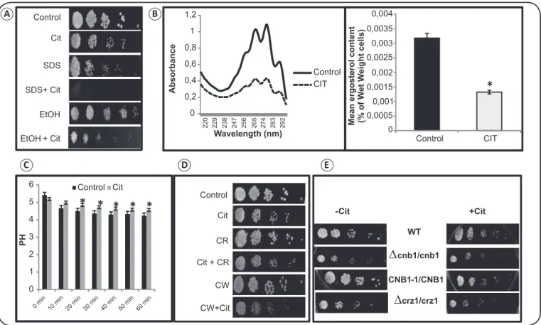
To fi nd out whether the antifungal mechanism of Cit is associated with any cell membrane disruption, phenotypic susceptibility assays were performed in the presence of SDS and ethanol (EtOH). SDS is a commonly used membrane disrupting anionic detergent. We observed that treatment with Cit led to hypersensitivity of the cells in the presence of both SDS and EtOH, which is suggestive of one mechanism by which Cit damages the cell membrane of C. albicans (Figure 2A). Further, we estimated the ergosterol level in the presence of Cit. Ergosterol is one of the main components of the cell membrane of C. albicans. We observed a marked decrease (p value < 0.05) in ergosterol levels by more than 50% in the presence of Cit (Figure 2B). Next, we determined whether the disrupted membrane homeostasis in the presence of Cit could lead to any reduction in plasma membrane ATPase activity. We observed that Cit signifi cantly (p < 0.05) delayed the glucose-induced reduction in the external medium of C. albicans (Figure 2C).
To test whether Cit affects cell wall integrity (CWI), we performed the phenotypic susceptibility assay in the presence of cell wall disrupting agents, namely CW and CR. We observed that the Candida cells were resistant to both CR and CW even in the presence of Cit (Figure 2D). To rule out the possibility of the indispensability of functional calcineurin signaling in the presence of Cit, we performed spot assays using calcineurin mutants such as Δcnb1 (regulatory B subunit) and Δcrz1, and a calcineurin strain having a hyperactive allele of CNB1. We observed that the growth of C. albicans cells was not affected by any of the tested mutants even in the presence of Cit (Figure 2E).
Cit induces oxidative and genotoxic stresses
Relative growth
100 % Growth No Growth
C. tropicalis
Conc. (µg/ml)
Control 1mg/ml
C. glabrata
C. krusei
C. parapsilosis
1.2mg/ml
SC5314 SC5314
(0.015-4000)
C.glabrata (0.015-4000)
C. krusei (0.015-4000)
C. parapsilosis (0.015-4000)
2 1.6 1.4 1.2 1 0.8 0.6 0.4 0.2 0
0.8mg/ml
FIGURE 1. Drug susceptibility assays against Candida albicans and non-albicans species of Candida in the presence of citronellal. (A) Broth microdilution assay to determine the MIC80 of C. albicans (reference strain, SC5314) and C. glabrata, C. krusei, and C. parapsilosis in the presence of Cit. Data are quantitatively displayed with color (see color bar), where each shade of color represents the relative optical density of a cell suspension. The numerical range of 0 to 2 corresponds to no growth to 100% growth, respectively. The minimum drug concentration that inhibits growth by 80% relative to the drug-free growth control is indicated as MIC80 for each strain. (B) Spot assay of C. albicans (reference strain, SC5314) and C. glabrata, C. krusei, C. parapsilosis, and C. tropicalis in the absence (control) and presence of Cit. C.: Candida; MIC: minimum inhibitory concentration; Cit: citronellal.
A
B
We observed hypersensitivity in the Cit-treated cells, which suggests that there may be some defects caused by Cit in the DNA repair machinery (Figure 3B).
Cit disrupts virulence traits
Yeast-to-hyphae transition: the effect of Cit on the hyphal
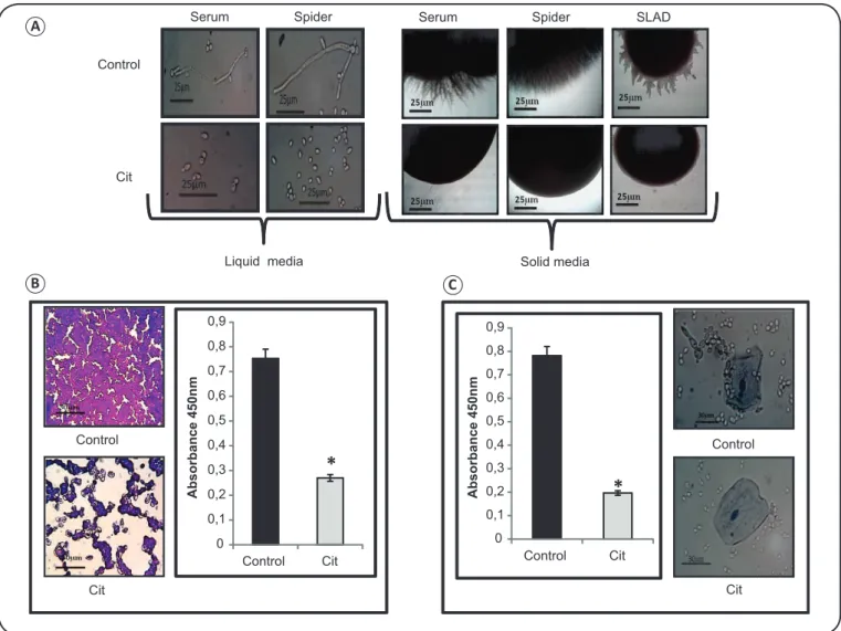
morphogenesis of C. albicans was tested in the absence and presence of Cit. Hyphal morphogenesis is an important virulence trait in fungi. We found that Cit effi ciently inhibited
morphological switching of Candida albicans in various hyphae-inducing media (Figure 4A), which confi rms that Cit is a potent inhibitor of yeast-to-hypha transition.
Biofilm formation: biofilm formation was visualized
Control
Cit
SDS
SDS+ Cit
EtOH
EtOH + Cit
0
220 229 238 247 256 265 274 283 292
0,2 0,4 0,6 0,8 1 1,2
Wavelength (nm)
Control
CIT
*
0 0,0005
0,001 0,0015 0,002 0,0025 0,003 0,0035 0,004
Control CIT
0 1 2 3 4 5 6
P
H
Control Cit
* *
*
*
*
ControlCit
CR
Cit + CR
CW
CW+Cit
∆
cnb1/cnb1CNB1-1/CNB1
∆
crz1/crz1 WT-Cit +Cit
Absorbance
Mean ergosterol content (% of W
et W
eight cells)
FIGURE 2. Effect of citronellal on cell membrane, cell wall, and calcineurin signaling. (A) Spot assays in the absence (control) and presence of Cit (250μg/ml) with SDS (0.02%) and EtOH (5%). (B) The left panel shows the ultraviolet spectrophotometric profi les of ergosterol in C. albicans. Absorbance was measured over a range of 220 to 300nm from a cell culture grown for 16h in the absence and presence of Cit (250μg/ml). The right panel shows the relative percentages of ergosterol in the absence (control) and presence of Cit (250μg/ml). The mean % ergosterol levels were normalized by considering the untreated control as 100 ± SD of 3 independent sets of experiments. *Depicts p value < 0.05. (C) Effect of Cit on the acidifi cation (pH) of the external medium of C. albicans cells. Data are expressed as mean ± SD of three independent sets of experiments. *Depicts p value < 0.05. (D) Spot assay showing no hypersensitivity of C.albicans cells to Cit (250μg/ml) in the presence of the following cell wall-perturbing agents: CR (10μg/ml) and CW (10μg/ml). (E) Spot assay depicting no growth defect in Δcnb1 mutant, Δcrz1 mutant, and CNB1-1/CNB1 (calcineurin overexpressing strain) in the presence of Cit (250μg/ml). Cit: citronellal; SDS: sodium dodecyl sulfate; EtOH: ethanol; CR: Congo red; CW: calcofl uor white; WT: wild type; CNB1: hyperactive allele of calcineurin subunit B; C.: Candida; SD: standard deviation.
A B
C D E
Control
Cit
EtBr
EtBr + Cit
Effect on genotoxicity
FIGURE 3. Effect of citronellal on reactive oxygen species production and genotoxicity in C. albicans. (A) Spot assays showing hypersensitivity of C.albicans cells in the presence of Cit at MIC80 (1mg/ml) alleviated by AA (20mM), which is an antioxidant. (B) Spot assays depicting inhibition of growth by Cit (250µg/ ml) in the presence of EtBr (30μg/ml), which is a DNA-damaging agent. ROS: reactive oxygen species; Cit: citronellal; AA: ascorbic acid; EtBr: ethidium bromide; C.: Candida; MIC: minimum inhibitory concentration; DNA: deoxyribonucleic acid.
B
Control
Cit
AA
AA + Cit
Effect on ROS
the results from the crystal violet staining. Biofi lm formation was considerably (p < 0.05) inhibited in C. albicans by more than 50% in the presence of Cit (Figure 4B).
Cell adhesion to polystyrene surface and human oral
epithelial cells: we observed that cell adherence was signifi cantly
(p < 0.05) reduced by more than 50% on a microtiter polystyrene plate in the presence of Cit (Figure 4C). In order to confi rm this, an adherence assay was performed using human oral epithelial cells with trypan blue dye. Figure 4C shows that C. albicans
treated with Cit presented a normal morphology with few or no adherence to the epithelial cells. In the control experiment,
C. albicans appeared adhered to the epithelial cells, with only a few free cells in the culture medium.
Control
Cit Control
0 0,1 0,2 0,3 0,4 0,5 0,6 0,7 0,8 0,9
Control Cit
*
0 0,1 0,2 0,3 0,4 0,5 0,6 0,7 0,8 0,9
Control Cit
Cit
Serum
Control
Cit
Spider Serum Spider SLAD
Liquid media Solid media
*
*
A
b
sor
b
ance 450n
m
A
b
sor
b
ance 450n
m
A
B C
FIGURE 4. Effect of citronellal on the virulence traits of C. albicans. (A) The left panel shows hyphal morphogenesis in liquid hyphal-inducing media (YEPD containing either 10% horse serum or spider medium) in the absence (control) and presence of Cit (250μg/ml) in C. albicans (SC5314) at 4h of incubation(magnifi cation 40×). The right panel shows hyphal morphogenesis in solid hyphal-inducing media (10% horse serum, spider, or SLAD) in the absence (control) and presence of Cit (250μg/ml) in C. albicans (SC5314) at 6 days of incubation (magnifi cation 4×). (B) The left panel displays crystal violet staining showing biofi lm formation in the absence (control) and presence of Cit (250μg/ml). The right panel shows biofi lm formation depicted as a bar graph and quantifi ed using MTT assay. Data are expressed as mean ± SD of three independent sets of experiments. *Depicts p value < 0.05. (C) The left panel shows the effect of Cit on the adhesion of C. albicans to a polystyrene surface, which was quantifi ed using MTT assay. Data are expressed as mean ± SD of three independent sets of experiments. *Depicts p value < 0.05. The right panel shows the effect of Cit on the adherence of C. albicans to human oral epithelial cells. The untreated cells displayed pseudohyphae formation and adhered to the human oral epithelial cells, whereas the Cit (250μg/ml)-treated cells existed only in the yeast form and did not adhere to the epithelial cells. SLAD: synthetic low ammonium dextrose; Cit: citronellal; C.: Candida; YEPD: yeast extract peptone dextrose;MTT: thiazolyl blue; SD: standard deviation.
DISCUSSION
In this study, we showed that Cit has a potent antifungal activity against C. albicans. We also tested the effi ciency of
Cit against other Candida species. Our fi ndings confi rmed that
Cit is equally effective against both albicans and non-albicans
species of Candida (Figure 1). Interestingly, we did not observe any appreciable susceptibility of C. tropicalis to Cit, which suggests that C. tropicalis may have intrinsic resistance to Cit.
Many antifungal drugs such as azoles, polyenes, allylamines, and echinocandins target the cell membrane or cell wall of
the fungal membrane composition closely. Our data showed that Cit caused a decrease in ergosterol levels (Figure 2B); however, whether the decreased ergosterol level was the causal factor of a faster entry of Cit across the fungal membrane remains to be validated. A functional plasma membrane ATPase activity is normally required to maintain pH equilibrium across the plasma membrane. When fungal cells are starved of a carbon source (e.g. glucose), they tend to take up glucose through a proton motive force upon their exposure to a glucose-containing medium. The proton motive force is generated by a proton gradient created by the pumping of intracellular protons out of the cell. This change results in acidifi cation of the external medium due to the change in pH(20). Our results confi rmed that there was an interruption in the pumping of protons to the external medium in the Cit-treated Candida cells (Figure 2C), which reinforced the hypothesis that Cit tampers with membrane homeostasis. The CWI regulates key cellular responses that are important for survival after the exposure of cells to antifungal drugs that target the cell wall and phenocopies compromised calcineurin signaling(21). Moreover, calcineurin signaling governs responses such as membrane stress in C. albicans(15). Thus, the membrane-damaging effect of Cit observed in this study and the existence of a crosstalk with CWI necessitated to confi rm any defect in calcineurin signaling due to Cit. However, based on the above observations (Figure 2D and Figure 2E) it can be hypothesized that the antifungal activity (membrane-damaging effect) of Cit is independent of the cell wall and the calcineurin signaling pathway.
Enhanced oxidative stress leads to the production of reactive oxygen species (ROS) in cells. ROS production is among the dominant mechanisms by which natural compounds cause cell damage in Candida species(22). For instance, monoterpene phenols like thymol and carvacrol increase ROS production in
C. albicans, which negatively affects the antioxidant system of the fungus(8). Similarly, retigeric acid B, which is a triterpene acid isolated from Lobaria kurokawa, exerts its antifungal effect against C. albicans through enhanced ROS production(23). Naphthoquinoidal compounds also exert their effects against
Candida species potentially via an increased intracellular ROS production(24). Therefore, we investigated whether Cit induces oxidative stress in C. albicans. The test was conducted in the presence of AA. Our results indicated that the antifungal action of Cit might be associated with an enhanced production of ROS (Figure 3A). ROS production due to oxidative stress leads to DNA damage and a defective repair machinery(25). Hence, the effect of Cit on DNA repair was tested in the presence of EtBr to confi rm if Cit targets DNA repair (Figure 3B). However, in order to conclude that Cit affects DNA repair machinery in
C. albicans, further studies would be required.
Hyphal morphogenesis, biofilm formation, and cell adherence are potential virulence attributes of C. albicans that govern pathogenicity. C. albicans undergoes yeast-to-hyphae transition, which is an important virulence factor for the pathogenicity of C. albicans(26). Terpenoids are known to inhibit this yeast-to-hypha transition(9) (27). Similarly, cells of Candida species form biofi lms on indwelling devices, which is the major cause of hospital acquired Candida infections(28). Biofi lms are
highly resistant to antifungals and are therefore a signifi cant trait for virulence. Biofi lms consists of extracellular matrix composed of enclosed microcolonies of yeasts and hyphae, which are arranged in a bilayer structure. The inhibitory effect of Cit on yeast-to-hypha transition that was observed in the present study (Figure 4A), as well as functional hyphal morphogenesis being a prerequisite for biofi lm formation(29) led us to examine the effect of Cit on biofi lm formation (Figure 4B). We therefore tested whether Cit inhibits biofi lm formation and whether it affects the adhesion of Candida cells to polystyrene surface and human oral epithelial cells. The adhesion of cells to a substrate is the foremost process in biofi lm formation(29). We observed a reduced in-vitro adhesion (Figure 4C) of Candida cells to a polystyrene surface. This prompted us to assess the adherence of the Candida cells to human oral epithelial cells (Figure 4C), which is required for the pathogenicity of Candida species in oral candidiasis(17).
Taken together, the data obtained from our study clearly depict Cit, with its diverse modes of action, as a promising antifungal agent. Therefore, the antifungal activity of Cit needs to be further explored and validated by conducting more research on it.
Acknowledgments
The authors are thankful to Joseph Heitman for providing the Candida reference and the calcineurin mutant strains as generous gifts. The authors are also thankful to Sumathi Muralidhar, the Regional Sexually Transmitted Disease Research Centre, and Safdarjung Hospital (New Delhi) for providing the clinical isolates of non-albicans strains of Candida. Finally, the authors thank Rajendra Prasad (Dean, Faculty of Science, Engineering, & Technology, Amity University Haryana) for his encouragement and allowing us access to the facilities required for this research.
Confl ict of interest
The authors declare that there is no confl ict of interest.
Financial support
This work was supported by the Science and Engineering Research Board (SERB), New Delhi; grant number SR/FT/LS-12/2012.
REFERENCES
1. Singh S, Fatima Z, Hameed S. Predisposing factors endorsing Candida infections. Infez Med 2015; 23:211-223.
2. Fidel Jr PL. Distinct protective host defenses against oral and vaginal candidiasis. Med Mycol 2002; 40:359-375.
3. Sanglard D, Coste A, Ferrari S. Antifungal drug resistance mechanisms in fungal pathogens from the perspective of transcriptional gene regulation. FEMS Yeast Res 2009; 9: 1029-1050.
4. Tanwar J, Das S, Fatima Z, Hameed S. Multidrug resistance: an emerging crisis. Interdiscip Perspect Infect Dis 2014; 2014:541340.
6. Negri M, Salci TP, Shinobu-Mesquita C, Capoci IRG, Svidzinski TIE, Kioshima ES. Early state research on antifungal natural products. Molecules 2014; 19:2925-2956.
7. Zore GB, Thakre AD, Jadhav S, Karuppayil SM. Terpenoids inhibit Candida albicans growth by affecting membrane integrity and arrest of cell cycle. Phytomed 2011; 18:1181-1190.
8. Khan A, Ahmad A, Ahmad Khan L, Padoa CJ, Van Vuuren S, Manzoor N. Effect of two monoterpene phenols on antioxidant defense system in Candida albicans. Microb Pathog 2015; 80:50-56.
9. Singh S, Fatima Z, Hameed S. Insights into the mode of action of anticandidal herbal monoterpenoid geraniol reveal disruption of multiple MDR mechanisms and virulence attributes in Candida albicans. Arch Microbiol 2016; 198:459-472.
10. Aguiar RWS, Ootani MA, Ascencio SD, Ferreira TPS, dos Santos MM, dos Santos GR. Fumigant antifungal activity of Corymbia citriodora and Cymbopogon nardus essential oils and citronellal against three fungal species. Scientifi c World J 2014; 2014:492138. 11. Trindade LA, de Araujo Oliveira J, de Castro RD, de Oliveira Lima
E. Inhibition of adherence of C. albicans to dental implants and cover screws by Cymbopogon nardus essential oil and citronellal. Clin Oral Investig 2015; 19:2223-2331.
12. Davis D, Edwards Jr JE, Mitchell AP, Ibrahim AS. Candida albicans RIM101 pH response pathway is required for host-pathogen interactions. Infect Immun 2000; 68: 5953-5959.
13. Blankenship JR, Wormley FL, Boyce MK, Schell WA, Filler SG, Perfect JR, et al. Calcineurin is essential for Candida albicans survival in serum and virulence. Eukaryot Cell 2003; 2:422-430.
14. Chen YL, Brand A, Morrison EL, Silao FG, Bigol UG, Malbas Jr FF, et al. Calcineurin controls drug tolerance, hyphal growth, and virulence in Candida dubliniensis. Eukaryot Cell 2011; 10: 803-819.
15. Cruz MC, Goldstein AL, Blankenship JR, Del Poeta M, Davis D, Cardenas ME, et al. Calcineurin is essential for survival during membrane stress in Candida albicans. EMBO J 2002; 21:546-559.
16. Clinical and Laboratory Standards Institute (CLSI). Reference method for broth dilution antifungal susceptibility testing of yeasts. Approved standard, 3rd M27-A3. National Committee for
Clinical and Laboratory Standards, Wayne: 2008.
17. Lopes G, Pinto E, Andrade PB, Valentao P. Antifungal activity of phlorotannins against dermatophytes and yeasts: approaches to the
mechanism of action and infl uence on Candida albicans virulence factor. PLoS One 2013; 8:e72203.
18. Karl WH, Cruz MC, Katiyar SK, Edlind TD. Antagonism of azole activity against Candida albicans following induction of multidrug resistance genes by selected antimicrobial agents. Antimicrob Agents Chemother 1999; 43:1968-1974
19. Song JC, Stevens DA. Caspofungin: pharmacodynamics, pharmacokinetics, clinical uses and treatment outcomes. Crit Rev Microbiol 2015; 15:1-34.
20. Manavathu EK, Dimmock JR, Vashishtha SC, Chandrasekar PH. Proton-pumping-ATPase-targeted antifungal activity of a novel conjugated styryl ketone. Antimicrob Agents Chemother 1999; 43:2950-2959.
21. LaFayette SL, Collins C, Zaas AK, Schell WA, Betancourt-Quiroz M, Gunatilaka AA, et al. PKC signaling regulates drug resistance of the fungal pathogen Candida albicans via circuitry comprised of Mkc1, calcineurin, and Hsp90. PLoS Pathog 2010; 6:e1001069.
22. Li Y, Chang W, Zhang M, Li X, Jiao Y, Lou H. Diorcinol D exerts fungicidal action against Candida albicans through cytoplasm membrane destruction and ROS accumulation. PLoS ONE 2015; 10:e0128693.
23. Chang WQ, Wu XZ, Cheng AX, Zhang L, Ji M, Lou HX. Retigeric acid B exerts antifungal effect through enhanced reactive oxygen species and decreased cAMP. Biochim Biophys Acta 2011; 1810:569-576.
24. A Neto JB, da Silva CR, S Neta MA, Campos RS, Siebra JT, Silva RAC, et al. Antifungal activity of naphthoquinoidal compounds in vitro against fl uconazole-resistant strains of different Candida species: a special emphasis on mechanisms of action on Candida tropicalis. PLoS One 2014; 9:e93698.
25. Ikner A, Shiozaki K. Yeast signaling pathways in the oxidative stress response. Mutat Res 2005; 569:13-27.
26. Ramage G, Saville SP, Thomas DP, López-Ribot JL. Candida biofi lms: an update. Eukaryot Cell 2005; 4:633-638.
27. Xie C, Sun L, Meng L, Wang M, Xu J, Bartlam M, et al. Sesquiterpenes from Carpesium macrocephalum inhibit Candida albicans biofi lm formation and dimorphism. Bioorg Med Chem Lett 2015; 25:5409-5411.
28. Douglas LJ. Candida biofi lms and their role in infection. Trends Microbiol 2003; 11:30-36.