Methotrexate for refractory
Hailey–Hailey disease
Editor
We would like to report the use of methotrexate to treat a female patient with refractory, long standing Hailey–Hailey disease (HHD). To our knowledge, this is only the second case report of methotrexate being used to treat this disease1.
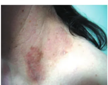
A 42-year-old woman presented to us with active skin lesions affecting both axillae, groins and lateral aspect of neck (Fig. 1) that had an ondulant course, with acute exacerbations and partial remissions for 10 years. A positive family history was present. Skin biopsy confirmed the clinical diagnosis of Hailey–Hailey disease (Fig. 2). Previous treatments included mainly topical corticosteroids, and topical and oral courses of antibiotics and antifungals. Considering disease extension and also standard treatment failure, we decided to start oral methotrexate, 15 mg per week. Lesions started to improve and were nearly clear 1 month later, leaving postinflammatory hyperpigmentation (Fig. 3). Methotrexate was tapered and finally stopped 2 months later. She remained asymptomatic since then with 2 years follow-up and topical preparation of betamethasone/fucidic acid is sufficient to abort discrete developing lesions.
C Vilarinho,* F Ventura, C Brito Hospital Sa˜o Marcos, Largo Carlos Amarante, Apartado 2242,
4701-965 Braga, Portugal *Correspondence:C Vilarinho.E-mail:[email protected]
Reference
1 Fairris GM, White JE, Leppard BJ, Goodwin PG. Methotrexate for intractable benign familial chronic pemphigus.Br J Dermatol1986;115:
640.
DOI: 10.1111/j.1468-3083.2009.03360.x
Severe palmar–plantar
erytrodysesthesia after
treatment with capecitabine
Editor
A 49-year-old woman was diagnosed of an invasive ductal carcinoma in 1999 and was treated with total mastectomy. She received six courses of a combination chemotherapy containing epirubicin, 5-fluoracil and cyclophosphamide plus tamoxifen for 5 years. In December 2007, she was diagnosed with metastatic disease consisting of liver metastasis and she was given six courses of Figure 2 Full-thickness acantholysis.
Figure 3 Post-inflammatory hyperpigmentation.
Figure 1 Microvesicules and crusts in the base of the neck.
106 LETTERS TO THE EDITOR
ª2009 The Authors