301 301 301 301 301 Mem Inst Oswaldo Cruz, Rio de Janeiro, Vol. 99(3): 301-306, M ay 2004
Genomic Characterization of a Brazilian TT Virus Isolate Closely
Related to SEN Virus-F
Leonardo D iniz-M endes, Sylvie D evalle, Christian Niel+
Departamento de Virologia, Instituto Oswaldo Cruz-Fiocruz, Av. Brasil 4365, 21045-900 Rio de Janeiro, RJ, Brasil
SEN virus (SENV) is a circular, single stranded DNA virus that has been first characterized in the serum of a human immunodeficiency virus type 1 (HIV-1)-infected patient. Eight genotypes of SENV (A-H) have been identified and further recognized as variants of TT virus (TTV) in the family Circoviridae. Here we describe the first genomic characterization of a SENV isolate (5-A) from South America. Using ‘universal’ primers, able to amplify most, if not all, TTV/SENV genotypes, a segment of > 3 kb was amplified by polymerase chain reaction from the serum of an HIV-1 infected patient. The amplicon was cloned and a 3087-nucleotide sequence was determined, that showed a high (85%) homology with the sequence of the Italian isolate SENV-F. Proteins encoded by open reading frames (ORFs) 1 to 4 consisted of 758, 129, 276, and 267 amino acids, respectively. By phylogenetic analysis, isolate 5-A was classified into TTV genotype 19 (phylogenetic group 3), together with SENV-F and TTV isolate SAa-38.
Key words: hepatitis human immunodeficiency virus type 1 nucleotide sequencing open reading frames 14 -SEN virus-F - TT virus
Recently, a new circular, single stranded DNA virus, related to the TT virus (TTV) prototype in the genus Anellovirus (Circoviridae family), was identified and des-ignated as SEN virus (SENV) after the initials of the in-fected patient, a human immunodeficiency virus (HIV) infected injection drug user (Fiordalisi et al. 2000). Phylo-genetic analysis of SENV isolates has demonstrated the existence of eight genotypes (A-H), all of which showing < 55% sequence homology at the nucleotide level and < 37% at the amino acid (aa) level with TTV prototype TA278 (Tanaka et al. 2001). However, TTV and SENV show a similar genomic organization. Analysis of the complete TTV nucleotide sequence (3.8 kb) has identified two ma-jor, partially overlapping open reading frames (ORFs), ORF1 and ORF2, which encode 770 and 120 aas, respec-tively (Okamoto et al. 1998). An alternative RNA splicing mechanism generates two additional coding regions, ORF3 (286 codons) and ORF4 (289 codons). The N-termi-nal portions of ORF3 and ORF4 products are common to ORF2 product, while the C-terminal moieties of ORF1, ORF3, and ORF4 products are encoded by the same ge-nome region, each in a different reading frame, without homology to each other (Kamahora et al. 2000, Okamoto et al. 2000).
Human circoviruses are ubiquitous and most humans are infected with at least one (and frequently more than one) genotype. These viruses show a very wide range of sequence divergence, and the number of recognized geno-types has increased continuously in the recent years. A comprehensive classification of the TTV-related viruses
into five major phylogenetic groups (1-5) has been pro-posed (Peng et al. 2002), in which the eight SENV geno-types were included in group 3.
Accumulated evidence suggests that many cases of viral hepatitis are not related to the well-characterized hepatitis viruses A to E (Mushahwar 2000). In this con-text, TTV was originally recovered from a patient with posttransfusion hepatitis of unknown etiology (Nishizawa et al. 1997). Moreover, a strong association has been found between acute infection with SENV-D or SENV-H and the development of transfusion associated non-A to E hepa-titis (Unemura et al. 2001). On the other hand, TTV group 4 has been recently associated with acute respiratory dis-eases in children (Maggi et al. 2003). However, whether TTV and SENV are causative agents of any disease has not been clearly demonstrated.
In the last two years, several studies, performed in Europe, Asia, and North America, have evaluated the prevalence, modes of transmission and clinical relevance of SENV (Shibata et al. 2001, Unemura et al. 2001, Kao et al. 2002, Mikuni et al. 2002, Pirovano et al. 2002a,b, Yoshida et al. 2002, Schröter et al. 2002, 2003). However, few data are available regarding the circulation of SENV strains in South America. The present study constitutes the first genomic characterization of a SENV isolate from South America.
MATERIALS AND METHODS
Twenty-four serum samples, collected from 25-35 years old, HIV-1 infected patients (18 male and 6 female) living in Rio de Janeiro, Brazil, were used in this study. Twelve of the 18 men were homosexual or bisexual and four oth-ers reported having multiple sexual partnoth-ers. All six women had a HIV-1 seropositive sexual partner. One TTV clone was sequenced in this study. It was derived from a 31-year-old bisexual man, who was in stage IV of acquired immunodeficiency syndrome (AIDS) the most advanced stage according to the classification of the US Centers for Disease Control and Prevention.
Financial support: CNPq and Faperj
+Corresponding author. Fax: + 55-21-22706397.
302 302 302 302
302 SEN Virus in Brazil • Leonardo D iniz-M endes et al.
Viral DNA was extracted using phenol/chloroform af-ter treatment of 250 µl of serum with 0.5 mg/ml of protein-ase K in the presence of 0.2 M NaCl, 0.25% SDS, for 4 h at 37oC. After precipitation with ethanol, the pellet was dried and resuspended in 30 µl of distilled water.
Polymerase chain reaction (PCR) assays were per-formed using 2.5 µl of DNA and 0.75 unit of a mixture of
Taq and Pyrococcus species GB-D thermostable DNA poly-merases (Elongase, Invitrogen, Carlsbad, CA) in a final volume of 25 µl. Oligonucleotide primers used in this study were T1S (external sense), T1A (external antisense), T2S (internal sense), and T2A (internal antisense), which have been designed at the 3’ (primers sense) and 5’ (primers antisense) ends of the conserved untranslated region, so that a large DNA segment (of approximately 3100 bp) was amplified, encompassing the most variable part (and all the ORFs) of the genome (Devalle & Niel 2004). After an initial DNA denaturation for 30 s at 94oC, first round am-plification was performed for 35 cycles at 94oC for 20 s, 59oC for 30 s, and 68oC for 3 min 30 s, followed by a final elongation of 10 min at 68oC. One microliter of amplifica-tion product was used in a second round PCR which was done in the same conditions, only decreasing annealing temperature to 53oC. PCR products (10 µl) were loaded on a 2% agarose gel, electrophoresed, and stained with ethidium bromide to visualize DNA bands.
PCR products were cloned into the pCR2.1-TOPO vec-tor (TOPO TA cloning kit, Invitrogen, San Diego, CA) according to the manufacturer’s instructions. Recombi-nant plasmids were purified and the presence of inserts was confirmed after digestion with EcoRI restriction en-donuclease, 2% agarose electrophoresis, and visualiza-tion of DNA bands under ultraviolet light. Nucleotide se-quencing of one clone (5-A) was performed using the Big Dye Terminator cycle-sequencing kit (Applied Biosys-tems, Foster City, CA). Cycle sequencing was carried out with an automatic DNA sequencer (ABI model 373; Ap-plied Biosystems) and performed in both orientations for confirmation. The nucleotide sequence is available in the DDBJ/EMBL/GenBank databases under the accession number AY449524.
The nucleotide sequence of clone 5-A was compared with previously characterized sequences using BLAST search (internet address http://www.ncbi.nlm.nih.gov/ BLAST/) to identify the samples most closely related to clone 5-A. The positions of the translation initiation codons, splice donor and acceptor sites, stop codons and polyadenylation site were determined by multiple se-quence alignment performed with PILEUP program (Wis-consin Sequence Analysis Package, GCG, Madison, WI). This program, which uses the unweighted pair-group method with arithmetic averages (UPGMA) procedure, was also used for the construction of a phylogenetic tree.
RESULTS
Viral DNAs were extracted from 24 serum samples col-lected from HIV-1 infected patients, and submitted to nested PCR amplification using oligonucleotide primers T1S, T2S, T1A, and T2A. These primers have been de-signed in conserved genome regions to be able to amplify most, if not all, TTV/SENV DNAs and to generate a
amplicon covering approximately 80% (and the whole cod-ing region) of the viral genome (Devalle & Niel 2004). Twenty-one (87%) of the 24 samples tested were success-fully amplified, and a DNA band of approximately 3.1 kb was observed after agarose gel electrophoresis. One of the amplicons was cloned and the entire nucleotide se-quence of one clone (5-A) was determined.
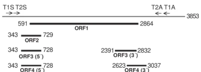
The sequence was 3087 bp in length (nucleotides 223-3309, numbering according to TTV prototype TA278). Fig. 1 shows the localization of the four ORFs of isolate 5-A. ORF1 extended from nt 591 to nt 2864, and encoded a protein of 758 aas that showed an arginine-rich domain at its amino terminus (49% Arg among residues 1-91). The translation initiation codon common to ORF2, ORF3, and ORF4 was located at nt 343 and the ORF2 stop codon at nt 730. ORF2 encoded therefore a putative protein of 129 aas. The donor site for alternative splice, the acceptor site for ORF3 and the acceptor site for ORF4 were situated at nt 728, 2391, and 2623, respectively, giving rise to an ORF3 of 276 aas and an ORF4 of 267 aas. The ORF4 stop codon (nts 3038-3040) and the unique polyadenylation signal (nts 3039-3044) were found to overlap in the motif TAATAAA.
Fig. 1: localization of the four open reading frames (1-4) on the genome of isolate 5-A. For convenience, the circular genome of the virus is represented as a linear DNA. Since the total genomic length of the 5-A isolate has not been determined, numbering was based on TTV prototype isolate TA278 (3853 nt), assuming that the genome segment of isolate 5-A located upstream oligonucle-otide T2S does not show any insertion or deletion. Note that the ORF2 stop codon was located one nucleotide downstream from the donor site for alternative splicing which generates ORF3 and ORF4.
Table I shows the percent identities (at nt and aa lev-els) between clone 5-A and TTV-related viruses belong-ing to the five major phylogenetic groups and whose ORFs 1 to 4 have been characterized. Clone 5-A showed a low (53.3-59.5%) sequence homology with the five isolates. Despite the great nucleotide sequence divergence exist-ing among the five groups, the sizes of the proteins ap-peared to be well conserved, with ORF1 product varying from 719 to 770 aas, ORF2 product from 115 to 130 aas, ORF3 protein from 260 to 303 aas, and ORF4 protein from 249 to 297 aas. When other TTV-related viruses were as-sessed (Table II), isolate 5-A showed a relatively high sequence homology with members of phylogenetic group 3, particularly with SENV-F and SAa-38 (82-85% homol-ogy) and, in a lesser extent, with SENV-D and TJN01 (76-77%).
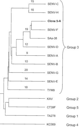
A phylogenetic analysis was performed with the nucle-otide sequences of clone 5-A and of 14 TTV/SENVs
avail-T1S T2S T2A T1A
303 303 303 303 303 Mem Inst Oswaldo Cruz, Rio de Janeiro, Vol. 99(3), M ay 2004
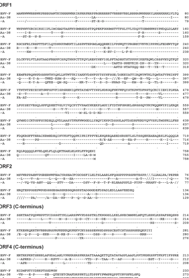
able in the DNA databanks, i.e. one from each of the phy-logenetic groups 1, 2, 4, and 5, as well as 10 representative isolates from group 3, including the eight SENV geno-types. Phylogenetic tree (Fig. 2) shows that clone 5-A was classifiable into genotype 19 (phylogenetic group 3), together with isolates SENV-F and SAa-38. The sequences of the four putative proteins of SENV-F, SAa-38, and clone 5-A were aligned (Fig. 3). The most conserved region was the C-terminal portion of ORF3 product, a
post-trans-lationally phosphorylated serine-rich region that has been suggested to modulate function of host cellular proteins and to play a role in maintaining persistent infection (Asabe et al. 2001). SENV-F ORF4 product appeared to be truncated when compared to the corresponding products of SAa-38 and clone 5-A. Of note, the tail of ORF4 of clone 5-A (positions 229 to 267) showed a high (82%) homology with the corresponding ORF4 region of isolate SAa-38.
TABLE I
Nucleotide and amino acid sequence identity between isolate 5-A and TTV-related viruses representative of the five major phylogenetic groups
Percent identity (amino acids) Number of amino acid residues Isolate Genomic Accession Percent identity
group number (nucleotides) ORF1 ORF2 ORF3 ORF4 ORF1 ORF2 ORF3 ORF
5-A AY449524 100 100 100 100 100 758 129 276 267
TA278 1 AB017610 56.1 47.3 50.4 46.9 50.6 770 120 286 289
KAV 2 AF435014 57.0 53.2 51.3 49.8 50.0 719 125 264 273
TYM9 3 AB050448 59.5 45.0 50.4 48.8 45.9 748 115 260 249
KC009 4 AB038621 53.3 45.7 47.6 44.4 47.1 733 128 303 297
CT39F 5 AB064604 56.3 47.5 54.1 42.7 47.8 743 130 272 266
TABLE II
Nucleotide sequence identity (near full-length genome) between isolate 5-A and TTV/SENV isolates from different origins
Isolate Accession number Genomic group Genotype Percent identity a
JA1 AF122916 1 2 55.1
T3PB AF247138 1 3 55.2
PMV AF261761 2 17 56.1
Kt-08F AB054647 2 22 56.1
Kt-10F AB054648 2 23 56.0
SAa-01 AB060597 3 27 59.3
SAa-10 AB060594 3 24 58.2
SAa-38 AB060593 3 19 82.4
SAa-39 AB060592 3 18 59.5
SAf-09 AB060596 3 26 58.9
SAj-30 AB060595 3 25 59.7
SANBAN AB025946 3 13 60.1
SENV-A AX025667 3 9 69.2
SENV-B AX025677 3 10 63.0
SENV-C AX025718 3 15 63.3
SENV-D AX025730 3 12 76.5
SENV-E AX025761 3 14 61.1
SENV-F AX025822 3 19 84.7
SENV-G AX025830 3 20 64.5
SENV-H AX025838 3 16 63.1
TJN01 AB028668 3 12 77.1
TJN02 AB028669 3 13 60.2
TUPB AF247137 3 11 66.4
TUS01 AB017613 3 11 67.1
TYM9 AB050448 3 18 59.5
CT23F AB064595 4 N.A. 52.7
CT25F AB064596 4 N.A. 54.9
JT14F AB064601 4 N.A. 53.1
CT44F AB064605 5 N.A. 55.9
JT33F AB064606 5 N.A. 56.1
JT34F AB064607 5 N.A. 55.4
304 304 304 304
304 SEN Virus in Brazil • Leonardo D iniz-M endes et al.
DISCUSSION
Evidence has accumulated that TTV viremia is ex-tremely common in the general population worldwide (Prescott & Simmonds 1998, Takahashi et al. 1998, Niel et al. 1999). TTV DNA has been detected at higher viral loads in HIV infected patients than in controls. It has thus been proposed that TTV load may reflect the degree of immune status of immunocompromised hosts (Christensen et al. 2000, Shibayama et al. 2001, Touinssi et al. 2001). Further-more, coinfection of single individuals with different TTV isolates has been found more frequently in HIV infected patients than in blood donors (Sherman et al. 2001, Devalle & Niel 2004). In this study, a large (3.1 kb) segment of the SENV genome was PCR amplified. Since effective amplifi-cation of long targets are sometimes difficult, a mixture of thermostable DNA polymerases was used to increase PCR efficiency, making possible the successful amplification of TTV/SENV genomes from 21/24 (87%) sera collected from HIV-1 infected patients. In a parallel experiment, how-ever, no amplification was obtained from 24 sera collected from blood donors (not shown). As the PCR assay used here was designed to amplify most, if not all, TTV/SENV genomes, it can be concluded that such a discrepancy between HIV infected patients and blood donors was likely due to a higher viral load (rather than a higher prevalence)
in HIV infected patients.
The detection of SENV in Brazilian patients with chronic hepatitis of unknown etiology has been mentioned previously (Chemin et al. 2002), but without any details. The present study constitutes the first characterization of a SENV isolate from South America. The first SENV isolate identified in the world, namely SENV-A, was ini-tially found in an Italian HIV infected patient (Tanaka et al. 2001). A high prevalence of genotype SENV-A has been later demonstrated in intravenous drug user HIV infected patients (Pirovano et al. 2002b). The virus characterized here was also found in an HIV infected patient, more pre-cisely a bisexual, 31 year-old man who was at an advanced stage of AIDS. The virus showed a high (84.7%) homol-ogy with SENV-F and was classified into genotype 19, together with isolates SENV-F and SAa-38. Further stud-ies will be necessary to determine whether the other SENV genotypes (A-E, G, H) circulate or not in South America, both in HIV infected individuals and in the general popu-lation.
All eight SENV genotypes have been recently classi-fied into TTV phylogenetic group 3, which is the group showing the major genetic diversity and including the largest number of genotypes (Okamoto & Mayumi 2001, Peng et al. 2002). Here we observed a low (59-70%) se-quence homology between isolate 5-A, a variant not very distant of SENV-F, and well-characterized viral strains from group 3, as SANBAN (Hijikata et al. 1999), TUS01 (Okamoto et al. 1999), and TYM9 (Okamoto et al. 2000). At this respect, it should be kept in mind that the present classification of TTVs/SENVs in five phylogenetic groups is provisional and may be modified in the future if new genotypes, whose existence has been recently suggested (Kojima et al. 2003, Maggi et al. 2003, Devalle & Niel 2004), are actually identified.
The natural history and pathogenic potential of TTVs/ SENVs are currently the subject of active investigation. These viruses have been proposed to be associated with hepatitis and acute respiratory diseases, although a caus-ative effect has not been established. A possibility exists that certain strains cause disease while most others are non-pathogenic. In this context, studies like the present one, which contribute to determine which genotypes cir-culate in defined geographic regions, should be of great relevance in the future, when the clinical significance of the various TTV/SENV genotypes is known.
REFERENCES
Asabe SI, Nishizawa T, Iwanari H, Okamoto H 2001. Phos-phorylation of serine-rich protein encoded by open reading frame 3 of the TT virus genome. Biochem Biophys Res Commun 286: 298-304.
Chemin I, Parana R, Trepo C 2002. A new viral agent, SEN virus (SENV), has been detected in patients from several countries: the pathogenic role of SENV in coinfections with hepatitis B virus or hepatitis C virus should be investi-gated. J Infect Dis 185: 710.
Christensen JK, Eugen-Olsen J, Sorensen M, Ullum H, Gjedde SB, Pedersen BK, Nielsen JO, Krogsgaard K 2000. Preva-lence and prognostic significance of infection with TT virus in patients infected with human immunodeficiency virus. J Infect Dis 181: 1796-1799.
305 305 305 305 305 Mem Inst Oswaldo Cruz, Rio de Janeiro, Vol. 99(3), M ay 2004
306 306 306 306
306 SEN Virus in Brazil • Leonardo D iniz-M endes et al.
Devalle S, Niel C 2004. Distribution of TT virus genomic groups 1 to 5 in Brazilian blood donors, HBV carriers, and HIV-1-infected patients. J Med Virol 72: 166-173.
Fiordalisi G, Bonelli M, Olivero P, Primi D, Vaglini L, Mattioli S, Bonelli F, Dal Corso A, Mantero GL, Sottini A 2000. Identification of SENV genotypes. International patent number WO 0028039 (international application published under the patent cooperation treaty). Internet address: http:/ /ep.espacenet.com/
Hijikata M, Takahashi K, Mishiro S 1999. Complete circular DNA genome of a TT virus variant (isolate name SANBAN) and 44 partial ORF2 sequences implicating a great degree of diversity beyond genotypes. Virology 260: 17-22. Kamahora T, Hino S, Miyata H 2000. Three spliced mRNAs of
TT virus transcribed from a plasmid containing the entire genome in COS1 cells. J Virol 74: 9980-9986.
Kao JH, Chen W, Chen PJ, Lai MY, Chen DS 2002. Prevalence and implication of a newly identified infectious agent (SEN virus) in Taiwan. J Infect Dis 185:389-392.
Kojima H, Kaita KD, Zhang M, Giulivi A, Minuk GY 2003. Genomic analysis of a recently identified virus (SEN virus) and genotypes D and H by polymerase chain reaction. An-tiviral Res 60: 27-33.
Maggi F, Pifferi M, Fornai C, Andreoli E, Tempestini E, Vatteroni M, Presciuttini S, Marchi S, Pietrobelli A, Boner A, Pistello M, Bendinelli M 2003. TT virus in the nasal secretions of children with acute respiratory diseases: Re-lations to viremia and disease severity. J Virol 77: 2418-2425.
Mikuni M, Moriyama M, Tanaka N, Abe K, Arakawa Y 2002. SEN virus infection does not affect the progression of non-A to -E liver disease. J Med Virol 67:624-629.
Mushahwar I 2000. Recently discovered blood-borne viruses: Are they hepatitis viruses or merely endosymbionts? J Med Virol 62: 399-404.
Niel C, de Oliveira JM, Ross RS, Gomes SA, Roggendorf M, Viazov S 1999. High prevalence of TT virus infection in Brazilian blood donors. J Med Virol 57: 259-263.
Nishizawa T, Okamoto H, Konishi K, Yoshizawa H, Miyakawa Y, Mayumi M 1997. A novel DNA virus (TTV) associated with elevated transaminase levels in posttransfusion hepa-titis of unknown etiology. Biochem Biophys Res Commun 241: 92-97.
Okamoto H, Nishizawa T, Kato N, Ukita M, Ikeda H, Iizuka H, Miyakawa Y, Mayumi M 1998. Molecular cloning and characterization of a novel DNA virus (TTV) associated with posttransfusion hepatitis of unknown etiology. Hepatol Res 10: 1-16.
Okamoto H, Nishizawa T, Ukita M, Takahashi M, Fukuda M, Iizuka H, Miyakawa Y, Mayumi M 1999. The entire nucle-otide sequence of a TT virus isolate from the United States (TUS01): Comparison with reported isolates and phyloge-netic analysis. Virology259: 437-448.
Okamoto H, Nishizawa T, Tawara A, Takahashi M, Kishimoto J, Sai T, Sugai Y 2000. TT virus mRNAs detected in the bone marrow cells from an infected individual. Biochem Biophys Res Commun279: 700-707.
Okamoto H, Mayumi M 2001. TT virus: virological and
ge-nomic characteristics and disease associations. J Gas-troenterol36: 519-529.
Peng YH, Nishizawa T, Takahashi M, Ishikawa T, Yoshikawa A, Okamoto H 2002. Analysis of the complete genomes of thirteen TT virus variants classifiable into the fourth and fifth genetic groups, isolated from viremic infants. Arch Virol147: 21-41.
Pirovano S, Bellinzoni M, Ballerini C, Cariani E, Duse M, Albertini A, Imberti L 2002a. Transmission of SEN virus from mothers to their babies. J Med Virol 66: 421-427. Pirovano S, Bellinzoni M, Matteelli A, Ballerini C, Albertini A,
Imberti L 2002b. High prevalence of a variant of SENV in intravenous drug user HIV-infected patients. J Med Virol 68: 18-23.
Prescott LE, Simmonds P 1998. Global distribution of transfu-sion-transmitted virus. New England J Med 339: 776-777. Schröter M, Laufs R, Zollner B, Knodler B, Schafer P, Sterneck M, Fischer L, Feucht HH 2002. Prevalence of SENV-H viraemia among healthy subjects and individuals at risk for parenterally transmitted diseases in Germany. J Viral Hepatol 9: 455-459.
Schröter M, Laufs R, Zollner B, Knodler B, Schafer P, Feucht HH 2003. A novel DNA virus (SEN) among patients on maintenance hemodialysis: prevalence and clinical impor-tance. J Clin Virol 27: 69-73.
Sherman KE, Rouster SD, Feinberg J 2001. Prevalence and ge-notypic variability of TTV in HIV-infected patients. Dig Dis Sci 46: 2401-2407.
Shibata M, Wang RYH, Yoshiba M, Shih JWK, Alter HJ, Mitamura K 2001. The presence of a newly identified in-fectious agent (SEN virus) in patients with liver disease and in blood donors in Japan. J Infect Dis 184:400-404. Shibayama T, Masuda G, Ajisawa A, Takahashi M, Nishizawa
T, Tsuda F, Okamoto H 2001. Inverse relationship between the titre of TT virus DNA and the CD4 cell count in pa-tients infected with HIV. AIDS 15: 563-570.
Takahashi K, Hoshino H, Ohta Y, Yoshida N, Mishiro S 1998. Very high prevalence of TT virus (TTV) infection in gen-eral population of Japan revealed by a new set of PCR primers. Hepatol Res 12:233-239.
Tanaka Y, Primi D, Wang RY, Umemura T, Yeo AE, Mizokami M, Alter HJ, Shih JW 2001. Genomic and molecular evolu-tionary analysis of a newly identified infectious agent (SEN virus) and its relationship to the TT virus family. J Infect Dis 183:359-367.
Touinssi M, Gallian P, Biagini P, Attoui H, Vialettes B, Berland Y, Tamalet C, Dhiver C, Ravaux I, De Micco P, De Lamballerie X 2001. TT virus infection: prevalence of el-evated viraemia and arguments for the immune control of viral load. J Clin Virol 21: 135-141.
Umemura T, Yeo AE, Sottini A, Moratto D, Tanaka Y, Wang RY, Shih JW, Donahue P, Primi D, Alter HJ 2001. SEN virus infection and its relationship to transfusion-associated hepa-titis. Hepatology33:1303-1311.