TEMPERATURE AND AIR HUMIDITY
Luiz Gustavo Gardinassi1, Paulo Vitor Marques Simas1, João Batista Salomão2, Edison Luiz Durigon3, Dirce Maria Zanetta Trevisan4, José Antonio Cordeiro5, Mauricio Nogueira Lacerda6, **Paula Rahal1, *Fátima Pereira de Souza7
1
Universidade Estadual Paulista, Instituto de Biociências, Letras e Ciências Exatas, Departamento de Biologia, Ribeirão Preto, SP,
Brasil; 2Faculdade de Medicina de São José do Rio Preto, Departamento de Pediatria e Cirurgia Pediátrica, Pediatria e
Puericultura, São José do Rio Preto, SP, Brasil; 3Universidade de São Paulo, Instituto de Ciências Biomédicas, Departamento de
Microbiologia, São Paulo, SP, Brasil; 4Faculdade de Saúde Pública, Universidade de São Paulo, São Paulo, SP, Brasil; 5Faculdade
de Medicina de São José do Rio Preto, Departamento de Epidemiologia e Saúde Coletiva, São José do Rio Preto, SP, Brasil; 6
Faculdade de Medicina de São José do Rio Preto, Departamento de Doenças Infecciosas e Parasitárias, Laboratório de Pesquisas
em Virologia, São José do Rio Preto, SP, Brasil; 7Universidade Estadual Paulista, Instituto de Biociências, Letras e Ciências
Exatas, Departamento de Física, São José do Rio Preto, SP, Brasil.
Submitted: March 21, 2010; Returned to authors for corrections: December 12, 2010; Approved: August 30, 2011.
ABSTRACT
Viruses are the major cause of lower respiratory tract infections in childhood and the main viruses involved
are Human Respiratory Syncytial Virus (HRSV), Human Metapneumovirus (HMPV), Influenzavirus A and B
(FLUA and FLUB), Human Parainfluenza Virus 1, 2 and 3 (HPIV1, 2 and 3) and Human Rhinovirus
(HRV). The purposes of this study were to detect respiratory viruses in hospitalized children younger than
six years and identify the influence of temperature and relative air humidity on the detected viruses. Samples
of nasopharyngeal washes were collected from hospitalized children between May/2004 and
September/2005. Methods of viral detection were RT-PCR, PCR and HRV amplicons were confirmed by
hybridization. Results showed 54% (148/272) of viral positivity. HRSV was detected in 29% (79/272) of the
samples; HRV in 23.1% (63/272); HPIV3 in 5.1% (14/272); HMPV in 3.3% (9/272); HPIV1 in 2.9%
(8/272); FLUB in 1.4% (4/272), FLUA in 1.1% (3/272), and HPIV2 in 0.3% (1/272). The highest detection
rates occurred mainly in the spring 2004 and in the autumn 2005. It was observed that viral respiratory
infections tend to increase as the relative air humidity decreases, showing significant association with
monthly averages of minimal temperature and minimal relative air humidity. In conclusion, viral respiratory
infections vary according to temperature and relative air humidity and viral respiratory infections present
major incidences it coldest and driest periods.
Key words: Viral Respiratory Infections, HRSV, HRV, Temperature, Relative Air Humidity.
*Corresponding Author. Mailing address: Department of Phisics – Instituto de Biociências, Letras e Ciências Exatas - University of São Paulo State – Cristóvão Colombo Street, 2265, 15054-000 São Jose do Rio Preto, SP, Brazil.; Tel.: +55-17-3221-2463.; E-mail: fatyssouza@yahoo.com.br
INTRODUCTION
Respiratory viruses are the most frequent cause of acute
respiratory infections (ARI) in infants and young children, and
important causes of hospitalization in developing countries (1).
The respiratory viruses, particularly Human Respiratory
Syncytial Virus (HRSV), Influenzavirus types A and B (FLUA
and B), Human Parainfluenzavirus 1, 2 and 3 (HPIV 1, 2 and
3), Human Rhinovirus (HRV) and Human Metapneumovirus
(HMPV) have been recognized as the most important
pathogens involved in lower respiratory tract infections (LRTI)
(32, 35, 38). Frequency of viral detection in LRTI depends on
many factors, such as sample management, disease severity,
diagnostic methods and climatic season (34).
Several studies have reported that viral respiratory
infections present seasonal patterns, which are more
accentuated in regions with temperate climate rather than
tropical climate (8). As an example, outbreaks of respiratory
syncytial virus and influenza viruses occur in the winter season
in regions with temperate climate(3).
Brazil is a country where several studies revealed viruses
as the main causes of respiratory infections in Fortaleza, in Rio
de Janeiro, in São Paulo and in Curitiba (18, 35, 36), as
reported by Arruda et al. (1). These viruses cause illness that
can range from a brief upper respiratory tract infection, as a
common cold, to a severe systemic illness, like bronchiolitis
and pneumonia, resulting in death(11).
Large countries like Brazil possess a wide range of
geographical areas, each one with unique climatic
characteristics. Epidemiological studies from tropical regions
indicate that factors as rainy seasons (34) or low air humidity
(3), in addition to temperature, may influence respiratory
viruses outbreaks. Global warming carries profound changes in
Earth´s climate, and major changes in the atmosphere and in
the climate have a vast impact on the biosphere and the human
environment (20). Consequently, climatic variations and
extreme weather events also cause profound impacts on
infectious diseases incidences. Infectious agents (such as
protozoa, bacteria and viruses) are devoid of thermostatic
mechanisms, and reproduction and survival rates are thus
strongly affected by fluctuations in temperature. Temperature
dependencies are seen in correlations between disease rates and
weather variations over weeks, months or years, as well as in
close geographic associations with key climatic variables and
the distributions of important infectious diseases (28).
Knowledge of the trends and seasonality of respiratory
viral infections in the community could be a first step to confer
information for health care providers, to facilitate the
implementation of strategies to prevent and minimize
transmission, and also introduce early therapeutic options to
high risk patients (13).
The aim of this study was to detect the presence of
respiratory viruses in clinical samples collected from children
with respiratory tract infection, and analyze the seasonal trends
and occurrence patterns of the identified viruses.
MATERIALS AND METHODS
Clinical Specimens
This study was conducted at the Genomic Studies
Laboratory of the Sao Paulo State University, located in Sao
Jose do Rio Preto, São Paulo State, whose inhabitants reach the
number of 402.770. Samples were collected at the Sao Jose do
Rio Preto Base Hospital, a tertiary health care facility. Patients
were children between one month and six years of age
attended between May 2004 and September 2005, suffering
from ARI. A single nurse was responsible for collecting the
samples during the whole study. Nasopharyngeal washes were
obtained after instillation of 0.5ml of sterile PBS (Phosphate
Buffered Saline - NaCl, Na2HPO4, NaH2PO4) into each nostril
with immediate aspiration through a sterile neonatal canula
inserted into the child’s nasopharynx. The sample was
transferred to a sterile vial and immediately transported to the
(Invitrogen, Carlsbad, CA) for later RNA extraction and
RT-PCR testing. This method was chosen because of its high
specificity and sensibility, besides being a rapid test for the
identification of many pathogens, including respiratory viruses.
This study was approved by Research Ethics Unesp/IBILCE by
opinion n° 062/2001 on June 11, 2001, in São José do Rio
Preto, Brazil.
RNA Extraction and cDNA Synthesis
Viral RNA was extracted from 250µl of clinical specimen
following the Trizol LS manufacturer’s instructions and
suspended in 20µl of MiliQ water treated with DEPC
(diethylpyrocarbonate, Sigma, St Louis, USA), cDNA was
synthesized using the High-Capacity cDNA Archive Kit
(Applied Biosystems, Foster City, CA) utilizing random
primers according to the manufacturer’s protocol, and stored at
-20oC.
PCR amplification
The amplification reaction of HMPV, FLU A and FLU B,
HPIV1, HPIV2 and HPIV3 and HRV was performed in a final
volume of 25µl, with 3µl of cDNA, 5µl of buffer (75mM
Tris-HCl pH 9.0, 50mM KCl, 20mM (NH4)2SO4), 3µl of 50 mM
MgCl2, 1µl of 10 mM dNTPs, 2.5µl of each primer (as per 6,
12, 14, 21 ) at 10 pmol (10), 0.5µl (2 U) of DNA polymerase
(Biotools), and MilliQ water treated with DEPC. The reaction
consisted of 40 amplification cycles with denaturation at 94°C
for 45 seconds, annealingat 54°C for 45 seconds; extension at
72°C for 45 seconds, and final extension at 72°C for 7 min. For
Human Rhinovirus identification, the amplicons produced by
PCR were confirmed through hybridization performed with
OLP and OLE oligonucleotide probes as previously published
(31).
HRSV amplification was made by a semi-nested PCR
method which involves two steps. The first step resulted in
final reaction volume of 50µl, using 5µl of cDNA and a
reaction composition identical to the one describe above for the
other viruses. The reaction started with a denaturation step at
95ºC for 5 minutes, followed by 40 cycles of 94ºC for 1
minute, 55ºC for 1 minute, 72ºC for 1 minute, and a final
extension step at 72ºC for 7 minutes. The samples which were
tested negative in this step were undergone a second round of
semi-nested PCR. For the second round, 5 µl of product from
the previous reaction were mixed with 5µl of buffer and the
same remaining components from the 1st round, except for the
use of 1.5µl of each primer at 10pmol (as per 20, 29, 30). The
reaction was preceded by a denaturation step at 95ºC for 5
minutes, followed by 40 cycles at 94ºC for 1 minute, 55ºC for 1
minute, 72ºC for 1 minute, and an extension step at 72ºC for 7
minutes.
The amplification products were analyzed in a 1.2 %
agarose gel stained with ethidium bromide, visualized under
UV light.
Statistical Analysis
Statistical analysis was performed using the Minitab
Statistical Software for Windows, version 12.22, and
differences were considered significant if p<0.05
Geographical and Meteorological Data
São José do Rio Preto is localized in Northwest of São
Paulo State, which is one of the states composing the Southeast
region from Brazil. The city is located at an average altitude of
489 meters, latitude of -20,81972º and longitude of -49,37944°.
São José do Rio Preto has a subtropical climate with an annual
average temperature of 23,6º C, composed by dry and cold
winters, and rainy summers with increased temperatures.
January and March generally are the warmer months of the
year, and the average temperature is 27 ºC. July is the coldest
month, with an average temperature of 19,9 ºC. The autumn
and spring are transition periods.
Data on temperature were supplied by the agriculture and
supply secretary - represented by the Integral Technical
temperature measures made by the Seeds, Seedlings and
Matrices Department of Seeds Production Nucleus from Sao
Jose do Rio Preto. Data on relative air humidity were obtained
from SOMAR Meteorology through daily measures of
REDEMET (Air Command Meteorological Network) at Sao
Jose do Rio Preto Airport.
RESULTS
During the period of study, 272 nasopharyngeal washes
were collected from 272 children aged from1 to 68 months
(average age of 29.5 months). From this total, 44.5% (121/272)
were younger than 24 months and 57% were male (Table 1).
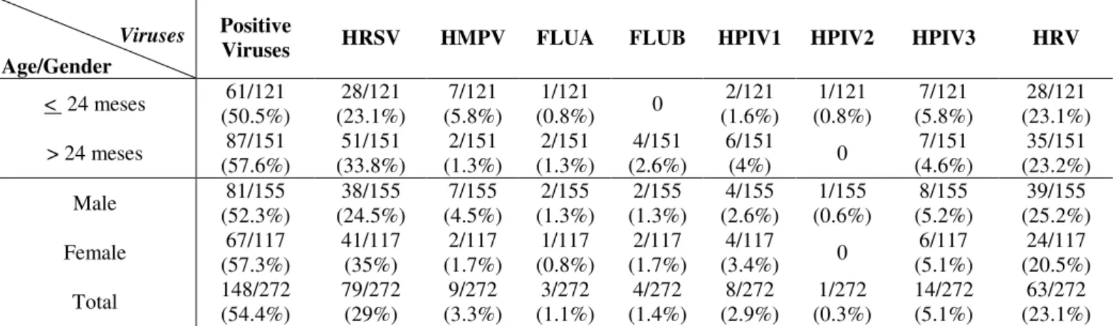
Respiratory viruses were detected in 54.4% (148/272) of the
samples, being HRSV the most detected consisting in 29%
(79/272) from the positive samples. HRV was detected in
23.1% (63/272), HPIV3 in 5.1% (14/272), HMPV in 3.3%
(9/272), HPIV1 in 2.9% (8/272), FLUB in 1.4% (4/272),
FLUA in 1.1% (3/272) and HPIV2 in 0.3% (1/272) (Table 1).
From the 148 positive samples, 29.7% (81/272) were collected
from male patients and 24.6% (67/272) were from female
(Table 1).
Table 1. Association between detected respiratory viruses with age and gender (%).
Viruses
Age/Gender
Positive
Viruses HRSV HMPV FLUA FLUB HPIV1 HPIV2 HPIV3 HRV
< 24 meses 61/121 (50.5%) 28/121 (23.1%) 7/121 (5.8%) 1/121
(0.8%) 0
2/121 (1.6%) 1/121 (0.8%) 7/121 (5.8%) 28/121 (23.1%)
> 24 meses 87/151 (57.6%) 51/151 (33.8%) 2/151 (1.3%) 2/151 (1.3%) 4/151 (2.6%) 6/151
(4%) 0
7/151 (4.6%)
35/151 (23.2%)
Male 81/155
(52.3%) 38/155 (24.5%) 7/155 (4.5%) 2/155 (1.3%) 2/155 (1.3%) 4/155 (2.6%) 1/155 (0.6%) 8/155 (5.2%) 39/155 (25.2%)
Female 67/117
(57.3%) 41/117 (35%) 2/117 (1.7%) 1/117 (0.8%) 2/117 (1.7%) 4/117
(3.4%) 0
6/117 (5.1%)
24/117 (20.5%)
Total 148/272
(54.4%) 79/272 (29%) 9/272 (3.3%) 3/272 (1.1%) 4/272 (1.4%) 8/272 (2.9%) 1/272 (0.3%) 14/272 (5.1%) 63/272 (23.1%) HRSV: Human Respiratory Syncytial Virus; HRV: Human Rhinovirus; HMPV: Human Metapneumovirus; HPIV3: Human Parainfluenzavirus Type 3; HPIV 1:
Human Parainfluenzavirus Type 1; FLUB: Influenzavirus Type B; FLUA: Influenzavirus Type A; HPIV 2: Human Parainfluenzavirus Type 2.
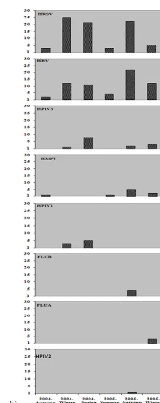
The total respiratory infections were detected mainly in
winter, spring and summer of 2004, and autumn and winter of
2005, as showed in Figure 1A, which also demonstrate the
seasonal distribution of the detected respiratory viruses.
Interestingly, the incidence of viral respiratory infections
occurred between late winter and late spring 2004, with
outbreaks occurring in spring. In contrast, the period of
incidence in 2005 occurred between early autumn and late
winter, with outbreaks in autumn.
Among all seasons that samples were collected, HRSV
was the most identified virus, except for the summer 2005.
Outbreaks were observed in winter and spring 2004, and
autumn 2005. Presenting similar results, HRV was also
detected during the whole period of sample collection.
Outbreaks were observed in winter and spring 2004, and
autumn 2005 (Figure 1B).
HPIV3 and HMPV were detected in four of all seasons
studied, being HPIV3 mainly detected in spring 2004 and
HMPV in autumn 2005. HPIV1 had an incidence in two
seasons, principally detected in spring 2004. FLUA, FLUB and
HPIV2 presented incidences only in one season of the year,
a)
b)
Figure 1. A: Distribution of total respiratory infections according to climatic season (total of collected samples of hospitalized children with
respiratory infections) (above) and viral respiratory infections between autumn/2004 to spring/2005, distributed according to climatic season
(below). B: Viral respiratory infections between autumn/2004 to winter/2005, demonstrating the seasonality of each isolated virus according to
the climatic season. HRSV: Human Respiratory SyncytialVirus; HRV: Human Rhinovirus; HMPV: Human Metapneumovirus; HPIV3: Human
Parainfluenzavirus Type 3; HPIV1: Human Parainfluenzavirus Type 1; FLUB: Influenzavirus Type B; FLUA: Influenzavirus Type A; HPIV2:
The Figure 2 illustrates viral respiratory infections
correlated to monthly average of relative air humidity and
monthly average of temperature. In the beginning of autumn
2004, relative air humidity was 75% and respiratory viruses
were detected in few samples. The results show that when
relative air humidity and temperature begin to decrease, viral
respiratory infections tend to increase; when the temperature
and the levels of relative air humidity increase and are
maintained relatively high (summer 2005), the detection rate of
respiratory viruses decreases, evidencing strong influence of
temperature and relative air humidity levels on the incidence of
viral respiratory infections in this period. The influence of
temperature and relative air humidity on the circulation pattern
of respiratory viruses was evidenced by statistical analysis.
Results present a significant association between viral
respiratory infections and monthly average of minimal relative
air humidity (p=0.046) as well as between the monthly average
of minimal temperature (p=0.044). Therefore, it was evidenced
that meteorological aspects may present influence one over the
other, thus temperature tends to fluctuate according to
characteristics of relative air humidity while the relative air
humidity also varies according to the temperature.
Figure 2. Correlation of viral respiratory infections to monthly average temperature and monthly average relative air humidity,
showing the variation of infections according to the variation of the meteorological factors [T (°C) – monthly average of
Through a specific analysis of the relation between the
positivity of each detected respiratory virus to the temperature
and relative air humidity, it is possible to note that the major
HRSV outbreak occurred in the period of lowest temperatures,
in the winter of 2004. This data also shows an association
statistically significant between HRSV circulation and monthly
average of minimal temperature (p=0.010) (Figure 3).
An interesting observation was made about data from
2005. There was an anticipation of HRSV infections, which
occurred at higher rates in March and April - a transition
between summer and autumn. In this period, there was a
fluctuation of the average temperature, which dropped a few
degrees, then increased for a short period of time and decreased
again. We can also observe that HRSV infections in 2004 were
more prevalent than in 2005, thus evidencing the influence of
temperature on infections by HRSV.
Figure 3. Association of HRSV infections to monthly average of minimal temperatures demonstrating the variation of infections
according to the climatic season and variation of relative air humidity.
DISCUSSION
Acute respiratory infections (ARI) constitute a major
factor in respiratory tract diseases, which are responsible for
high rates of hospitalization and mortality. ARI accounts
approximately for 20% of mortality in children up to 5 years
old (30).
Respiratory viruses have been reported as the main cause
of low respiratory tract infections in children (3, 11, 38,39)
living in emerging countries, and despite advances in the
epidemiological knowledge of these infections, researches
involving respiratory viruses features that circulate in countries
as Brazil are still necessary.
From 272 samples collected from hospitalized children
because of ARI, 54.4% presented viral etiology. These data are
viruses as the main cause of ARI, consisting of rates averaging
from 30 to 75.5% (7, 11, 26).
There is knowledge that ARI, caused by viral infections,
are influenced by a diversity of factors (34) and the mainly one
observed on the present study was the relation between the
detection of respiratory virus and climatic season, which
presents differences in the meteorological factors of each
season, including the variation of temperature and relative air
humidity.
Before the prospect of anthropogenic climatic change
emerged, epidemiologists were not greatly interested in
climate-health relation (32). Recently, the discussions over the
impacts of global warming on the terrestrial environment have
awoken the interest of the association between climatic
variations and health problems. Studies showed that the
temperature-mortality relation varies greatly according to
latitude and climatic zone. People living in warmer cities are
more affected by lower temperatures, and people in colder
cities are more affected by higher temperatures. In contrast, in
the UK and some other northern high latitude countries,
seasonal death rates and illness events are higher in the winter.
(23).
In Brazil circulation of respiratory viruses shows different
patterns according to region (7). In the Southeast region, which
possesses a predominant subtropical climate, characterized by
dry winters with moderate low temperatures and rainy
summers, respiratory viruses have higher incidences in the
coldest and driest months. Our results showed a significant
association between viral respiratory infections and monthly
average of minimal relative air humidity (p=0.046), and
between monthly average of minimal temperature (p=0.044).
Therefore, it is observed that meteorological aspects may
present influence one over the other, occurring variations on
temperature according to fluctuations on relative air humidity,
as relative air humidity varies according to temperature. These
data reveal concomitant influences of climatic factors on viral
respiratory infections. Consequently, the low temperatures and
low relative air humidity could have effects on children
respiratory tracts and immunological system, increasing their
susceptibility to pathogens. Also these climatic profiles could
cause an improvement of viral particles dissemination, leading
to a higher probability of infection.
In the United States, the HRSV season has been
documented to start in late December and end in late March
with regional variability in average start time – ranging from
late November and early January in the South and the Midwest,
respectively (26). Compared to several other respiratory viruses
which are primarily spread by droplets, the mode of
transmission for HRSV is direct or indirect contact (17, 18). It
has been suggested that human social behavior related to
weather may increase person-to-person contact and may play a
part in the seasonality of HRSV epidemics (4, 34). Crowding,
large family size, multiple birth, and crowded homes are
known risk factors for HRSV infection (2).
In Singapore rates of HRSV disease were associated with
higher temperature, lower relative humidity and higher
maximal day-to-day temperature variation (3). Weber et al.
(1998) described a peak in HRSV-associated ALRI during the
rainy season in The Gambia (39); however, earlier studies did
not find such an association (15). Chan and colleagues reported
HRSV disease to be directly associated with number of days of
rainfall in a month and inversely associated with monthly mean
temperature in Malaysia (4). The study, however, did not find
an association between HRSV disease and total monthly
precipitation (4).
On our study HRSV outbreaks presented defined
seasonality, occurring in winter, early spring and late autumn
periods, being these data similar to previous obtained results
(3, 22, 27). It is observed that there is a moderate fluctuation of
temperature over the period of study (about 10 ° C). Thus, it
was verified a statistical significant association (p=0.010)
between HRSV identification and monthly average of minimal
temperature, showing that in Southeast Brazil this virus
contrast to our observations, Omer et al. (2008) observed an
association of higher HRSV incidence with the increase of
rainfall and temperature; however, it is necessary to consider
that this study was performed on an island population, at sea
level in a tropical equatorial region.
Concerning HRV, outbreaks were observed during spring,
autumn and winter, agreeing to previous obtained results (7,
10), that detected HRV mainly in coldest months. Associating
HRSV with HRV outbreaks, it was possible to verify that in
2004 HRSV had a major detection rate in late winter and
spring; and comparing with the same period in 2005 it was
verified that HRV had a higher prevalence, almost occurring an
inversion on the incidences of these viruses in this period. An
explanation for it would be the genotypes of HRSV circulating
in 2004 and 2005. Children infected by HRSV in 2004 could
obtain a better immune response to this virus in 2005, therefore
leaving better conditions for HRV infections.
Knowledge about HMPV seasonality is limited in Brazil.
Despite our results show a low percentage of HMPV detection,
they agree with previous reported data (5, 16) showing this
virus detection mainly in autumn; however, no significant
statistical correlation to variations on temperature or relative air
humidity was obtained.
The only positive sample to HPIV2 was collected during
winter. HPIV1 and HPIV3 detection occurred mainly in late
winter and spring. Similar results were reported (5, 26)
showing this virus presence in samples collected in spring,
autumn and winter. According to literature, HPIV3 are the
most frequent viruses from this family, being type 1 and 2
viruses barely detected or even detected, which shows
agreement with obtained results to literature data.
FLUA and FLUB were detected in few samples; however,
these samples were collected during autumn and winter,
agreeing to previous studies that showed Inlfuenza outbreaks
occurring between late summer and early winter (11, 24, 26).
The occurrence of these infections showed associations
with the coldest and driest periods, represented by autumn,
winter and spring seasons; however, the minimal temperatures
are high when compared to those observed in countries of
temperate climate where the seasonality is defined. Also there
is a small variation on temperature throughout the seasons in
regions of subtropical climate.
In conclusion, results obtained by the present study show
that these pathogens have distinct seasonal patterns in São José
do Rio Preto, which are especially influenced by
meteorological factors, such as variations on temperature and
relative air humidity. This is the first report showing those
seasonal patterns of circulating respiratory viruses and their
relation to climatic events in this city. Therefore, the results
demonstrate the importance of epidemiological surveillance of
respiratory viruses in all regions from Brazil, suggesting that
future studies correlating clinical aspects to climatic variations
are needed, principally concerning viral respiratory infections
on Southeast region of Brazil.
ACKNOWLEDGEMENTS
To Fundação de Amparo à Pesquisa do Estado de São
Paulo for the financial support (02/08461-2 and 04/06883-2
process) and Juliana Ribeiro for the samples collect.
REFERENCES
1. Arruda, E.; Hayden, F.G.; Mcauliffe, J.F.; Souza, M.A.; Mota, S.B.; Macauliffe, M.I.; Geist, F.C:, Carvalho, E.P:, Fernandes, M.C:, Guerrant, RL:, Jack, M.; Gwatelney, Jr. (1991). Acute respiratory viral infections in ambulatory children of urban northeast Brazil. J Infect Dis 164, 252-8. 2. Bulkow LR, et al. Risk factors for severe respiratory syncytial virus infection among Alaska native children. (2002) Pediatrics 109: 210–216. 3. Chew, F.T.; Doraisingham, S.; Ling, A.E.; Kumarasinghe, G.; Lee, B.W. (1998). Seasonal trends of viral respiratory tract infections in the tropics. Epidemiol Infect 121, 121-8.
4. Chan PK, et al. (1999) Epidemiology of respiratory syncytial virus infection among paediatric patients in Hong Kong: seasonality and disease impact. Epidemiology and Infection 123: 257–262.
infections causes by subgroup A and B respiratory syncytial virus in children in southeast Brazil. J Med Virol 65, 408-12.
6. Class, E.C.J.; Sprenger, M.J.W.; Kleter, G.E.M.; van Beek, R.; Quit, W.G.V.; Masurel, N. (1992) Type-specific identification of influenza viruses A, B and C by the polymerase chain reaction. J Virol Methods, v. 39, p. 1-13.
7. Collins, P.I.; Chanock, R.M.; Murphy, B.R. (2001). Respiratory syncytial virus. In: Knipe, D.M.; Howley, P.M. Fields Virology. Philadelphia: Lippincott Williams & Wilkins, p. 1443-86.
8. Costa, L.F.; Yokosawa, J.; Mantese, O.C.; Oliveira, T.F.M.; Silveira, H.L.; Nepomuceno, L.L.; Moreira, LS;, Dyonisio,G;, Rossi, LMG;, Oliveira, RC;, Ribeiro, LZG;, Queiroz;, DAO. (2006). Respiratory viruses in children young than five years old with acute respiratory disease from 2001 to 2004 in Uberlândia, MG, Brazil. Mem Inst Oswaldo Cruz 101, 301-6.
9. D’amato, G.; Cecchi, L. (2008). Effects of climate change on environmental factors in respiratory allergic deseases. Clin Exp Allergy
38, 1264-74.
10. Donaldson, G.C. (2006). Climate change and the end of the respiratory syncytial virus. Clin Infec Dis 42, 677-9.
11. Druce, J.; Tran, T.; Kelly, H.; Kaye, M.; Chibo, D. Kostecki, R.; Amiri, A., Catton, M.; Birch, C. 2005. Laboratory Diagnosis and Surveillance of Human Respiratory Viruses by PCR in Victoria, Australia, 2002-2003. J Med Virology 75: 122-9.
12. Echevarria, J.E.; Erdman, D.D.; Swierkosz, E.M.; Holloway, B.P.; Anderson, L.J. (2003). Simultaneous detection and identification of human parainfluenza viruses 1, 2 and 3 from clinical samples by multiplex PCR. J Clin Microbiol v. 41, p. 4298-4303.
13. Estrada, B. Carter.; M, Barik, S.; Vidal, R.; Herbert, D.; Ramsey, K M. (2007). Humam Metapneumovírus Infection. Clin Ped (Phila), 46(3), 258-62.
14. Falsey, A.R.; Erdman, D.; Anderson, L.J.; Walsh, E.E. (2003). Human metapneumovirus infections in young and elderly adults. J. Infect. Dis.
187(5), 785-90.
15. Forgie IM, et al. (1991) Etiology of acute lower respiratory tract infections in Gambian children Pediatric Infectious Disease Journal 10: 33–341.
16. Galiano, M.; Videla, C.; Puch, S.S.; Martinez, A.; Echavarria, M.; Carballal, G. (2004). Evidence of human metapneumovirus in children in Argentina. J Med Virol 72(2), 299-303.
17. Goldmann DA. (2000). Transmission of viral respiratory infections in the home. Pediatric Infectious Disease Journal 19 (10 Suppl.): S97–102. 18. Hall CB, Douglas Jr. RG. (1981) Modes of transmission of respiratory
syncytial virus. Journal of Pediatrics 99: 100–103
19. Kuiken, T.; Fouchier, R.; Rimmelzwaan, G.; Osterhaus, A. (2003). Emerging viral infections in a rapidly changing world. Curr Opin Biotechnol 14, 641-6.
20. Lee, J.T.; Chang, L.Y.; Wang, L.C.; Kao, C.L.; Shao, P.L.; Lu, C.Y.; Lee, P.I.; Chen, J.M.; Lee, C.Y.; Huang, L.M. (2007). Epidemiology of respiratory syncytial virus infection in northern Taiwan, 2001-2005 — seasonality, clinical characteristics, and disease burden. J Microbiol Immunol Infect 40, 293-301.
21. Mazzuli, T.; Peret, T.C.; Mcgeer, A.; Cann, D.; Macdonald, K.S.; Chua, R.; Erdman D.D.; Anderson, L.J. (1999) Molecular characterization of a nosocomial outbreak of human respiratory syncytial virus on an adult leukemia/lymphoma ward. J Infect Dis, v. 180, p. 1686-1689.
22. Mcmichael, A.J.; Woodruff, R.E.; Halles, S. (2006). Climate change and human health: present and future risks. Lancet 367, 859-69.
23. Miyao, C.R.; Gilio, A.E.; Vieira, S.; Hein, N.; Pahl, M.M.C.; Betta, S.L.; Durigon E.L, Stewien K.E., Queiroz, D.A.O.; Botoso, V.F.; Gomes, M.C.S.; Lopes, C.L.B.C.; Ejzenberg, B.; Okay, Y. (1999). Infecções respiratórias em crianças internadas por doença aguda do trato respiratório inferior. J Pediatr 73, 334-44.
24. Moura, F.E.; Borges, L.C.; Portes, S.A.; Ramos, E.A.; Siqueira, M.M. (2003). Respiratory syncytial virus infections during an epidemic period in Salvador, Brazil. Viral antigenic group analysis and description of clinical and epidemiological aspects. Mem Inst Oswaldo Cruz 98, 739-43.
25. Mullins JA, et al. (2003) Substantial variability in community respiratory syncytial virus season timing. Pediatric Infectious Disease Journal; 22: 857–862.
26. Nascimento, J.P.; Siqueira, M.M.; Sutmoller, F.; Krawczuk, M.M.; Farias, V.; Ferreira, V.M.J.; Rodrigues. (1991). Longitudinal study of acute respiratory diseases in Rio de Janeiro: occurrence of respiratory viruses during four consecutive years. Rev Inst Med Trop S Paulo 33,
287-96.
27. Omer SB, Sutanto A, Sarwo H, Linehan M, Djelantik IGG, Mercer D, Moniaga V, Moulton LH, Widjaya A, Muljati P, Gessner BD, Steinhoff MC. (2008). Climatic, temporal, and geographic characteristics of respiratory syncytial virus disease in a tropical island population.
Epidemiol. Infect. 136, 1319–1327.
28. Patz, J.A.; Campbell-Lemdrum, D.; Holloway, T.; Foley, J.A. (2005). Impact of regional climate change on human health. Nature 438,310-7. 29. Peret, T.C.; Hall, C.B.; Hammond, G.W.; Piedra, P.A.; Storch, G.A.;
Sullender, W.M.; Tsou, C.; Anderson, LJ. (2000). Circulation patterns of group A and B human respiratory syncytial virus genotypes in 5 communities in North America. J. Infect. Dis 18, 1891-6.
30. Peret, T,C.T.; Hall, C.B.; Schnabel, K.C.; Golub, J.A.; Anderson, L.J. (1998). Circulation patterns of genetically distinct group A and B strains of Human Respiratory Syncytial Sirus in a community. J. Gen Vir 79, 2221-9.
Chain Reaction. Pediatrics 102(2), 191-5.
32. Souza, L.S.F.; Ramos, E.A.G.; Carvalho, F.M.; Guedes, V.M.C.R.; Souza, L.S.; Rocha, C.M.; Soares, A B.; Velloso, LF.; Macedo, ISF.; Moura, EA.; Siqueira, M., Fortes;S.; Jesus,CC.; Santiago, CMG.; Carvalho, AMS.; Arruda, E. (2003). Viral respiratory infections in young children attending day care in urban northeast Brazil. Pediatr Pulmonol
35, 184-91,
33. Stensballe LG, Devasundaram JK, Simoes EA. (2003). Respiratory syncytial virus epidemics: the ups and downs of a seasonal virus.
Pediatric InfectiousDisease Journal; 22 (2 Suppl.): S21–S32.
34. Straliotto, S.M.; Siqueira, M.M.; Machado, V.; Maia, T.M.R. (2004). Respiratory viruses in the pediatric intensive care unit: prevalence and clinical aspects. Mem Inst Oswaldo Cruz 99,883-7.
35. Tsuchiya, L.R.R.V.; Costa, L.M.D.; Raboni, S.M.; Nogueira, M.B.; Pereira, L.A.; Rotta, I.; Takahashi, G.R.A.; Coelho, M.; Siqueira, M.M (2005). Viral respiratory infections in Curitiba, Southern Brazil. J Infection 51, 401-7.
36. Vieira, S.E.; Stewien, K.E.; Queiróz, D.A.O.; Durigon, E.L.; Torok, T.J.; Anderson, L.J.; Miyao, C.R.; Hein, N.; Botosso, V.F.; Pahl, M.M.; Alfredo. E.G.; Ejzenberg, B.; Okay, Y. (2001). Clinical patterns and seasonal trends in respiratory syncytial virus hospitalizations in São Paulo, Brazil. Rev Inst Med Trop São Paulo 43, 125-31.
37. Weber MW, Mulholland EK, Greenwood BM. (1998) Respiratory syncytial virus infection in tropical and developing countries. Tropical Medicine & International Health; 3: 268–280.
38. Wolf, D.G.; Greenberg, D.; Kalkstein, D.; Shemer-Anvi, Y.; Givon-Lavi, N.; Saleh Kalkstein, D.; Goldberg, M.; Dagan, R. (2006). Comparison of Human Metapneumovirus, Respiratory Syncytial Virus and Influenza A Viurs Lower Respiratory Tract Infections in Hospitalized Young Children. Pediatr Infect Dis J 25, 320-4.
39. World Health Organization. Acute respiratory infections in children. [updated 2007 Sep 20] Available from: http://www.who.int/fch/depts/ cah/resp_infections/en