131
I-induced changes in rat
thyroid gland function
1Department of Nuclear Medicine, 2Department of Pathology, Clinical Hospital Split, Split, Croatia
3Department of Medical Biology, 4Department of Pharmacology, 5Department of Biochemistry, School of Medicine,
6Department of Chemistry, Faculty of Natural Sciences, University of Split, Split, Croatia
V. Torlak1, T. Zemunik3, D. Modun4, V. „apkun1, V. PeÓutiƒ-Pisac2, A. Markotiƒ5, M. Pavela-Vran…iƒ6 and A. Stani…iƒ1†
Abstract
Therapeutic doses of 131I administered to thyrotoxic patients may
cause thyroid failure. The present study used a rat model to determine thyroid function after the administration of different doses of 131I
(64-277 µCi). Thirty male Fisher rats in the experimental group and 30 in the control group (untreated) were followed for 6 months. The animals were 4 months old at the beginning of the experiment and were sacrificed at an age of 9 months. Hormone concentration was deter-mined before 131I administration (4-month-old animals) and three
times following 131I administration, when the animals were 7, 8, and 9
months old. The thyroid glands were removed and weighed, their volume was determined and histopathological examination was per-formed at the end of the experiment. Significant differences in serum triiodothyronine and thyroid-stimulating hormone concentration, measured at the age of 7, 8, and 9 months, were found in the experimental group. During aging of the animals, the concentration of thyroxin fell from 64.8 ± 8.16 to 55.0 ± 6.1 nM in the control group and from 69.4 ± 6.9 to 25.4 ± 3.2 nM in the experimental group. Thyroid gland volume and weight were significantly lower in the experimental than in the control group. Thyroid glands from the experimental group showed hyaline thickness of the blood vessel wall, necrotic follicles, a strong inflammatory reaction, and peeling of necrotic cells in the follicles. In conclusion, significant differences in hormone levels and histopathological findings indicated prolonged hypothyroidism after 131I administration to rats, which was not 131I
dose dependent.
Correspondence
T. Zemunik Department of Biology School of Medicine University of Split
Šoltanska 2 21000 Split Croatia
Fax: +38-5-21-557-625 E-mail: [email protected]
Research supported by the Ministry of Science, Education, and Sports of the Republic of Croatia (Grant No. 0216011).
Received July 31, 2006 Accepted April 3, 2007
Key words
•Thyroid gland •131I
•Hormones •Histopathology •T3
•T4 •TSH
Introduction
Hyperthyroidism, or thyrotoxicosis, is a clinical state that results from hypersecre-tion of thyroid hormones, principally triio-dothyronine (T3) and thyroxin (T4). The
most common cause of hyperthyroidism is toxic diffuse goiter, or Graves’ disease. Less common causes are toxic nodular goiter, or autonomous functioning thyroid nodules, and thyroid cancer. The use of iodine-131 (131I)
back to 1942. Now, after more than 60 years and over a million hyperthyroid patients treated, treatment with 131I is recognized as
the simplest, safest, least expensive, and most effective therapy for most patients, which has largely replaced surgery as the final treatment of this disorder. The biologi-cal basis for radioiodine therapy of hyper-thyroidism lies in high thyroidal concentra-tion of iodine and in radiaconcentra-tion-induced dam-age that inhibits thyroid follicle cell function (1). The effectiveness of a particular dose of radioiodine in controlling hyperthyroidism depends on several factors: iodine uptake, gland weight, effective half-life of iodine in the gland, tissue distribution of radioactiv-ity, and radiosensitivity of the follicle cells. Since only some of these factors can be easily evaluated, no single formula can be applied in determining the optimal therapeu-tic dose (1).
Therapeutic doses of 131I administered to
thyrotoxic patients may cause some patients to undergo thyroid failure and some to ex-hibit recurrent thyrotoxicosis, although most return to a euthyroid state. Evaluation of the functional thyroid state soon after 131I
thera-py is often difficult. Correlations between clinical status and various laboratory param-eters are variable. Diminished serum T4 does not necessarily signify hypothyroidism be-cause a preferential secretion of T3 may occur. On the other hand, some patients who are clinically hypothyroid may have normal or elevated serum T4 levels. A substantial number of these patients later become hy-perthyroid, and they must be under careful surveillance. Only 9% of hyperthyroid pa-tients develop transient hypothyroidism within 8 months of 131I administration.
Al-though thyroid-stimulating hormone (TSH) is usually a reliable indicator of thyroid hor-mone production, occasionally it can be found at elevated levels in euthyroid pa-tients, probably because of a reduced thy-roid hormone reserve following therapy (1,2). We used an animal model in order to
determine thyroid function after administer-ing different doses of 131I (64-277 µCi),
fol-lowed by serum T3, T4, and TSH analyses. We also analyzed the morphological param-eters of the thyroid gland 5 months after 131I
administration. The doses of 131I were
calcu-lated according to a known thyroid volume (using thyroid glands of three 4-month-old sacrificed animals) and were in the range from the lowest to the highest therapeutic doses.
In the present study, we followed serum T3, T4, and TSH after 131I administration in
rats during a longer period of time (5 months). This long-term follow-up is important in order to understand the tendency of hor-mone-status retention following therapy with
131I. At the same time, we followed the
hor-mone status in a controlled untreated group of animals to evaluate the effect of aging on thyroid activity in rats.
Material and Methods
Male Fisher rats were kept under con-trolled conditions (temperature of 22-25ºC, a 14-h light and 10-h dark cycle, and 60-70% humidity) receiving food and water without iodine. Three days before testing iodine intake, the animals were not fed. The animals (30 in the experimental group, and 30 in the control untreated group, were 4 months old at the beginning of the experi-ment and were sacrificed at the age of 9 months. 131I was given to rats with a gastric
tube after intraperitoneal anesthesia with thio-pental (50 µg/g animal weight). Due to the small size of the thyroid glands it was not possible to perform determination by ultra-sound. Therefore, the volume of the thyroid gland needed to calculate the dose of 131I to
be given to the animals was determined us-ing thyroid glands of three 4-month-old sac-rificed animals. Immediately after applica-tion, the 131I activity in the syringe was
40 cm above the center of the crystal to reproduce constant geometry. The activity remaining in the syringe after 131I
adminis-tration to the animals was also measured. The doses of 131I were calibrated so as to
deliver radiation-absorbed doses to the thy-roid of the order of those applied in radioio-dine therapy of diffuse toxic goiter in hu-mans. Approximation of the activities to be administered was based on previous meas-urements of thyroid gland weight and iodine turnover in rats. Control measurements were repeated after 24 h.
Blood samples for hormone analyses were obtained from the jugular vein prior to 131I
administration and three times following 131I
administration, when the animals were 7, 8, and 9 months old. T3, T4, and TSH were determined by radioimmunoassay using the following kits: Cat #3423, Immunotech, Marseille, France, for T3 and T4 (intra-as-say coefficient of variation for T3 was 2.5-4%, and for T4 2-4%), and rat TSH Cat #AH R001, Biocode S.A., Salvay, Liege, France (intra-assay coefficient of variation for TSH was 4-5.1%). Body weight was determined at the beginning of the experiment and be-fore each blood collection. The control un-treated group was used in order to evaluate thyroid function during aging.
Animals were sacrificed at the age of 9 months. The thyroid gland was removed and weighed, and its volume was determined. His-topathological examination of the thyroid gland was performed by hemalaun-eosin staining.
Hormone measurements and body weight (determined four times during the experi-ment) are reported as means ± SD. Analysis of variance for repeated measurements and the post hoc Fisher LSD test were used for statistical evaluation of the data, using the SPSS 12.0 program. Correlation between hormone concentration and animal age, or hormone concentration and the dose of 131I
in combination with animal age, was deter-mined by regression and multiple-regres-sion analysis. Correlation coefficients were
calculated using a linear regression analysis. The weight of the thyroid gland and its vol-ume were compared between treated ani-mals and controls using the t-test.
Results
At the beginning of the experiment, there were no statistically significant differences between the experimental and the control groups regarding hormone levels: T3, 1.3 ± 0.14 vs 1.26 ± 0.16 nM; T4, 68.5 ± 6.9 vs 64.8 ± 8.2 nM; TSH, 4.78 ± 2.9 vs 3.79 ± 1.01 ng/mL) an or body weight (309.8 ± 35.2 vs 315.4 ± 28.9 g).
The volume of the thyroid glands of 4-month-old sacrificed animals ranged from 0.0269 to 0.0282, and that value was used to calculate the 131I to be given to the
experi-mental group of animals.
The doses of 131I per animal in the
experi-mental group ranged from 64 to 277 µCi, and in relation to a rat thyroid volume, they were between the lowest and the highest human therapeutic doses (3).
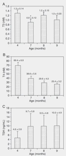
The concentrations of T3, T4, and TSH measured at the ages of 4, 7, 8, and 9 months are shown in Figure 1. Significant differ-ences in serum T3 concentration measured at the ages of 4, 7, 8, and 9 months were found in the experimental group of animals (Figure 1A, T3: F = 30.0, P = 0.000), while no difference was found in the control group (data not shown). Serum T3 concentration was significantly reduced at the age of 7 months in the experimental group (P < 0.05) compared to control animals of the same age (see Figure 1A).
A significant difference in serum TSH concentration measured at the age of 4, 7, 8, and 9 months was found in the experimental group (Figure 1C, T3: F = 30.0, P = 0.000, TSH: F = 18.9, P = 0.0000), while no differ-ence was found in the control group (data not shown). Serum TSH concentration was significantly elevated at the age of 7 months (3 months after 131I administration) in the
experimental group (P < 0.05) compared to control animals of the same age.
Regression analysis showed a correla-tion between serum T3, T4, and TSH con-centrations and age in control animals (T3: F = 21.9, P = 0.000, age - ß = -0.40, P = 0.000; T4: F = 1063, P = 0.000, age - ß = -0.95, P = 0.000; TSH: F = 23.9, P = 0.000, age - ß = 0.42, P = 0.000).
Multiple regression analysis showed a correlation between serum T3 and TSH con-centrations and 131I dose, but no correlation
with the age in the experimental animals (T3: F = 28.6, P = 0.000, age - ß = 0.12, P = 0.34; dose - ß = -0.67, P = 0.000; TSH: F = 40.6, P = 0.000, age - ß = 0.196, P = 0.089; dose - ß = 0.789, P = 0.000). On the other hand, serum T4 concentration showed a cor-relation with the age of the experimental animals, but not with the dose of 131I
admin-istered (F = 551, P = 0.000, age - ß = -0.87, P = 0.000, dose - ß = -0.098, P = 0.033). The time-course of hormone levels during the follow-up period according to 131I
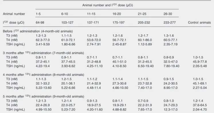
adminis-tration is shown in Table 1. The animals were divided into 6 groups of 5 animals each according to the absorbed doses of 131I.
Volume and weight of the thyroid gland
The thyroid gland volume was signifi-cantly lower in the experimental group than in the control group at the age of 9 months, i.e., 5 months after 131I administration
(ex-perimental vs control = 0.0184 ± 0.039 vs 0.0279 ± 0.0063, t = 6.1, P = 0.0000). The weight of the thyroid gland was significantly lower in the experimental group than in the
Figure 1. Mean values of serum triiodothyronine (T3, nM; panel A), thyroxine (T4, nM; panel B), and thyroid-stimulating hormone (TSH, ng/mL; panel C) concen-trations in the experimental group at the age of 4, 7, 8, and 9 months. The animals received
131I at the age of 4 months.
Sta-tistical difference was found among all experimental groups. Asterisk indicates that the val-ues for the animals of the same age differed statistically between experimental and control group (P < 0.05 for the post hoc Fisher LSD test performed after analy-sis of variance for repeated measurements).
control group at the age of 9 months (experi-mental vs control = 0.0173 ± 0.0026 vs 0.020 ± 0.0033, t = 4.0, P = 0.0002).
Regression analysis showed no correlation between the volume and weight of the thyroid gland and the dose of 131I (volume: F = 0.7, P
= 0.41, dose - ß = -0.16, P = 0.41; weight: F = 1.4, P = 0.24, dose - ß = -0.224, P = 0.24).
Body weight
Animals of both groups entered the ex-periment at a similar body weight. At the age of 7 months there was no difference in body weight between the control and the experi-mental groups (t = 0.79, P = 0.43 (366.6 ± 30.9 vs 372.6 ± 30.0 g)). Statistically signifi-cant differences were found at the age of 8 months (t = 2.25, P = 0.03) when the
experi-mental group of animals had a higher body weight than the controls (374.0 ± 29.4 vs 391.5 ± 29.8 g), and disappeared at the age of 9 months (t = 1.8, P = 0.08 (377.8 ± 33.9 vs 391.8 ± 29.0 g)).
Histopathological analysis of the thyroid gland
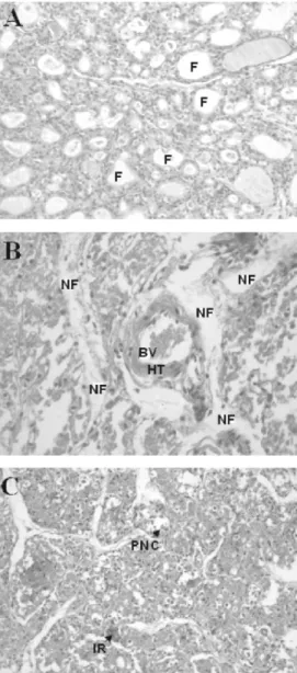
A histological image of the thyroid gland of the control group of animals is shown in Figure 3A. Histopathological analysis of thy-roid glands from the experimental group of animals showed hyaline thickness of the blood vessel walls, necrotic follicles (Figure 3B), a strong inflammatory reaction, and peeling of necrotic cells in the follicles (Fig-ure 3C). There were no differences between the thyroid glands of animals receiving dif-ferent doses of 131I.
Table 1. Effect of 131I dose on the time-course of serum T3, T4, and TSH in the experimental group of animals.
Animal number and I131 dose (µCi)
Animal number 1-5 6-10 11-15 16-20 21-25 26-30
I131 dose (µCi) 64-98 103-127 137-171 175-197 205-232 233-277 Control animals
Before I131 administration (4-month-old animals)
T3 (nM) 1.2-1.3 1.1-1.5 1.2-1.3 1.2-1.6 1.2-1.7 1.3-1.6 T4 (nM) 62.3-77.0 61.0-72.1 53.6-72.0 56.7-72.1 60.1-86.0 60.5-77.1 TSH (ng/mL) 3.41-9.59 1.80-6.66 2.74-7.91 2.45-8.87 1.12-5.89 2.35-7.19
3 months after 131Iadministration (7-month-old animals)
T3 (nM) 0.8-1.1 0.9-1.1 0.7-1.1 0.7-1.1 0.8-1.1 0.8-0.9 1.0-1.5 T4 (nM) 37.2-45.1 37.7-45.5 31.2-48.8 40.1-51.0 31.2-45.5 32.5-47.0 45.9-77.8 TSH (ng/mL) 4.22-19.4 3.93-6.62 4.25-11.10 4.10-8.50 6.50-19.40 7.80-19.40 2.05-5.48
4 months after 131I administration (8-month-old animals)
T3 (nM) 1.1-1.3 1.2-1.5 1.1-1.2 1.1-1.4 1.1-1.5 0.9-1.5 1.0-1.5 T4 (nM) 32.1-33.2 20.1-38.1 21.4-32.9 27.3-30.6 23.7-32.8 24.2-30.5 45.1-69.1 TSH (ng/mL) 5.22-13.60 5.22-6.66 4.48-11.4 4.66-10.50 7.40-17.0 8.90-17.0 2.27-5.04
5 months after 131I administration (9-month-old animals)
T3 (nM) 1.2-1.3 1.2-1.4 0.9-1.3 0.8-1.1 0.7-0.9 0.8-1.0 1.2-1.4 T4 (nM) 22.4-26.9 22.0-25.7 18.9-27.5 19.9-29.1 22.2-31.9 24.7-29.3 37.6-64.5 TSH (ng/mL) 4.99-15.50 5.23-7.20 4.20-11.60 4.88-8.82 7.65-17.0 12.3-17.0 2.04-4.70
Data are reported as range for 5 rats in each group and time-course of serum T3, T4, and TSH in the control group. T3 = triiodothyronine; T4 = thyroxine; TSH = thyroid-stimulating hormone.
Statistical differences between groups: 7-month-old animals: T3, P < 0.05; T4, P < 0.05; TSH, P < 0.05; 8-month-old animals: T3, non-significant (NS); T4, P < 0.05; TSH, NS; 9-month-old animals: T3, NS; T4, P < 0.05; TSH, NS (analysis of variance for repeated measures and the post hoc
Discussion
In this study rat model was used in order to determine the thyroid function after appli-cation of different doses of 131I (64-277 µCi).
Concentration of T3, T4 and TSH was deter-mined before 131I administration
(4-month-old animals) and when rats were 7, 8, and 9 months old. Nine-month-old animals were sacrificed, thyroid glands were removed for histophatological examination.
Serum T3, T4, and TSH levels of control animals measured at 4, 7, 8, and 9 months of age agreed with those described in the
litera-ture. We found a decrease in serum T4 con-centration between the age of 4 and 8 months, in accordance with the results of da Costa et al. (4). Serum T4 entirely originates from the thyroid gland, while more than 80% of T3 is produced by deiodination of T4 in other tissues, especially the liver and kidneys. Decreased serum T4 concentration stimu-lates TSH secretion through the pituitary-thyroid feedback mechanism. However, there are only two reports of increased TSH with aging in rats (5,6). Our finding of a constant TSH level corresponds to the most recent studies (4,7-9). No feedback response seems to have occurred, and the serum TSH re-mained unchanged in old animals despite a decreased serum T4. This may have been due to the unchanged serum T3 levels (4). Furthermore, da Costa et al. (4) explained the increased serum T4 levels by low ex-pression of the gene for thyroperoxidase, the enzyme responsible for thyroglobulin iodi-nation in old males, suggesting that the gene expression of androgens might increase (10, 11). The timing and/or degree of the decline may differ between rat strains (5,10,12). There is also the possibility that the aged murine thyroid becomes less responsive to circulating TSH.
One possible explanation for this lack of response to TSH during thyroid gland aging was given by Studer et al. (13) and Gerber et al. (14). These investigators suggested that a gradual failure of endocytosis in response to normal TSH stimulation would occur in the aging thyroid. At first, thyroglobulin exocy-tosis and iodination would be unaffected, resulting in gradual distension of the follicu-lar lumen that would impair the normal api-cal membrane function (13,14).
Since thyroid hormone reserves are ex-hausted slowly from the follicles, the result of therapy with 131I should be evaluated two
or three months after administration (15). Therefore, we evaluated the thyroid hor-mone status 3, 4, and 5 months after 131I
administration. We found decreased serum
T4 concentrations, while TSH was signifi-cantly increased. These findings were in ac-cordance with an earlier report by Reilly et al. (16). However, Railly et al. showed no change in T3 concentration 85 days after administration of 131I in their Wistar rat
mo-del, our Fisher rat model showed a signifi-cant decrease of T3 concentration 3 and 5 months after 131I administration. These
dif-ferences in behavior of serum T3 are prob-able the result of the doses applied, the fol-low-up period, as well as the mechanism of defense against radioactivity in different rat models. Reilly et al. (16) applied a constant dose of 150 µCi, while the doses applied in the present study were between 64 and 277 µCi. In the present study, we showed that serum T3 concentrations significantly de-creased as the 131I dose increased. The
low-est concentration of T3 was detected 3 months after administration of 131I, followed by a
slow increase. However, the serum T4 con-centration showed a decrease throughout the study period.
It is known that thyroid gland hormones enhance catabolic reactions in the organism. As a result of hormone shortage, catabolism slows down, with a consequent increase in body weight. The latest investigations have shown that the rat thyroid gland expresses receptors of leptin, an adipose tissue-secreted hormone which decreases the caloric intake and increases energy expenditure (17). There-fore, our finding of a significant increase in body weight 4 months after radiotherapy could be caused not only by hypothyroid-ism, but also by a deficit of leptin receptors in radioiodine-treated animals. The absence of statistically significant differences in body weight between experimental and control animals 5 months after radiotherapy indi-cates that hypothyroidism became less se-vere during that period.
The low thyroid weight and volume of the experimental group of animals is in ac-cordance with the findings of Agote et al. (18). The histopathological findings of
hya-line thickness of blood vessels after expo-sure to radioactivity correspond to changes described after therapy of thyroid hyper-function by injection of ethanol through the skin (19). The balance between thyroid re-generation and fibrosis appears to determine in part whether hypothyroidism will occur (1). Abnormal distribution of hyaline prob-ably disrupts thyroid hormone secretion, as shown by Li et al. (20). Therefore, despite an increased TSH concentration during the monitoring period, T3 concentration was low but close to the minimum normal level. This finding indicates that T3 occurs in non-thy-roid tissues by T4 deiodination and this is the reason why its concentration, contrary to the concentration of T4, does not depend on tissue status.
We have described the state of rats fol-lowing 131I administration (the doses they
received were in human relations from the lowest to the highest therapeutic doses) which resembles the state clinically observed in some human subjects receiving therapeutic doses of 131I. There are only few previously
published long-term follow-up studies re-garding radioiodine treatment of hyperthy-roidism in patients (15,21,22). It was ob-served that hypothyroidism will develop in 82% of patients with Graves’ disease and in 32% of patients with multinodular goiter treated with 131I within 25 years (15). The
higher rate of hypothyroidism in patients with Graves’ disease than in patients with toxic multinodular goiter might result from protection of the suppressed normal extra-nodular tissue by its inability to concentrate
131I in patients with toxic multinodular goiter
(15,21). Metso et al. (15) did not find a dose-response relationship between the radioac-tive dose and the rate of hypothyroidism or a positive correlation between the cure rate and hypothyroidism. Because the develop-ment of hypothyroidism seems to be inevi-table and unpredicinevi-table by any clinical fac-tor, the objective of 131I treatment should be
hyperthyroid-References
1. Harbert JC. Nuclear medicine therapy. New York: Thieme Medical Publisher Inc.; 1987.
2. Simmonds MJ, Howson JM, Heward JM, Cordell HJ, Foxall H, Carr-Smith J, et al. Regression mapping of association between the human leukocyte antigen region and Graves disease. Am J Hum Genet 2005; 76: 157-163.
3. Reinhardt MJ, Brink I, Joe AY, Von Mallek D, Ezziddin S, Palmedo H, et al. Radioiodine therapy in Graves’ disease based on tissue-absorbed dose calculations: effect of pre-treatment thyroid volume on clinical outcome. Eur J Nucl Med Mol Imaging 2002; 29: 1118-1124.
4. da Costa V, Moreira DG, Rosenthal D. Thyroid function and aging: gender-related differences. J Endocrinol 2001; 171: 193-198. 5. Pekary AE, Knoble M, Garcia N. Thyrotropin-releasing hormone
(TRH)-Gly conversion to TRH in rat ventral prostate is inhibited by castration and aging. Endocrinology 1989; 125: 679-685.
6. Console GM, Gomez Dumm CL, Goya RG. Immunohistochemical and radioimmunological assessment of thyrotrophs in the pituitary of aging rats. Acta Anat 1995; 152: 28-32.
7. Klug TL, Adelman RC. Age-dependent accumulation of an immu-noreactive species of thyrotropin (TSH) which inhibits production of thyroid hormones [proceedings]. Adv Exp Med Biol 1978; 97: 259-264.
8. Greeley GH Jr, Lipton MA, Kizer JS. Serum thyroxine, triiodothyro-nine, and TSH levels and TSH release after TRH in aging male and female rats. Endocr Res Commun 1982; 9: 169-177.
9. Donda A, Lemarchand-Beraud T. Aging alters the activity of 5'-deiodinase in the adenohypophysis, thyroid gland, and liver of the male rat. Endocrinology 1989; 124: 1305-1309.
10. Wang C, Sinha Hikim AP, Lue YH, Leung A, Baravarian S, Swerdloff RS. Reproductive aging in the Brown Norway rat is characterized by accelerated germ cell apoptosis and is not altered by luteinizing hormone replacement. J Androl 1999; 20: 509-518.
11. Gruenewald DA, Naai MA, Marck BT, Matsumoto AM. Age-related decrease in hypothalamic gonadotropin-releasing hormone (GnRH) gene expression, but not pituitary responsiveness to GnRH, in the male Brown Norway rat. J Androl 2000; 21: 72-84.
12. Lu CC, Tsai SC, Chien EJ, Tsai CL, Wang PS. Age-related differ-ences in the secretion of calcitonin in male rats. Metabolism 2000;
49: 253-258.
13. Studer H, Forster R, Conti A, Kohler H, Haeberli A, Engler H. Transformation of normal follicles into thyrotropin-refractory “cold” follicles in the aging mouse thyroid gland. Endocrinology 1978; 102: 1576-1586.
14. Gerber H, Peter HJ, Studer H. Age-related failure of endocytosis may be the pathogenetic mechanism responsible for “cold” follicle formation in the aging mouse thyroid. Endocrinology 1987; 120: 1758-1764.
15. Metso S, Jaatinen P, Huhtala H, Luukkaala T, Oksala H, Salmi J. Long-term follow-up study of radioiodine treatment of hyperthyroid-ism. Clin Endocrinol 2004; 61: 641-648.
16. Reilly CP, Symons RG, Wellby ML. A rat model of the 131I-induced
changes in thyroid function. J Endocrinol Invest 1986; 9: 367-370. 17. Nowak KW, Kaczmarek P, Mackowiak P, Ziolkowska A, Albertin G,
Ginda WJ, et al. Rat thyroid gland expresses the long form of leptin receptors, and leptin stimulates the function of the gland in euthyroid non-fasted animals. Int J Mol Med 2002; 9: 31-34.
18. Agote M, Viaggi M, Kreimann E, Krawiec L, Dagrosa MA, Juvenal GJ, et al. Influence of nicotinamide on the radiosensitivity of normal and goitrous thyroid in the rat. Thyroid 2001; 11: 1003-1007. 19. Pomorski L, Bartos M. Histologic changes in thyroid nodules after
percutaneous ethanol injection in patients subsequently operated on due to new focal thyroid lesions. APMIS 2002; 110: 172-176. 20. Li M, Carcangiu ML, Rosai J. Abnormal intracellular and
extracellu-lar distribution of basement membrane material in papilextracellu-lary carcino-ma and hyalinizing trabecular tumors of the thyroid: implication for deregulation of secretory pathways. Hum Pathol 1997; 28: 1366-1372.
21. Holm LE, Lundell G, Israelsson A, Dahlqvist I. Incidence of hypothy-roidism occurring long after iodine-131 therapy for hyperthyhypothy-roidism.
J Nucl Med 1982; 23: 103-107.
22. Ahmad AM, Ahmad M, Young ET. Objective estimates of the prob-ability of developing hypothyroidism following radioactive iodine treatment of thyrotoxicosis. Eur J Endocrinol 2002; 146: 767-775. 23. Allahabadia A, Daykin J, Sheppard MC, Gough SC, Franklyn JA.
Radioiodine treatment of hyperthyroidism-prognostic factors for out-come. J Clin Endocrinol Metab 2001; 86: 3611-3617.
ism with an easily manageable treatment scheme with minimal costs. Some authors recommend administration of empirical (the same fixed dose for every patient regardless to thyroid volume) doses of 131I for the
treat-ment of hyperthyroidism (15,23). There are very few data on animal models regarding a prolonged follow-up of thyroid status after administration of different doses of 131I. The
present study clearly showed that
hypothy-roidism develops anyway, when either low or high I131 doses have been administered,
similarly to the findings described for hu-mans. Therefore, our findings support the administration of empirical rather than cal-culated doses of 131I. This raises the
possibil-ity of avoiding the need to measure the size and the 131I uptake of the thyroid gland and