Morphological changes of the jejunal
mucosa in protracted diarrhea and
their correlation with disease duration,
weight loss and serum albumin levels
1Departamento de Pediatria and 2Departamento de Anatomia Patológica,
Faculdade de Medicina, Universidade Federal de Minas Gerais, Belo Horizonte, MG, Brasil
L.A. Péret-Filho1,
G. Brasileiro-Filho2
and F.J. Penna1
Abstract
The pathogenesis of protracted diarrhea is multifactorial. In develop-ing countries, intestinal infectious processes seem to play an important role in triggering the syndrome. Thirty-four children aged 1 to 14 months, mean 6.5 months, with protracted diarrhea were studied clinically and in terms of small intestinal mucosal morphology. Mild, moderate or severe hypotrophy of the jejunal mucosa was detected in 82% of cases, and mucosal atrophy was observed in 12%. The intensity of the morphological changes of the jejunal mucosa corre-lated negatively with serum albumin levels. No correlation was de-tected between mucosal grading and duration of diarrhea or between mucosal grading and weight reported as percentile. After nutritional support was instituted, serial jejunal biopsies were obtained from 12 patients: five patients submitted to parenteral nutrition for 7 to 38 days, mean 17 days, and 7 patients receiving a hypoallergenic oral diet (semi-elemental formula, 3; chicken formula, 3; human milk, 1). In seven cases (58%) a progressive increase in villus height and a decrease in the number of inflammatory cells were noted. Recovery of the morphologic pattern was accompanied by clinical improvement in all patients.
Correspondence
L.A. Péret-Filho Departamento de Pediatria Faculdade de Medicina, UFMG Av. Alfredo Balena, 190, 4º andar 30130-100 Belo Horizonte, MG Brasil
Fax: 55 (031) 224-7041 Research supported by CPq-UFMG, CNPq and FAPEMIG.
This work is part of a Master’s thesis presented by L.A. Péret-Filho to the Curso de Medicina Tropical, Faculdade de Medicina, UFMG, Belo Horizonte, MG, Brasil.
Received January 20, 1997 Accepted July 21, 1997
Key words
•Protracted diarrhea •Biopsy
•Jejunal mucosa •Serum albumin
Introduction
Protracted diarrhea is a clinical syndrome lasting 14 days or more which mainly affects infants during the first months of life, caus-ing water-electrolyte lability and progressive nutritional deterioration.
Structural lesions of the small intestinal mucosa are important components of the syndrome and seem to contribute in a signifi-cant manner to the worsening of clinical signs and symptoms. These lesions are caused
Greene etal. (3) reported jejunal changes in 16 patients with protracted diarrhea, all of them with significant mucosal villus hypo-trophy. After parenteral feeding and/or an enteral diet consisting of semi-elemental for-mulas, the lesions were fully reversed within 18 days, on average, from the beginning of nutritional treatment. In contrast, in a study of jejunal biopsies from children with the
same condition, Rossi et al. (4) detected
varying degrees of jejunal mucosa hypotro-phy that persisted for 6 months in 16 (70%) of their 23 patients. Goldegard and Vander-hoof (5) did not detect a correlation between severity of intestinal mucosal lesions and prognosis in 19 children.
Few studies have been conducted in tropical countries with the objective of analyzing the reversal of jejunal mucosal abnormalities by the treatment instituted in infants with protracted diarrhea. In a study of 20 children with persistent diarrhea in Gambia, Sullivan et al. (6) did not observe an improvement in villus architecture or re-duction of the inflammatory infiltrate after one month of treatment with a high protein-calorie diet, despite the occurrence of weight gain and normalization of serum albumin levels.
The objective of the present study was to investigate the morphological aspects of the jejunal mucosa of infants with protracted diarrhea from low socioeconomic level fami-lies and to correlate serum albumin levels, body weight and duration of diarrhea with the extent of jejunal mucosal damage.
Patients and Methods
Thirty-four children with protracted di-arrhea, characterized by three or more daily evacuations, lasting two weeks or more and with water-electrolyte lability were admitted to the Pediatric Ward of the University Hos-pital, UFMG, from January 1980 to Decem-ber 1993. A total of 49 jejunal biopsies were obtained from 34 patients (18 girls and 16
boys aged 1 to 14 months; mean, 6.5 months) (Table 1).
Most of the patients were from the out-skirts of the city of Belo Horizonte, where they lived under precarious basic sanitation conditions. All patients were transferred to our Hospital from others after failure of conservative treatment.
The jejunal biopsies were obtained 15 to 127 days after the onset of diarrhea (mean duration, 53 days), after the parents gave informed consent. The 34 patients were equilibrated in terms of water-electrolyte balance and acid-base balance and had been fasting for at least 3 h. Crosby-Kugler (pedi-atric model, 1975), Carey or Watson cap-sules were used. Patients were intubated as described by Toccalino and O’Donnell (7). When a signal of the presence of the capsule in the duodenum was obtained in terms of bile flow or spontaneous progression of the tube, a single abdominal X-ray was obtained to localize it. The biopsy was performed when the capsule was close to the Treitz angle. The fragment obtained was stretched on porous paper previously soaked in 10% formalin, with the villi looking up. After fixation for 6 to 18 h, the fragment was examined with a dissection microscope for mesoscopic evaluation and selection of the most representative areas for histological analysis, and routinely processed for histo-pathology. Histological sections (6 µm thick) perpendicular to the mucosal surface were stained with hematoxylin-eosin (HE) and periodic acid Schiff (PAS).
present in the lamina propria.
Grade I or mild hypotrophy: villus height is slightly decreased, corresponding to 1.5 to 2 times the crypt length. A discrete flattening of enterocytes and a larger number of mono-nuclear cells are visible in the lamina pro-pria.
Grade II or moderate hypotrophy: the villi are even shorter and their height does not exceed the depth of the crypts. The enterocytes are more flattened and the brush border may be absent throughout the entire mucosa. Marked infiltration of inflamma-tory cells is often observed in the lamina propria. The number of intraepithelial lym-phocytes may be increased.
Grade III or severe hypotrophy: there is an inversion of the villus/crypt ratio due to pronounced shortening and widening of the villi, which appear only as rudimentary struc-tures. The mononuclear infiltrate in the lamina propria is increased.
Grade IV or atrophic pattern: this stage corresponds to the maximum degree of mu-cosal atrophy, characterized by the absence of villi. The enterocytes become flattened and lose the brush border. A large number of mononuclear cells are frequently detected in the lamina propria, generally coexisting with increased numbers of intraepithelial lym-phocytes. Edema of the lamina propria is sometimes observed.
Serum albumin levels were determined in 26 patients. Samples were collected 24 h before, on the same day, or at most on the day after the jejunal biopsy. Albumin was measured by the biuret method and the re-sults are reported as g/dl. The duration of diarrhea (three or more evacuations daily) was determined on the basis of information provided by the parents or persons respon-sible and recorded as number of days. Body weight was evaluated in terms of percentile for age using the computer software devel-oped by Jordan (11).
The total parenteral nutrition was com-posed of amino acids and lipids, which were
provided initially at 1.0 g kg-1 day-1 and
pro-gressively increased for 7 days to 2.5 g kg-1
day-1 and 3.0 g kg-1 day-1, respectively. All solutions were mixed in 10% dextrose. Elec-trolytes and other additives were as follows:
30 mEq/l NaCl, 25 mEq/l K+, 2 mEq/l Mg2+,
15 mEq/l Ca2+, 10 mEq/l KH
2PO4,
multivita-min preparation (Polivit A® - 10 ml) and
oligoelements (Politrace® - 0.2 ml/kg). The hypoallergenic diet was composed
of semi-elemental formula (Alfaré®), 15%
dilution, 52 g hydrolized whey protein, lip-ids (17.2 g triglycerides, 10.3 g milk fat, 6.9 g corn oil, 1.5 g biotin), carbohydrates (67.0 g dextrin-maltose, 9.0 g potato wheat, 1.5 g lactose), vitamins, minerals and oligoele-ments, and water added to make 1000 ml. Calories: 72/100 ml.
The chicken formula was composed of 150 g cooked chicken, 40 g corn oil, 80 g
dextrin-maltose, 0.5 g NaCl, 2 g CaCO3,
vitamins (Protovit® - 15 drops) and cold
water to make 1000 ml. Calories: 70/100 ml. The study was approved by the Hospital Ethics Committee and consent was obtained from the parents.
Data were analyzed statistically by the chi-square test for comparison of the fre-quencies observed, by the Pearson correla-tion for comparison between variables, by simple concrete adjusted linear regression, and by the regression determination coeffi-cient R2, with the level of significance set at P<0.05.
Results
usually irregular, with no changes in enterocytes or in cellularity of the lamina propria. Villus hypotrophy was moderate in 12 cases (35.3%) and severe in 4 cases (11.8%). Two patients presented areas of moderate hypotrophy side by side with areas of marked hypotrophy. In four cases (11.8%) there was atrophy of the villi. In these last three groups of patients (a total of 20), in
addition to villous changes there were loss of the brush border, decreased enterocyte height, crypt elongation relative to the villus height and increased cellularity of the lamina pro-pria, all of these phenomena being more marked in the last group. The brush border was usually absent or showed irregular dis-tribution in the mucosa with marked hypo-trophy or ahypo-trophy, always in the presence of
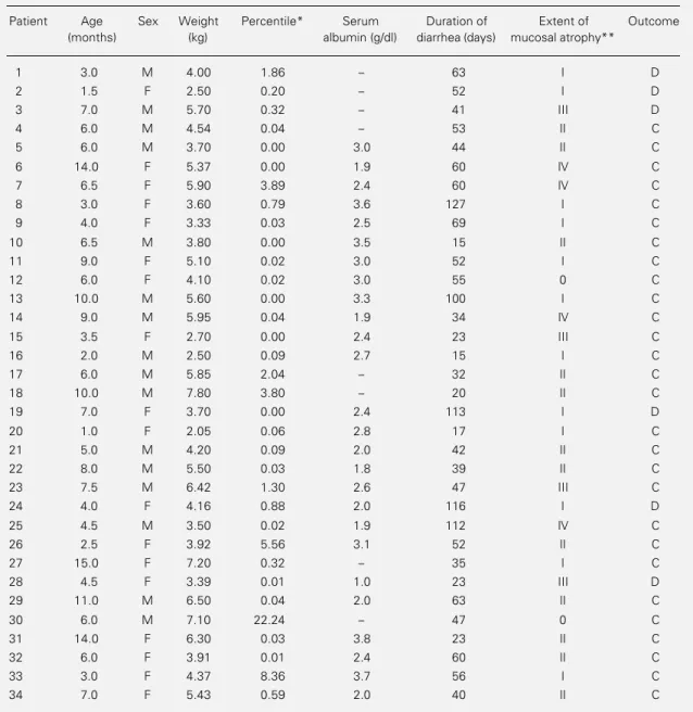
Table 1 - Clinical and laboratory characteristics of patients with protracted diarrhea.
*According to Jordan (11). **Modified from Shenk and Klipstein (9). D, Death; C, cure.
Patient Age Sex Weight Percentile* Serum Duration of Extent of Outcome
(months) (kg) albumin (g/dl) diarrhea (days) mucosal atrophy**
1 3.0 M 4.00 1.86 - 63 I D
2 1.5 F 2.50 0.20 - 52 I D
3 7.0 M 5.70 0.32 - 41 III D
4 6.0 M 4.54 0.04 - 53 II C
5 6.0 M 3.70 0.00 3.0 44 II C
6 14.0 F 5.37 0.00 1.9 60 IV C
7 6.5 F 5.90 3.89 2.4 60 IV C
8 3.0 F 3.60 0.79 3.6 127 I C
9 4.0 F 3.33 0.03 2.5 69 I C
10 6.5 M 3.80 0.00 3.5 15 II C
11 9.0 F 5.10 0.02 3.0 52 I C
12 6.0 F 4.10 0.02 3.0 55 0 C
13 10.0 M 5.60 0.00 3.3 100 I C
14 9.0 M 5.95 0.04 1.9 34 IV C
15 3.5 F 2.70 0.00 2.4 23 III C
16 2.0 M 2.50 0.09 2.7 15 I C
17 6.0 M 5.85 2.04 - 32 II C
18 10.0 M 7.80 3.80 - 20 II C
19 7.0 F 3.70 0.00 2.4 113 I D
20 1.0 F 2.05 0.06 2.8 17 I C
21 5.0 M 4.20 0.09 2.0 42 II C
22 8.0 M 5.50 0.03 1.8 39 II C
23 7.5 M 6.42 1.30 2.6 47 III C
24 4.0 F 4.16 0.88 2.0 116 I D
25 4.5 M 3.50 0.02 1.9 112 IV C
26 2.5 F 3.92 5.56 3.1 52 II C
27 15.0 F 7.20 0.32 - 35 I C
28 4.5 F 3.39 0.01 1.0 23 III D
29 11.0 M 6.50 0.04 2.0 63 II C
30 6.0 M 7.10 22.24 - 47 0 C
31 14.0 F 6.30 0.03 3.8 23 II C
32 6.0 F 3.91 0.01 2.4 60 II C
33 3.0 F 4.37 8.36 3.7 56 I C
cuboidal or flattened enterocytes. The num-ber of intraepithelial lymphocytes increased with decreasing villus size. A jejunal biopsy
was intensely parasitized with
Cryptospori-dium sp. No other parasites were detected upon histological examination.
Twelve patients were submitted to serial biopsies at intervals of 7 to 20 days, with a mean of 13 days and a median of 14 days. All of them showed clinical improvement on the occasion of the second biopsy. Five of them had intense mucosal lesions (grade III or IV) and were submitted to total parenteral nutri-tion. Four of these patients presented a marked improvement of the morphological picture after approximately 15 days, but one continued to present the same lesions after 38 days of follow-up (Figure 1). After 14 months of follow-up, this last patient, 15.5 months old at the time, started to tolerate cow’s milk. An additional jejunal biopsy was not obtained because the parents re-fused it. The remaining seven patients were treated with an oral diet (semi-elemental formula, human milk or chicken formula). Although their lesions were less severe than those of infants treated with parenteral nutri-tion, 4 of them presented a less marked improvement in the morphological pattern within the same period of time (a mean period of 13 days of treatment) (Figure 2). The patients submitted to parenteral nutri-tion received a caloric amount similar to that of the patients receiving an oral diet (80 kcal kg-1 day-1 and 2.5 g amino acids kg-1 day-1 at the end of the first week). Improvement of the histological pattern of the jejunal mucosa was accompanied by clinical improvement in all patients.
There was a negative correlation between serum albumin levels and mucosal grading (r = -0.501, P<0.005) (Figure 3). No correla-tion was detected between mucosal grading and duration of diarrhea (r = -0.131, P>0.05) or between mucosal grading and patient weight reported as percentile (r = -0.100, P>0.05).
Figure 1 - Evolution of jejunal mucosal morphology in 5 in-fants with protracted diarrhea submitted to total parenteral nu-trition (TPN).
Degree of atrophy
IV
III
II
I
0
5 10 15 20 38
Duration of TPN (days)
Degree of atrophy
IV
III
II
I
0
5 10 15 20
Duration of feeding (days)
Figure 2 - Evolution of jejunal mucosal morphology in 7 in-fants with protracted diarrhea treated with an oral diet.
Albumin
4
0 I I I I I I I I I II II II II II II II II III III III III IV IV IV IV 3
2
1
0
Degree of atrophy
Figure 3 - Serum albumin level vs degree of jejunal mucosal atrophy in 26 infants with
protracted diarrhea. r = -0.501, P<0.005. Discussion
The present study confirms that protracted diarrhea is a severe clinical condition caus-ing profound changes in the nutritional sta-tus of the patients. Of the 34 cases described, only one had normal weight, whereas the remaining 33 presented a considerably marked nutritional deficiency.
Analysis of the jejunal biopsies showed morphological alterations in most cases, with
Chicken formula
only two patients (5.0%) showing a normal histological pattern. Twenty patients (58.8%) presented moderate or severe morphological changes. The lesions were nonspecific in all cases and were similar to those reported for marasmus patients (8) and for children with kwashiorkor (12).
Total villus atrophy similar to that occur-ring in celiac disease was detected in four patients, a finding similar to that observed in patients with kwashiorkor (13). A more de-tailed comparative examination, however, showed that normal mucosal thickness is preserved in patients with celiac disease, whereas the mucosa is thinner in malnour-ished children with protracted diarrhea. Only one of the patients with complete villus atro-phy and with all of the other characteristics of celiac disease described above was later confirmed to have celiac disease during fol-low-up.
The dietary treatment of these patients (parenteral nutrition or semi-elemental di-ets) resulted in an improved morphological pattern of the intestinal mucosa in most of them. The vitamins and trace elements (zinc in particular) provided by parenteral nutri-tion may explain, at least in part, the better response of these patients compared to those receiving only the semi-elemental diet.
However, the claim that parenteral nutri-tion produces a better response than a semi-elemental formula cannot be made from our data since at the beginning of the study the children receiving the two treatments had different degrees of histological abnormal-ity.
The negative correlation between degree of intestinal mucosal lesion and serum
albu-min levels was predictable. Numerical fac-tors may explain the lack of statistical corre-lation between body weight and mucosal grading. Since almost all patients had suf-fered a considerable weight loss (all but one were below the 3rd percentile), the differ-ence in nutritional deficiency among them was quite small, since all were severely ema-ciated. Thus, the numerical interval among them was small, making it impossible to detect a statistical relationship between these two variables, although it was clear that 94% of the infants presented morphological mu-cosal changes and 97% were nutritionally deficient.
The absence of a correlation between duration of diarrhea and intensity of mor-phological lesions of the intestinal mucosa indicates once again that functional intesti-nal changes are not always related to the degree of structural involvement of the mu-cosa. In this respect, it is possible that mod-erate or severe morphological changes of the mucosa did not impair nutrient digestion or absorption in some patients, whereas mild or absent changes were accompanied by pro-found functional modifications in other pa-tients, resulting in water, electrolyte and nu-trient malabsorption. This possibility may also explain in part the lack of correlation between weight loss and intensity of mu-cosal changes.
The intensity of intestinal mucosal atro-phy explained the low serum albumin levels
detected in 25% of the patients (R2). The
References
1. Fagundes Neto U, Trabulsi LR & Patrício FRS (1988). Diarréia protraída: a importân-cia dos agentes enteropatogênicos na sua
gênese e fisiopatologia. Jornal de
Pediatria, 64: 237-241.
2. Bhan MK, Khoshoo V, Sommerfelt H, Pushker R, Sorawal S & Srivastava R (1989). Enteroaggregative “Escherichia coli” and “Salmonella” associated with nondysenteric persistent diarrhea. Pediat-ric Infectious Disease Journal, 8: 499-502. 3. Greene HL, McCabe DR & Merenstein GB (1975). Protracted diarrhea and malnu-trition in infancy: changes in intestinal morphology and disaccharidase activities during treatment with total intravenous nutritions or oral elemental diets. Journal of Pediatrics, 87: 695-704.
4. Rossi MT, Lebenthal E, Nord KS & Fazili RR (1980). Extent and duration of small intestinal mucosal injury in intractable di-arrhea of infancy. Pediatrics, 66: 730-735.
5. Goldegard CM & Vanderhoof JA (1986). Lack of correlation of small bowel biopsy and clinical course of patients with
intrac-table diarrhea of infancy.
Gastroenterol-ogy, 90: 527-531.
6. Sullivan PB, Lum PG, Northrop-Clews C, Crouce PT, Marsh MN & Neale G (1992). Persistent diarrhea and malnutrition - the impact of treatment on small bowel struc-ture and permeability. Journal of Pediatric Gastroenterology and Nutrition, 14: 208-215.
7. Toccalino H & O’Donnell JO (1962). Tecnica para la introdución de la sonda -Capsula de Crosby en niños. Revista del Hospital de Niños, 12: 29-30.
8. Barbieri D (1971). Mucosa jejunal na má nutrição protéica primária grave da criança. Doctoral thesis, Escola Paulista de Medicina, São Paulo.
9. Shenk EA & Klipstein FA (1972). A proto-col for the evaluation of small bowel
biop-sies. American Journal of Clinical
Nutri-tion, 25: 1108-1117.
10. Penna FJ, Hill ID, Kingston D, Robertson K, Slaving E & Shiner M (1981). Jejunal mucosal morphometry in children with and without gut symptoms and in normal
adults. Journal of Clinical Pathology, 34:
386-392.
11. Jordan JR (1986). Anthropometric
Soft-ware Package. Department of Health and Human Service, Public Health Service, Centers of Disease Control, Atlanta, GA. 12. Stanfield JP, Hutt MSR & Tunicliffe R
(1965). Intestinal biopsy in kwashiorkor. Lancet, ii: 519-523.
13. Schnneider RE & Viteri FE (1972). Mor-phological aspects of the duodenojejunal mucosa in protein-calorie malnourished
children and during recovery. American