Mo le cular characte rizatio n o f D D X2 6 ,
a hum an D EAD -bo x RNA he licase ,
lo cate d o n chro m o so m e 7 p1 2
1Instituto Ludwig de Pesquisa sobre o Câncer, São Paulo, SP, Brasil 2Department of Histopathology, University of Cambridge,
Addenbrooke’s Hospital, Cambridge, UK A.A. Camargo1, D.N. Nunes1,
H.B. Samaia1, L. Liu2,
V.P. Collins2, A.J.G. Simpson1
and E. Dias-Neto1
Abstract
DEAD-box proteins comprise a family of ATP-dependent RNA heli-cases involved in several aspects of RNA metabolism. Here we report the characterization of the human DEAD-box RNA helicase DDX26. The gene is composed of 14 exons distributed over an extension of 8,123 bp of genomic sequence and encodes a transcript of 1.8 kb that is expressed in all tissues evaluated. The predicted amino acid se-quence shows a high similarity to a yeast DEAD-box RNA helicase (Dbp9b) involved in ribosome biogenesis. The new helicase maps to 7p12, a region of frequent chromosome amplifications in glioblasto-mas involving the epidermal growth factor receptor (EGFR) gene. Nevertheless, co-amplification of DDX26 with EGFR was not de-tected in nine tumors analyzed.
Co rre spo nde nce A.A. Camargo
Instituto Ludwig de Pesquisa
sobre o Câncer
Rua Prof. Antônio Prudente, 109 4º andar
01509-010 São Paulo, SP Brasil
Fax: + 55-11-270-7001 E-mail:
anamaria@ compbio.ludwig.org.br
D.N. Nunes and H.B. Samaia are recipients of FAPESP fellowships.
The sequence described was deposited in GenBank under
accession No. AF247666.
Received February 1, 2001 Accepted June 28, 2001
Ke y wo rds
·DDX26
·DEAD-box RNA helicase
·7p12
·Ribosome biogenesis
Intro ductio n
Helicases, including DNA and RNA heli-cases, are grouped into two major superfami-lies of proteins (SFI and SFII) on the basis of the occurrence of conserved motifs (1). RNA helicases are mostly of the SFII super-family and can be further classified into families on the basis of particular consensus sequences in the conserved ATP hydrolysis motif. In contrast to DNA helicases, which processively unwind long regular dsDNA structures, most RNA helicases modulate only short duplex regions in RNA molecules in a one-step reaction in the presence of ATP (1).
RNA helicases are involved in various steps of RNA metabolism including
tran-scription, pre-mRNA splicing, ribosome bio-genesis, cytoplasmic transport, translation, initiation/elongation, and mRNA decay (1). DEAD-box RNA helicases are characterized by the presence of eight conserved motifs including an RNA interaction domain and an ATP hydrolysis motif containing the core amino acid sequence DEAD (2). In addition to the conserved motifs, RNA helicases con-tain variable N and C terminal extensions that might confer substrate specificity and/or contain information directing subcellular lo-calization (3).
report the chromosomal localization, genom-ic organization and expression of DDX26, a human DEAD-box RNA helicase, with high similarity to an essential yeast RNA helicase (Dbp9b) involved in the assembly of early pre-ribosomal particles. The new helicase maps to 7p12, a region of frequent chromo-some amplifications in glioblastomas involv-ing the epidermal growth factor receptor (EGFR) gene. Nevertheless, co-amplifica-tion of DDX26 with EGFR was not detected in nine tumors analyzed.
Mate rial and Me tho ds
Co mputatio nal pre dictio n o f ge ne structure
Gene structure was predicted by tBlastN analysis using the yeast orthologous protein Dbp9b as a query. Proximal and distal exons were then predicted by BlastN analysis us-ing the PAC sequence (AC004938) as a query against human ESTs available in dbEST (AW250279, AI242741). Oligonu-cleotides corresponding to the 5 and 3 ends of the putative transcript (Figure 1A) were designed and used to amplify cDNAs de-rived from colon and breast tumors by PCR. Intron/exon boundaries were determined by aligning the cDNA sequence with the ge-nomic sequence from the PAC clone.
RT-PCR and se que ncing
Total RNA was extracted from breast and colon tumors using Trizol reagent (Gibco-BRL, Gaithersburg, MD, USA) following manufacturers instructions. Two micro-grams of total RNA was reverse-transcribed into first-strand cDNA using SuperScript II and oligo dT in a final volume of 20 µl. PCR was carried out in a 10-µl reaction mixture containing 1 µl of cDNA, 1X Taq DNA polymerase buffer, 200 µM dNTPs, 2 pmol of each primer and 1 U Taq DNA poly-merase (Gibco-BRL). Cycling conditions were 94o
C for 5 min, followed by 40 cycles
of 94o
C for 1 min, 60o
C for 1 min, 72o C for 2 min and a final extension at 72oC for 10 min. PCR products were analyzed on ethi-dium bromide-stained agarose gels before sequencing. PCR products were used di-rectly in sequencing reactions with Big Dye terminator mix on an ABI377 sequencer ac-cording to manufacturers instructions. Se-quencing reactions were performed with the same primers used for RT-PCR and internal primers as indicated in Figure 1.
No rthe rn and do t blo t hybridizatio n
A commercial Human Master Blot (Clontech, Palo Alto, CA, USA) containing normalized loading of polyA+
RNA from 75 different human tissues and a Cancer Cell Lines Northern blot (Clontech) containing 2 µg of polyA+ RNA was used for evaluating DDX26 expression. A 32
P-labeled probe cor-responding to the 3 end of the transcript was hybridized in 5 ml ExpressHyb solution (Clontech) at 65o
C overnight. The mem-branes were washed twice with 2X SSC/ 0.1% SDS at room temperature for 5 min and then twice with 2X SSC/0.1% SDS for 20 min at 65o
C before exposure at -70o C for 24 h.
So uthe rn blo tting and de nsito me try
Allele dosage at the DDX26 locus was quantitated by Southern blotting in a panel of nine glioblastomas with known 7p12 am-plifications. Southern blotting, hybridization and PhosporImage analysis were performed as previously described (8). Probes used for hybridizations were: WI-15640, SGC34528, D7S2542, 6548, EGFR, SGC32574, WI-18503, WI-16990, SHGC11828 and a 1.2-kb fragment from the 3 end of DDX26 transcript.
Re sults
Cancer Genome Project (9), we identified an EST with significant similarity to a DEAD-box RNA helicase from yeast and various other organisms as well as to a finished human genomic sequence corresponding to the PAC clone DJ0971C03 (AC004938). Matches with previously known human helicases, however, were found to be only marginal, indicating that this EST is a new member of the DEAD-box family not anno-tated in the PAC sequence.
We have used the yeast orthologous gene to search for the human genomic sequence and to predict gene structure. Primers for RT-PCR amplification were designed for validation and sequencing of the predicted transcript. As expected from the computa-tional prediction, a 1,889-bp fragment was obtained by RT-PCR and fully sequenced by primer walking (Figure 1A). This gene was named DDX26 after consulting the HUGO Genome Nomenclature Committee and de-posited in GenBank (AF247666). The gene consists of 14 exons distributed over an extension of 8,123 bp of genomic se-quence. Exons and introns vary in size from 74 to 275 bp and from 92 to 1,594 bp, respectively (Figure 1B). Computer anal-ysis of the PAC sequence using Genscan failed to predict the correct gene structure due to the apparent existence of other genes in the same region and to the small size of exon 13 that was not detected by using this program.
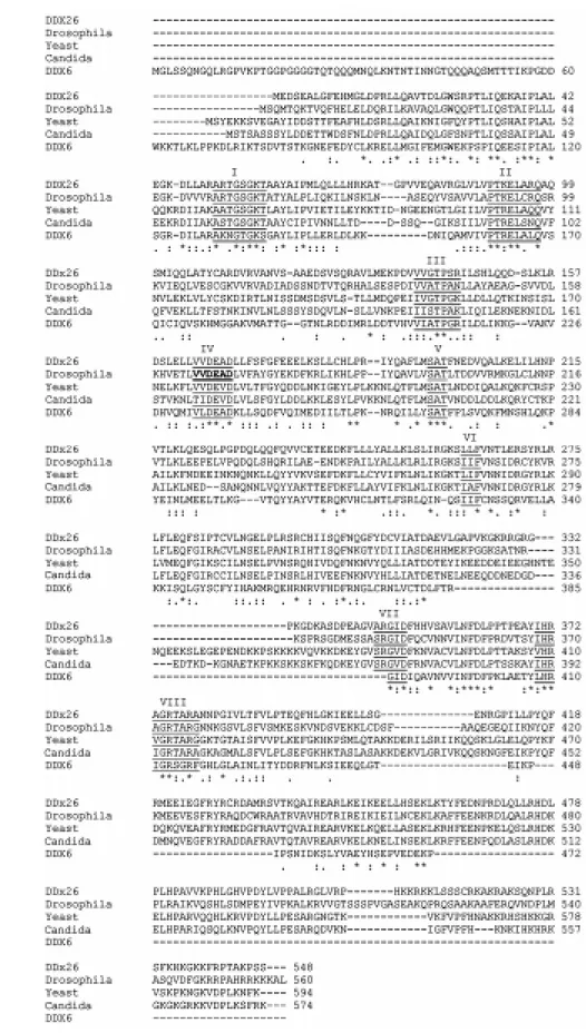
Complete sequencing of this helicase re-vealed a single putative open reading frame coding for a protein of 548 amino acids (Figure 1A). Analysis of the predicted pro-tein sequence using Pfam (http://pfam. wustl.edu/) revealed the presence of two highly conserved domains corresponding to a DEAD/DEAH-box helicase domain (20-234 amino acids) and a helicase C-terminal conserved domain (272-380 amino acids). Figure 2 shows the alignment of the pre-dicted amino acid sequence of DDX26 with RNA helicases from Drosophila, yeast,
Can-dida and the human DDX6 using CLUSTAL W (10). As for the first EST, a high similarity was observed with an essential yeast RNA helicase (Dbp9b) involved in the assembly of early pre-ribosomal particles (43% iden-tity and 64% similarity). The putative func-tion of this protein in ribosome biogenesis was recently confirmed by Zirwes and col-leagues (11) during the preparation of this manuscript.
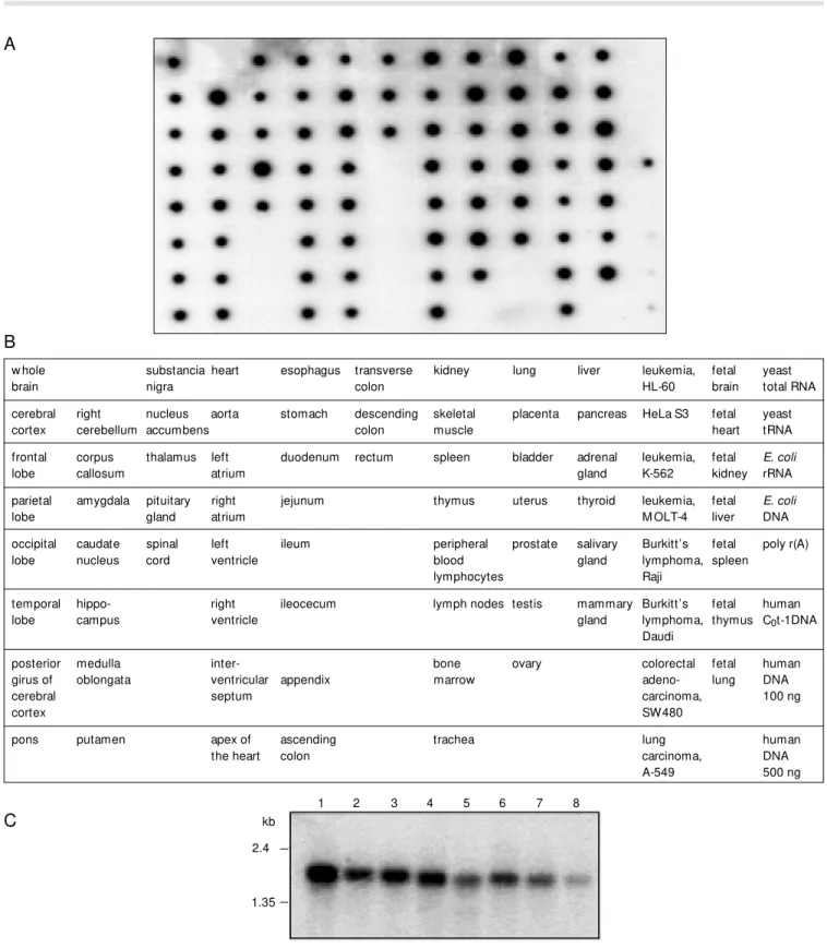
To determine the pattern of expression of DDX26 in different human tissues, a 1.5-kb cDNA fragment corresponding to the 3 por-tion of the mRNA was hybridized against commercial filters (Clontech) (Figure 3). While dot blot analysis indicated a ubiqui-tous expression of DDX26 in all tissues analyzed, a more detailed analysis showed a higher expression in cerebellum, pituitary gland, fetal kidney and fetal lung (Figure 3A). The specificity of the dot blot signal was confirmed by Northern blot analysis which showed the presence of a band of the expected size in all tumor cell lines tested. The highest expression level was observed in the melanoma cell line G-361 (Figure 3C). No alternative splicing forms were evident by Northern blot or Blast analysis against human ESTs.
A
B
Figure 2. Alignment of the predicted amino acid sequence of DDX26 and RNA helicases f rom Drosophila
(AF017777), yeast (NP013378), Can-dida (AL033391) and the hum an DDX6 RNA helicase (NM 004397). Conserved DEAD-box RNA helicase motifs are underlined and marked from I to VIII in the amino acid se-quences. Numbers on the right cor-respond to residue number of the protein. *Identical or conserved resi-dues; conserved amino acid substi-tution; semi-conserved amino acid substitution.
Figure 3. The expression pattern of DDX26 in normal tissues and tumor cell lines. A 32P-labeled probe w as hybridized under stringent conditions to a Human M aster Blot (Clontech) containing normalized loadings of polyA+ RNA from 75 different human tissues (A) as w ell as to a Cancer Cell Lines Northern blot (Clontech) containing 2 µg of polyA+ RNA (C). B, Location of the various RNAs on the dot blot filter is show n schematically. C, For the Northern blot membrane, the cancer cell lines are: 1, melanoma, G-361; 2, lung carcinoma, A-549; 3, colorectal adenocarcinoma, SW480; 4, Burkitt’s lymphoma, Raji; 5, lymphoblastic leukemia, M OLT-4; 6, chronic myelogenous leukemia, K-562; 7, HeLa S3, and 8, promyelocytic leukemia, HL-60. M olecular markers (kb) are indicated.
A
1 2 3 4 5 6 7 8
2.4
1.35 kb
C
w hole substancia heart esophagus transverse kidney lung liver leukemia, fetal yeast
brain nigra colon HL-60 brain total RNA
cerebral right nucleus aorta stomach descending skeletal placenta pancreas HeLa S3 fetal yeast cortex cerebellum accumbens colon muscle heart tRNA
frontal corpus thalamus left duodenum rectum spleen bladder adrenal leukemia, fetal E. coli
lobe callosum atrium gland K-562 kidney rRNA
parietal amygdala pituitary right jejunum thymus uterus thyroid leukemia, fetal E. coli
lobe gland atrium M OLT-4 liver DNA
occipital caudate spinal left ileum peripheral prostate salivary Burkitt’s fetal poly r(A) lobe nucleus cord ventricle blood gland lymphoma, spleen
lymphocytes Raji
temporal hippo- right ileocecum lymph nodes testis mammary Burkitt’s fetal human lobe campus ventricle gland lymphoma, thymus C0t-1DNA
Daudi
posterior medulla inter- bone ovary colorectal fetal human girus of oblongata ventricular appendix marrow adeno- lung DNA
cerebral septum carcinoma, 100 ng
cortex SW480
pons putamen apex of ascending trachea lung human
the heart colon carcinoma, DNA
A-549 500 ng
D iscussio n
DDX26, a human RNA helicase from the DEAD-box family, was cloned and charac-terized based on similarity to a yeast ortholo-gous gene Dbp9b. Cross-species sequence comparison was shown to be a useful tool for identification of exons due to their strong conservation during evolution when com-pared with random genomic sequences. Ac-cordingly, we have used the yeast ortholo-gous gene to search the human genomic sequence and to predict gene structure. Prim-ers for RT-PCR amplification were designed for validation and sequencing of the pre-dicted transcript.
Complete sequencing of the 1,889-bp PCR fragment revealed a single putative open reading frame coding for a protein of 548 amino acids with high similarity to an
essen-tial yeast RNA helicase (Dbp9b) involved in the assembly of early pre-ribosomal par-ticles. The corresponding transcript is ex-pressed in a broad range of tissues in agree-ment with the putative functional role in ribosome biogenesis.
DDX26 is located on chromosome 7p12, a region of frequent chromosome rearrange-ments in glioblastomas (12). The EGFR gene is located in this same region which is ampli-fied in 40% of glioblastomas together with extensive amplicons including a number of adjacent loci in some of these tumors. Re-cently, Wang et al. (13) described a new gene coding for a putative tyrosine kinase substrate (GBAS) that was co-amplified with EGFR in 2 of 12 tumors and in 2 of 3 cell lines analyzed.
Chromosome rearrangements, amplifica-tions and overexpression of different DEAD-Table 1. Amplification of different markers located on chromosome 7p12 in glioblastoma tumors.
box RNA helicases have already been re-ported for different tumors (4-7). Neverthe-less, co-amplification of DDX26 was not detected in eight tumors with amplification at the EGFR locus, excluding the involve-ment of this helicase in glioblastoma devel-opment.
Ackno wle dgm e nts
We thank Dr. Ricardo R. Brentani for continuous support and for a critical reading of the manuscript. We also thank Elaine Pereira Guimarães for technical assistance.
Re fe re nce s
1. de La Cruz J, Kressler D & Linder P (1999). Unw inding RNA in Saccharomyces cerevi-siae: DEAD-box proteins and related fami-lies. Trends in Biochemical Sciences, 24: 192-198.
2. Schmid SR & Linder P (1992). D-E-A-D protein family of putative RNA helicases.
M olecular M icrobiology, 6: 283-391. 3. Wang Y & Guthrie C (1998). PRP16, a
DEAH-box RNA helicase, is recruited to the spliceosome primarily via its noncon-served N-terminal domain. RNA, 4: 1216-1229.
4. Akao Y, Seto M , Yamamoto K, Iida S, Nakazaw a S, Inazaw a J, Abe T, Takahashi T & Ueda R (1992). The RCK gene associ-ated w ith t(11;14) translocation is distinct from the M LL/ALL-1 gene w ith t(4;11) and t(11;19) translocations. Cancer Re-search, 52: 6083-6087.
5. Amler LC, Schurmann J & Schw ab M (1996). The DDX1 gene maps w ithin 400 kbp 5' to M YCN and is frequently coam-plified in human neuroblastoma. Genes, Chromosomes and Cancer, 15: 134-137. 6. Arai Y, Hosoda F, Kobayashi H, Arai K,
Hayashi Y, Kamada N, Kaneko Y & Ohki M
(1997). The inv(11)(p15q22) chromosome translocation of de novo and therapy-re-lated myeloid malignancies results in fu-sion of the nucleoporin gene, NUP98, w ith the putative RNA helicase gene, DDX10. Blood, 89: 3936-3944.
7. Ishiguro T, Nakajima M , Naito M , M uto T & Tsuruo T (1996). Identification of genes differentially expressed in B16 murine melanoma sublines w ith different meta-static potentials. Cancer Research, 56: 875-879.
8. Liu L, Ichimura K, Pettersson EH & Collins VP (1998). Chromosome 7 rearrange-ments in glioblastomas; loci adjacent to EGFR are independently amplified. Jour-nal of Neuropathology and Experimental Neurology, 57: 1138-1145.
9. Dias-Neto E, Garcia CR, Verjovski-Almeida S, Briones M R, Nagai M A, da Silva WJ, Zago M A, Bordin S, Costa FF, Goldman GH, Carvalho AF, M atsukuma A, Baia GS, Simpson DH, Brunstein A, de Oliveira PS, Bucher P, Jongeneel CV, O’Hare M J, Soares F, Brentani RR, Reis LF, de Souza SJ & Simpson AJ (2000). Shotgun se-quencing of the human transcriptome
w ith ORF expressed sequence tags. Pro-ceedings of the National Academy of Sci-ences, USA, 97: 3491-3496.
10. Thompson JD, Higgins DG & Gibson TJ (1994). CLUSTAL W: improving the sensi-tivity of progressive multiple sequence alignment through sequence w eighting, position-specific gap penalties and w eight matrix choice. Nucleic Acids Research, 22: 4673-4680.
11. Zirw es RF, Eilbracht J, Kneissel S & Schmidt-Zachmann M S (2000). A novel helicase-type protein in the nucleolus: pro-tein NOH61. M olecular Biology of the Cell, 11: 1153-1167.
12. Chaffanet M , Chauvin C, Laine M , Berger F, Chedin M , Rost N, Nissou M F & Benabid AL (1992). EGF receptor amplifi-cation and expression in human brain tu-mours. European Journal of Cancer, 28: 11-17.