D iffe re ntiatio n o f auto no m ic re fle x
co ntro l be gins with ce llular m e chanism s
at the first synapse within the nucle us
tractus so litarius
Department of Physiology and Pharmacology,
O regon Health and Science University, Portland, O R, USA M.C. Andresen,
M.W. Doyle, T.W. Bailey and Y.-H. Jin
Abstract
Visceral afferents send information via cranial nerves to the nucleus tractus solitarius (NTS). The NTS is the initial step of information processing that culminates in homeostatic reflex responses. Recent evidence suggests that strong afferent synaptic responses in the NTS are most often modulated by depression and this forms a basic principle of central integration of these autonomic pathways. The visceral afferent synapse is uncommonly powerful at the NTS with large unitary response amplitudes and depression rather than facilita-tion at moderate to high frequencies of activafacilita-tion. Substantial signal depression occurs through multiple mechanisms at this very first brainstem synapse onto second order NTS neurons. This review highlights new approaches to the study of these basic processes featuring patch clamp recordings in NTS brain slices and optical techniques with fluorescent tracers. The vanilloid receptor agonist, capsaicin, distinguishes two classes of second order neurons (capsai-cin sensitive or capsai(capsai-cin resistant) that appear to reflect unmyelinated and myelinated afferent pathways. The differences in cellular proper-ties of these two classes of NTS neurons indicate clear functional differentiation at both the pre- and postsynaptic portions of these first synapses. By virtue of their position at the earliest stage of these pathways, such mechanistic differences probably impart important differentiation in the performance over the entire reflex pathways. Co rre spo nde nce
M.C. Andresen
Department of Physiology and Pharmacology
O regon Health and Science University Portland, O R 97239-3098 USA
Fax: + 1-503-494-4352 E-mail: andresen@ O HSU.edu
This review was based in part on a symposium presentation at the kind invitation of the Brazilian Federation of Societies of Experimental Biology, Salvador, BA, Brazil, August 29-31, 2002.
Research supported by the National Institutes of Health (HL-41119 (M.C. Andresen), HL-56460 (M.C. Andresen), HL-70433 (T.W. Bailey)).
The present address of M.W. Doyle is Department of Biology, George Fox University, Newberg, O R 97132-2697, USA.
Received July 24, 2003 Accepted January 21, 2004
Ke y words
•Sensory •Vanilloid •Glutamate
•Presynaptic modulation •Autonomic
•Visceral
•Potassium currents
Intro ductio n
Rapid adjustments of the cardiovascular system depend on neural reflexes and the pathways for these networks are dominated by brainstem neurons (1,2). The effector mechanisms are primarily within the auto-nomic nervous system and together the
trac-tus solitarius (NTS). The NTS and these primary visceral afferent synapses thus con-stitute the first step of information process-ing. Recent findings suggest that this affer-ent synapse is the site of a surprising degree of differentiation associated with visceral afferent subtypes. Thus, NTS pathways are distinctly different on the basis of afferent myelination and these differences begin at the afferent terminal (presynaptic character-istics) and are also related to the properties of the NTS neurons receiving those afferents (i.e., the second order NTS neurons). The present review will focus on the basic mechanisms and organization of these dif-ferences within the NTS and how such early differentiation in the NTS may alter the sim-plest views of the brain stem integration. The review will concern, in particular, the
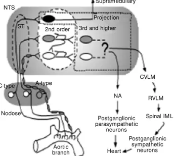
ex-ample of cardiovascular regulation (Figure 1).
Core brainste m circuits for autonomic pathways
The most basic schemes representing au-tonomic reflex circuits can be relatively sim-ply constructed as point-to-point sketches of central pathways that connect afferent in-puts to efferent autonomic outflows (Figure 1). Lumping each region for the moment as a single functional station, the NTS receives information directly from visceral afferents (e.g., nodose ganglia) and this information ultimately exits the NTS to separate sympa-thetic and parasympasympa-thetic pathways to the heart. The general pattern of central connec-tions for the sympathetic and parasympa-thetic systems is distinctly different (Figure 1). Data that support such a generalized con-ceptual diagram derive from multiple exper-imental sources (e.g., ranging from anatomi-cal point-to-point tracing to functional as-says of blood pressure responses to local drug microinjection) (1). Note that, in gen-eral, the consensus view represented in this scheme (Figure 1) identifies core paths for autonomic circuits as residing within the brainstem. Clearly, these region-to-region representations of even these core pathways are simplifications that do not incorporate important details of organization (cell to cell contacts or interactions) or information about the cellular mechanisms regulating that path-way. Much of this detail of the mechanisms by which these general areas interact re-mains to be established. From a circuitry or pathway perspective, fundamental questions of organization for the NTS might include addressing whether second order neurons in the NTS are interneurons (with axons lim-ited to the NTS), projection neurons or a mixture of both (Figure 1). NTS second or-der neurons are known to follow a loose viscerotopic distribution pattern based on afferent innervation (3). These second order NTS neurons, however, are likely to overlap
NTS
Supramedullary
CVLM
RVLM
Spinal IM L
Postganglionic sympathetic
neurons Postganglionic parasympathetic
neurons
Heart NA A-type
Nodose C-type
ST 2nd order 3rd and higher Projection
Aortic branch
in function so that NTS neurons may contri-bute to the circuitry of multiple defined ma-jor functions (e.g., cardiovascular and respi-ratory) (3).
One of the structurally simplest auto-nomic reflex pathways may be the cardiac parasympathetic arc (4). Thus, in concept, this simplest pathway for control of the heart would have only two central neurons, both of which are located in the brainstem. The second order neurons are found in the NTS and the efferent motor neurons in the nucleus ambiguus and/or dorsal motor nucleus of the vagus (5,6). The cardiac preganglionic para-sympathetic neurons are diffusely distrib-uted and are intermixed with adjacent neu-rons projecting elsewhere (6-9). In addition to central “cardiac” neurons, minimal para-sympathetic circuits include the primary vis-ceral afferent neurons that feed into the NTS (2) plus postganglionic efferent neurons within the heart (10). Evidence for such a four-neuron full reflex loop includes calcu-lations of reflex latencies (11) and the time course of distributions of retrograde viral tracers (5). Much more complicated and in-direct pathways are possible and cardiac parasympathetic control is likely to include additional, more convoluted processing ways. Nonetheless, the parasympathetic path-ways appear distinctly more compact than the sympathetic network (Figure 1). Even at the level of the brainstem, sympathetic path-ways appear to be anatomically diffuse and are incompletely understood (see, e.g., Ref. 12). The remainder of this review will focus on the earliest stage of autonomic pathways at NTS second order neurons (shaded box, Figure 1). New details on the nature of pro-cessing of afferent inputs to the NTS indi-cate sharp differentiation well before reflex responses emerge to the broader brainstem.
Barore ce ptor re fle xe s and use -de pe nde nt be havior
Baroreceptor reflexes are particularly
experi-mental animals are substantially modified by periods of sustained hemodynamic alter-ations such as bed rest or zero gravity (21). Little is known about the mechanisms re-sponsible or site(s) of alteration that such plasticity suggests. Since the most basic core pathways may involve relatively few neu-rons and synapses, understanding vagal heart rate control, for example, may focus on a surprisingly limited number of potential sites of action. The second order NTS neurons represent an interesting common “gateway” for these reflex pathways - no matter whether these paths contain very few neurons and are relatively direct or are part of more convo-luted paths (Figure 1).
The case for the strong synapse
The synapses from cranial visceral affer-ents within the medial portions of the NTS are unusual since they contact these second order neurons close to the soma and the resulting unitary synaptic currents are un-commonly large with low failure rates (22,23). Together, these characteristics cre-ate a strong synapse and a particularly high fidelity of transmission of excitation for that first afferent action potential through the next neuron in the pathway. NTS afferent transmission with large unitary responses from relatively few contacts is quite differ-ent from the classical view of cdiffer-entral synap-tic transmission as resulting from integration of large numbers of individually weak events (24).
The precise mechanisms that underlie the transfer and transformation of informa-tion from visceral afferent into discharge of NTS second order neurons are often difficult to resolve in vivo for many technical rea-sons. To better examine these mechanisms, the in vitro patch clamp approach has
impor-tant advantages in experimental control and much improved resolution (22). Horizontal slices containing the NTS effectively isolate afferent solitary tract (ST) axons and
pro-vide for selective electrical activation of ST axons to carefully examine synaptic timing. The large amplitude of the ST-evoked syn-aptic currents (commonly 300-600 pA) is a key characteristic of visceral afferents with important implications for synaptic integra-tion (22). Addiintegra-tional hallmarks include the substantial depression of ST transmission at physiologically important frequencies (above 5 Hz) and, more controversially, the reliance of second order neurons on non-NMDA glu-tamate receptors (22,25) (for more on this, see below). Since synaptic transmission op-erates close to maximum effectiveness (e.g., very high glutamate release probability), in-hibitory mechanisms and/or depression of glutamate release are a critical target of modu-lation of this afferent synapse.
In vitro experiments arose from a rich
history of work using varied approaches de-voted to discerning the neurotransmitter(s) responsible for transmission of afferent ex-citation at the NTS level. Glutamate is clearly the primary neurotransmitter (1). The earli-est NTS experiments elicited several inter-esting and controversial puzzles about the broader role of glutamate and specific recep-tors in the NTS. Early experiments used then newly developed selective drugs and sug-gested that both non-NMDA and NMDA glutamate receptors participate in CNS re-sponses such as the baroreceptor reflex. Mi-croinjection studies lead the way by placing glutamate selective agents directly into the NTS region (26). Subtype specific ionotrop-ic glutamate receptor antagonists blocked baroreflex responses but paradoxically not responses to injected glutamate (27). Vari-ous in vivo and cellular NTS studies
fol-lowed and confirmed this broad, ionotropic glutamatergic receptor participation (28-33). Unanswered questions remained, however, as to what was the site of action of these drugs and whether all neurons in the NTS were the same.
experimental work. For example, evidence from intracellular recordings in horizontal slices (22,34) does not support a substantial or direct role for NMDA receptors in ST afferent transmission. In most identified sec-ond order NTS neurons, glutamate - whether ST released or exogenous - fails to activate NMDA receptors even in the absence of extracellular Mg2+ (22,34,35). The
excep-tion is a small populaexcep-tion of second order NTS neurons with apparently myelinated ST afferent innervation (36). Methods of ap-proach are critical considerations for inter-preting results and, clearly, alternative ex-planations are needed to account for NMDA receptor responses in the NTS. For example, in transverse NTS slice preparations, mono-synaptic inhibitory mono-synaptic responses (GABAa mediated) are commonly found with electrical shocks in the vicinity of the ST (see, e.g., Ref. 37). The close proximity of the stimulating electrode appears to re-cruit local inhibitory interneurons and thus non-ST responses are clearly evident in such circumstances. Interestingly, NTS interneu-rons are activated by powerful NMDA syn-aptic components (38) and thus are pharma-cological targets during microinjection that are intermixed with second order neurons. Thus, the net effect on blood pressure or reflex function reflects a summation of all direct and indirect actions at multiple recep-tor subtypes and locations. Understanding the contributions of this heterogeneity will require a concerted effort and focused ex-perimental design.
He te roge ne ity in affe re nt ne urons to the NTS
Second order NTS neurons are, by defi-nition, driven by ST excitatory synaptic in-puts from visceral primary afferents arriving through the cranial nerves. The cellular prop-erties of visceral afferent neurons such as those in the nodose ganglion are highly var-ied (3). Relatively direct access to the
pe-ripheral cell bodies and sensory terminals shows that mechanoreception and chemore-ception are dominant sensory modalities among these cranial visceral primary affer-ents (3,39). Cardiovascular afferaffer-ents includ-ing arterial baroreceptors provide an inter-esting example since their physiological ac-tivation is now being translated into cellular mechanisms in increasing detail (40,41). A dominating, if under-appreciated, facet of the characteristics of cranial visceral affer-ent neurons, however, is their intrinsic vari-ability (42-44). Cranial visceral afferent neu-rons belong to two fundamental subdivi-sions (Figure 2), those with myelinated axons (A-type) and those with unmyelinated axons (C-type). A-type baroreceptors tend to have very regular discharge patterns and encode arterial pressure with great fidelity while C-type baroreceptors tend to have lower, often
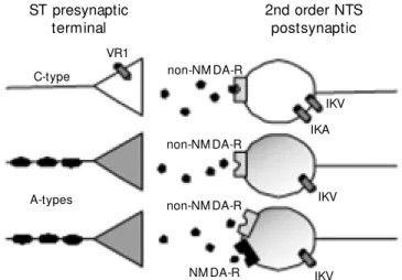
Figure 2. Solitary tract (ST) afferents fall into tw o broad classes based on the presence of the vanilloid receptor (VR1). Capsaicin-sensitive afferent axons are considered to be unmyelinated, C-type. A-type, myelinated ST axons as w ell as postsynaptic neurons are devoid of VR1. The anesthetic ketamine blocks NM DA receptors of a subset of A-type ST innervated neurons. Thus, presynaptic A- and C-type ST afferent terminals are highly differentiated on the basis of ligand- and voltage-dependent ion channel expression. Differential expression of pre- and postsynaptic effector proteins is likely to extend to particular ion channels as w ell as receptors (ionotropic and metabotropic). This offers multiple sites of potential action even for a single transmitter. These differences may provide the mechanistic basis for differential processing unique to each pathw ay. IKA = A-type potassium current; IKV = voltage-dependent potassium current; NM DA-R = N-methyl-D-aspartic acid postsynaptic receptors; NTS = nucleus tractus solitarius.
ST presynaptic terminal
2nd order NTS postsynaptic
C-type
A-types
VR1
non-NM DA-R
non-NM DA-R
non-NM DA-R
NM DA-R
IKV
IKA
IKV
sporadic discharge patterns that are only roughly proportional to pressure (45). Ob-servations of the reflex responses evoked by selective activation of these neuron classes suggest that these two pools of sensory affer-ent neurons give rise to reflexes with inter-esting differences in overall performance (46-48). For example, substantial cardiovas-cular reflex responses are evoked by as few as one action potential per second for C-type aortic baroreceptors, whereas reflex re-sponses emerge from A-type baroreceptor activation only at 10 action potentials per second or higher (46,47). A-type and C-type neurons differ in the properties at the cell soma (49). However, new results suggest that important basic cellular differences also separate the central terminations into phar-macologically and functionally distinct groups (Figure 2). Together, this heteroge-neity importantly shapes afferent central transmission.
A hallmark of the neurons within the NTS is broad cellular heterogeneity. Clearly, only a portion of the neurons within the NTS act as second order neurons - that is they receive afferent ST synaptic contacts. Other neurons in the NTS serve as either interneu-rons with contacts contained within the NTS or as projection neurons that send axons out of the NTS to other CNS areas. Anatomic studies have provided information about the wide spectrum of point-to-point connections between NTS and other CNS structures (3,50) although very little specific information about the nature of these interconnections is estab-lished. Anatomical subdivisions of the NTS have been described using histological and anatomical criteria by different investigators (51,52), but it is not clear what are the broader functional or mechanistic ramifications of such NTS diversity. Neurotransmitters and their receptors range tremendously with to-pographically varied expression within the NTS, but most classification groups are also without defined function or target (2,53). Neurons have been divided into different
classes of discharge patterns or ion channel expression in both caudal and rostral NTS and these key characteristics are linked to potassium channels (54,55). Thus, a major challenge in this area of the brainstem is to link these cellular characteristics with func-tion and/or identified pathways.
Cellular basis of NTS neurotransmission
The work summarized here considers the idea that, despite basic similarities of gluta-matergic transmission, other prominent dif-ferences in pre- and postsynaptic neurons shape an important differentiation of second order NTS neurons. We began with the premise that peripheral cellular differences in cranial afferent neurons (e.g., at the no-dose ganglion) are present in the central, presynaptic terminals of these same sensory neurons. The expectation (Figure 2) is that pathways through the NTS would be divided on the basis of afferent class (34,36,56).
Second order neurons in the NTS were selected by both electrophysiological and/or anatomical criteria, e.g., visualization of sen-sory boutons on the NTS neuron soma (22). ST shocks activated excitatory postsynaptic currents (EPSCs) with a range of latencies (22). Among the neurons recorded from such slices, a subset possesses ST-evoked EPSCs with nearly invariant latencies (low jitter) reflecting monosynaptic ST contacts identi-fying them as second order NTS neurons (22). These fast EPSCs activate non-NMDA postsynaptic receptors (Figure 2) in nearly all cases, confirming that the primary trans-mitter is glutamate (Figure 2).
including a high conductance for calcium ions (57,58). Capsaicin (CAP), the active ingredient in pungent peppers, is a selective ligand for VR1. CAP painted onto the aortic depressor nerve completely and selectively blocks the conduction of C-type axons and their cardiovascular reflex responses (46). Thus, we decided to test this mode of chem-ical modulation on brain stem slices contain-ing the NTS. In close correspondence to the peripheral action of CAP, we found two types of responses in second order NTS neurons. Although both types of neurons responded to ST stimulation with very simi-lar, low jitter EPSCs (<150 microsec jitter), the synaptic transmission to one set of neu-rons (CAP-sensitive) was blocked by CAP and the other neurons were unaffected by CAP (CAP-resistant). CAP applied to such slices triggered a burst of spontaneous syn-aptic currents that declined in the continued presence of CAP (34). CAP responses were prevented by application of selective non-NMDA receptor antagonists and unaltered by application of NMDA receptor antago-nists. CAP appears to trigger a massive re-lease of glutamate containing synaptic vesicles. Thus, pharmacologically, CAP re-sponses mimic ST afferent synaptic activa-tion. The blockade of ST synaptic transmis-sion by CAP appears to be a presynaptic action since postsynaptic glutamate recep-tors remain responsive to exogenous ago-nists during CAP block of electrically trig-gered ST synaptic transmission. Together, such results are consistent with specific CAP actions on VR1 receptors present only on the terminals of C-type ST afferent axons (Fig-ure 2). Interestingly, no partial blockades were found - an observation that suggests that A-type and C-type afferent axons do not converge on the same second order neuron in the NTS. The mechanism of C-type ST axon blockade is unclear. Intensive activa-tion of C-type axons might deplete the neu-rotransmitter. Depolarization block of presyn-aptic terminals could contribute to synpresyn-aptic
blockade during CAP. Although multiple mechanisms might contribute, the time course of onset of inhibition and its very slow rever-sal are consistent with depletion of synaptic vesicles from presynaptic terminals. These experiments provide one example of what is likely to be a broad presynaptic heterogene-ity of primary afferent innervation of the NTS (Figure 2).
whether release characteristics of neurotrans-mission are matched to specific postsynaptic properties and how those impact efficacy of overall translation of input to output. Given the differences in neurotransmitters ex-pressed by sensory afferents, the differences in postsynaptic NTS neurons could repre-sent an important component mechanism of differential afferent processing. For cardio-vascular regulation, future work should be targeted specifically toward aortic barore-ceptor terminals on NTS neurons (22) to see if this subtype matching exists in cardiovas-cular pathways.
In summary, detailed studies of synaptic transmission and ionic currents in second order NTS neurons reveal an unexpected differentiation of reflex pathways at these first CNS neurons. Such findings have strong implications for our understanding of the determinants of the overall reflex perfor-mance. Not only are the afferent discharge
patterns different in myelinated and unmy-elinated afferents, the reception of the re-leased neurotransmitter at the second order neurons is shaped in fundamental ways by the differences in ion channel expression of the central neurons. This early segregation of pathways produces substantially different reflex paths with performance characteris-tics that are tuned differentially to these dis-tinct afferent inputs. The differential expres-sion of VR1 across presynaptic afferent ST axons is likely to presage other presynaptic patterns that hold the prospect of chemical coding or selective interventional strategies that affect subsets of reflex pathways. Many details relating to afferent modality (e.g., cardiovascular, respiratory or gastrointesti-nal) remain largely unknown so that much work remains ahead before the potential im-pact of these heterogeneities can be fully appreciated or exploited.
Re fe re nce s
1. Pilow sky PM & Goodchild AK (2002). Baroreceptor reflex pathw ays and neurotransmitters: 10 years on. Journal of Hypertension, 20: 1675-1688.
2. Andresen M C & Kunze DL (1994). Nucleus tractus solitarius: gate-w ay to neural circulatory control. Annual Review of Physiology, 56: 93-116.
3. Loew y AD (1990). Central autonomic pathw ays. In: Loew y AD & Spyer KM (Editors), Central Regulation of Autonomic Functions. Oxford University Press, New York, 88-103.
4. Standish A, Enquist LW & Schw aber JS (1994). Innervation of the heart and its central medullary origin defined by viral tracing. Sci-ence, 263: 232-234.
5. Standish A, Enquist LW, Escardo JA & Schw aber JS (1995). Central neuronal circuit innervating the rat heart defined by transneuronal transport of pseudorabies virus. Journal of Neuroscience, 15: 1998-2012.
6. Irnaten M , Neff RA, Wang J, Loew y AD, M ettenleiter TC & M endelow itz D (2001). Activity of cardiorespiratory netw orks re-vealed by transsynaptic virus expressing GFP. Journal of Neuro-physiology, 85: 435-438.
7. M endelow itz D (2000). Superior laryngeal neurons directly excite cardiac vagal neurons w ithin the nucleus ambiguus. Brain Research Bulletin, 51: 135-138.
8. M endelow itz D (1998). Nicotine excites cardiac vagal neurons via three sites of action. Clinical and Experimental Pharmacology and Physiology, 25: 453-456.
9. M endelow itz D (1999). Advances in parasympathetic control of heart rate and cardiac function. New s in Physiological Sciences, 14: 155-161.
10. Thompson GW, Collier K, Ardell JL, Kember G & Armour JA (2000). Functional interdependence of neurons in a single canine intrinsic cardiac ganglionated plexus. Journal of Physiology, 528: 561-571. 11. Kunze DL (1972). Reflex discharge patterns of cardiac vagal efferent
fibres. Journal of Physiology, 222: 1-15.
12. M orrison SF (2001). Differential control of sympathetic outflow .
American Journal of Physiology, 281: R683-R698.
13. Thoren PN & Jones J (1977). Characteristics of aortic baroreceptor C-fibers in the rabbit. Acta Physiologica Scandinavica, 99: 448-456. 14. Hayw ard LF & Felder RB (1995). Cardiac rhythmicity among NTS neurons and its relationship to sympathetic outflow in rabbits.
American Journal of Physiology, 269: H923-H933.
15. Lambertz M , Kluge W & Langhorst P (1993). Discharge pattern of neurons in the nucleus tractus solitarii (NTS): its cardiac rhythm is modulated by firing rate of the neurons. Journal of the Autonomic Nervous System, 44: 137-150.
16. Brow n DL & Guyenet PG (1985). Electrophysiological study of car-diovascular neurons in the rostral ventrolateral medulla in rats.
Circulation Research, 56: 359-369.
17. Brunner M J, Sussman M S, Greene AS, Kallman CH & Shoukas AA (1982). Carotid sinus baroreceptor reflex control of respiration. Cir-culation Research, 51: 624-636.
Rapid resetting of the aortic baroreceptors in the rabbit and its implications for short-term and longer term reflex control. Circula-tion Research, 50: 428-439.
19. Ricketts JH & Head GA (1999). A five-parameter logistic equation for investigating asymmetry of curvature in baroreflex studies.
American Journal of Physiology, 277: R441-R454.
20. Kunze DL (1986). Acute resetting of baroreceptor reflex in rabbits: a central component. American Journal of Physiology, 250: H866-H870.
21. Eckberg DL & Fritsch JM (1992). Influence of ten-day head-dow n bedrest on human carotid baroreceptor-cardiac reflex function. Acta Physiologica Scandinavica Supplement, 604: 69-76.
22. Doyle M W & Andresen M C (2001). Reliability of monosynaptic transmission in brain stem neurons in vitro. Journal of Neurophysi-ology, 85: 2213-2223.
23. Andresen M C & Yang M (1995). Dynamics of sensory afferent synaptic transmission in aortic baroreceptor regions of nucleus tractus solitarius. Journal of Neurophysiology, 74: 1518-1528. 24. Trussell LO (1999). Synaptic mechanisms for coding timing in
audi-tory neurons. Annual Review of Physiology, 61: 477-496.
25. Andresen M C, Doyle M W, Jin Y-H & Bailey TW (2001). Cellular mechanisms of baroreceptor integration at the nucleus tractus soli-tarius. Annals of the New York Academy of Sciences, 940: 132-141. 26. Guyenet PG, Filtz TM & Donaldson SR (1987). Role of excitatory amino acids in rat vagal and sympathetic baroreflexes. Brain Re-search, 407: 272-284.
27. Leone C & Gordon FJ (1989). Is L-glutamate a neurotransmitter of baroreceptor information in the nucleus of tractus solitarius? Jour-nal of Pharmacological and Experimental Therapeutics, 250: 953-962.
28. Drew e JA, M iles R & Kunze DL (1990). Excitatory amino acid receptors of guinea pig medial nucleus tractus solitarius neurons.
American Journal of Physiology, 259: H1389-H1395.
29. M iller BD & Felder RB (1988). Excitatory amino acid receptors intrinsic to synaptic transmission in nucleus tractus solitarii. Brain Research, 456: 333-343.
30. Seagard JL, Dean C & Hopp FA (2003). Activity-dependent role of NM DA receptors in transmission of cardiac mechanoreceptor input to the NTS. American Journal of Physiology, 284: H884-H891. 31. Zhang J & M ifflin SW (1998). Differential roles for NM DA and
non-NM DA receptor subtypes in baroreceptor afferent integration in the nucleus of the solitary tract of the rat. Journal of Physiology, 511: 733-745.
32. Zhang W & M ifflin SW (1995). Excitatory amino-acid receptors contribute to carotid sinus and vagus nerve evoked excitation of neurons in the nucleus of the tractus solitarius. Journal of the Autonomic Nervous System, 55: 50-56.
33. M achado BH & Bonagamba LGH (1992). M icroinjection of L-gluta-mate into the nucleus tractus solitarii increases arterial pressure in conscious rats. Brain Research, 576: 131-138.
34. Doyle M W, Bailey TW, Jin Y-H & Andresen M C (2002). Vanilloid receptors presynaptically modulate visceral afferent synaptic trans-mission in nucleus tractus solitarius. Journal of Neuroscience, 22: 8222-8229.
35. Andresen M C & Yang M (1990). Non-NM DA receptors mediate sensory afferent synaptic transmission in medial nucleus tractus solitarius. American Journal of Physiology, 259: H1307-H1311. 36. Jin Y-H, Bailey TW, Doyle M W, Li BY, Chang KSK, Schild JH,
M endelow itz D & Andresen M C (2003). Ketamine differentially blocks sensory afferent synaptic transmission in medial nucleus tractus solitarius (mNTS). Anesthesiology, 98: 121-132.
37. Glaum SR, Brooks PA, Spyer KM & M iller RJ (1992). 5-Hydroxytryp-tamine-3 receptors modulate synaptic activity in the rat nucleus tractus solitarius in vitro. Brain Research, 589: 62-68.
38. Titz S & Keller BU (1997). Rapidly deactivating AM PA receptors determine excitatory synaptic transmission to interneurons in the nucleus tractus solitarius from rat. Journal of Neurophysiology, 78: 82-91.
39. Jänig W & M cLachlan EM (1992). Characteristics of function-specif-ic pathw ays in the sympathetfunction-specif-ic nervous system. Trends in Neuro-sciences, 15: 475-481.
40. Chapleau M W, Li Z, M eyrelles SS, M a X & Abboud FM (2001). M echanisms determining sensitivity of baroreceptor afferents in health and disease. Annals of the New York Academy of Sciences, 940: 1-19.
41. Hay M , Hoang CJ & Pamidimukkala J (2001). Cellular mechanisms regulating synaptic vesicle exocytosis and endocytosis in aortic baroreceptor neurons. Annals of the New York Academy of Sci-ences, 940: 119-131.
42. Schild JH, Clark JW, Hay M , M endelow itz D, Andresen M C & Kunze DL (1994). A- and C-type nodose sensory neurons: M odel interpre-tations of dynamic discharge characteristics. Journal of Neurophysi-ology, 71: 2338-2358.
43. Schild JH & Kunze DL (1997). Experimental and modeling study of Na+ current heterogeneity in rat nodose neurons and its impact on
neuronal discharge. Journal of Neurophysiology, 78: 3198-3209. 44. Seagard JL, Van Brederode JFM , Dean C, Hopp FA, Gallenberg LA
& Kampine JP (1990). Firing characteristics of single-fiber carotid sinus baroreceptors. Circulation Research, 66: 1499-1509. 45. Kunze DL & Andresen M C (1991). Arterial baroreceptors: Excitation
and modulation. In: Zucker IH & Gilmore JP (Editors), Reflex Control of the Circulation. CRC Press, Boca Raton, FL, USA, 141-166. 46. Fan W & Andresen M C (1998). Differential frequency-dependent
reflex integration of myelinated and nonmyelinated rat aortic barore-ceptors. American Journal of Physiology, 275: H632-H640. 47. Fan W, Schild JH & Andresen M C (1999). Graded and dynamic
reflex summation of myelinated and unmyelinated rat aortic barore-ceptors. American Journal of Physiology, 277: R748-R756. 48. Seagard JL, Hopp FA, Drummond HA & Van Wynsberghe DM
(1993). Selective contribution of tw o types of carotid sinus barore-ceptors to the control of blood pressure. Circulation Research, 72: 1011-1022.
49. Law son SN (1992). M orphological and biochemical cell types of sensory neurons. In: Scott SA (Editor), Sensory Neurons: Diversity, Development, and Plasticity. Oxford University Press, New York, 27-59.
50. Spyer KM (1990). The central nervous organization of reflex circula-tory control. In: Loew y AD & Spyer KM (Editors), Central Regulation of Autonomic Functions. Oxford University Press, New York, 168-188.
51. Kalia M & Sullivan JM (1982). Brainstem projections of sensory and motor components of the vagus nerve in the rat. Journal of Com-parative Neurology, 211: 248-264.
52. Ruggiero DA, Pickel VM , M ilner TA, Anw ar M , Otake K, M tui EP & Park D (1994). Viscerosensory processing in nucleus tractus solita-rii: structural and neurochemical substrates. In: Barraco RA (Editor),
Nucleus of the Solitary Tract. CRC Press, Boca Raton, FL, USA, 3-34.
53. Law rence AJ & Jarrott B (1996). Neurochemical modulation of cardiovascular control in the nucleus tractus solitarius. Progress in Neurobiology, 48: 21-53.
alters efficacy of tw o cardiorespiratory reflexes in vivo. American Journal of Physiology, 274: R677-R685.
55. Dekin M S & Getting PA (1984). Firing pattern of neurons in the nucleus tractus solitarius: modulation by membrane hyperpolariza-tion. Brain Research, 324: 180-184.
56. Bailey TW, Jin Y-H, Doyle M W & Andresen M C (2002). Vanilloid sensitive afferents activate neurons w ith prominent A-type potas-sium currents in nucleus tractus solitarius. Journal of Neurosci-ence, 22: 8230-8237.
57. Caterina M J & Julius D (2001). The vanilloid receptor: a molecular gatew ay to the pain pathw ay. Annual Review of Neuroscience, 24:
487-517.
58. Szallasi A & Blumberg PM (1999). Vanilloid (Capsaicin) receptors and mechanisms. Pharmacological Review s, 51: 159-212. 59. Oz M , Kolaj M & Renaud LP (2001). Electrophysiological evidence
for vasopressin V(1) receptors on neonatal motoneurons, premotor and other ventral horn neurons. Journal of Neurophysiology, 86: 1202-1210.