Infection with human papillomaviruses
of sexual partners of women having
cervical intraepithelial neoplasia
1Laboratório de Diagnóstico Molecular, Instituto de Biotecnologia, 2Laboratório de Patologia Médica, Departamento de Ciências Biomédicas, 3Ambulatório de Patologia do Trato Genital Feminino, Departamento de Clínica
Médica, 4Curso de Biologia, Universidade de Caxias do Sul, Caxias do Sul, RS, Brasil 5Instituto Ludwig de Pesquisa Sobre o Câncer, São Paulo, SP, Brasil
R.L. Rombaldi1,3,
E.P. Serafini1,2, L.L. Villa5,
A.C. Vanni4, F. Baréa4,
R. Frassini4, M. Xavier4
and S. Paesi1
Abstract
Epidemiological studies show that human papillomaviruses (HPV) are strongly related to cervical cancer and cervical intraepithelial neoplasias (CIN). Unlike the case for women, there are no consistent data on the natural history of HPV in the male population even though these viruses are prevalent in males. We carried out a prospective study to assess the prevalence of HPV in males as well as the factors that determine such infections in 99 male sexual partners of women with CIN. The genitalia of the males were physically examined and subjected to peniscopy for the collection of scrapings which were subjected to the polymerase chain reaction and restriction fragment length polymorphism to detect HPV. Of the 99 males sampled, 54 (54.5%) were positive for HPV DNA, 24% of whom presented normal peniscopy, 28% presented evident clinical lesions and 48% isolated lesions consistent with subclinical infection. In the HPV-negative group, 53% showed normal peniscopy, 4% presented evident clinical lesions and 42% isolated lesions consistent with subclinical infection. The study detected a statistically significant association (P < 0.02, Pearson chi-square test) between HPV infection and both the mean number of sexual partners which a male had during his life and the mean number of sexual partners in the year prior to testing. Viral types 6 and 11 were most frequently encountered. The study shows that infection with HPV was frequent in male sexual partners of women with CIN.
Correspondence
R.L. Rombaldi
Rua Humberto de Campos, 1055/402 95084-440 Caxias do Sul, RS Brasil
Fax: +55-54-221-5677
E-mail: rl_rombaldi@brturbo.com.br
Publication supported by FAPESP.
Received February 28, 2005 Accepted September 16, 2005
Key words
•Cervical intraepithelial
neoplasia
•Epidemiology
•Human papillomaviruses •Polymerase chain reaction
Introduction
Infection with human papillomavirus (HPV), a non-culturable DNA virus of the family Papillomaviridae (1) has gained great importance during the last decade due to the recognition that this virus participates in the genesis of cervical cancer (2) and has high worldwide prevalence (3). More than 118
his-tory of HPV infection in males. A previous study (8) found that the prevalence of HPV in males ranged from 3.6 to 84% depending on the population and the methodology used, while it is also known (9-11) that the highest incidence (63 to 84%) occurs in men attend-ing clinics for the treatment of sexually trans-mitted diseases. There is a high prevalence (10-85%) (12-17) of HPV lesions in the sexual partners of women with genital condy-loma or cervical intraepithelial neoplasias (CIN) as compared with the 10% prevalence found in the general male population (18).
The present study determined the preva-lence and the factors of HPV infection in an epidemiological survey of 99 male partners of women known to have CIN. Genital scrapings were submitted to the polymerase chain reaction (PCR) and restriction frag-ment length polymorphism (RFLP) to iden-tify HPV carriers.
Material and Methods
Population
A prospective study was carried out at the University of Caxias do Sul (UCS, Caxias do Sul, RS, Brazil) between February 2003 and July 2004 on 99 male sexual partners of women with CIN. The study was approved by the UCS Ethics Research Committee. There was no conflict of interest for any of the authors. All subjects signed an informed consent form to participate in the study. The external genitalia of each male were exam-ined physically before applying 5% acetic acid for 5 min and examining the genital area and collecting scrapings using penis-copy. Peniscopic biopsies were obtained from areas suggestive of HPV infection.
Epidemiological survey
In the epidemiological survey we con-sidered the following variables: age, age at first intercourse, circumcision, condom use
with female sex workers, condom use with sexual partners, educational level, marital status, marital stability (number of years married), race, sexual intercourse with fe-male sex workers, sexual partners up to the date of the survey, sexual partners in the year preceding the survey, sexually transmitted diseases, and tobacco smoking.
Peniscopy
Peniscopic images were classified as condylomatous lesions (acuminated, pig-mented or non-pigpig-mented warts), lesions suggestive of HPV infection (aceto-white areas, erythematous or macular lesions, pap-illomas or pearled papules) or normal.
Sampling methods
For each of the males studied a Urotest®
hyper-keratinosis, hyperplasia, keratiniza-tion, or papillomatosis. Biopsy samples were checked by submitting them to a second evaluation by the same pathologist, who was aware of the result of the first diagnosis.
Detection of HPV DNA
Total viral DNA was extracted from the samples using a commercial kit (Puregene™ from buccal cells; Gentra Systems Inc., Min-neapolis, MN, USA) and tested for HPV DNA using a PCR protocol which amplified a 450-bp segment of a conserved region of the L1 viral gene delineated by the MY9 and MY11 primers (19,20). The reaction mix-ture contained 2 µL of non-quantified extract-ed sample DNA in a final volume of 51 µL containing 10 mM Tris-HCl, pH 8.3, 5 mM MgCl2, 50 µM of each dNTP, 0.4 µg of each
primer, and 1.75 units of Taq DNA polymer-ase (Invitrogen, Carlsbad, CA, USA). Be-fore amplifying the sample DNA the quality of the host-cell DNA was evaluated using the PCO4 and GH20 primers (21) to amplify
a 268-bp segment of the human ß-globulin gene. Amplification was carried out in a PTC100 thermocycler (MJ Research, Wa-tertown, MA, USA) with 5 min for denatur-ation followed by 40 annealing cycles of 94ºC for 30 s, 55ºC for 1 min and 72ºC for 1 min, and a final extension at 72ºC for 5 min. All amplifications included a positive con-trol consisting of HPV 16 DNA extracted from SiHa cells (Ludwig Cancer Research Institute, São Paulo, SP, Brazil) and a nega-tive control consisting of a reaction mixture minus the genomic sample DNA. Amplifi-cation products were separated on 1% (w/v) agarose gel using Tris/boric acid buffer (TBE, containing 50 mM Tris, 50 mM boric acid, and 2.5 mM EDTA), pH 8.4, and a constant volt (3 V/cm), with a molecular mass marker (4 µg φX174 restriction fragment cleaved with HaeIII) being run simultaneously with the samples. After electrophoresis the gels were stained with ethidium bromide (4.62
ng/µL) and HPV or ß-globulin fragments visualized under ultraviolet light.
Viral typing
HPV-positive samples were typed using RFLP according to the method described by Bernard et al. (22). The product of the MY9/ MY11 amplifications was digested with a mixture containing 50 mM Tris-HCl, 10 mM MgCl2, and 10 units/µL of a restriction
en-zyme in a separate reaction (BamHI, DdeI,
HaeIII, HinfI, PstI, RsaI, or Sal3AI (Gibco-BRL, Gaithersburg, MD, USA). Fragments of different molecular masses were sepa-rated on 7% polyacrylamide gel (20.3% acrylamide, 0.7% bis-acrylamide, 0.07% ammonium persulfate and 0.7 µL/mL TBE 10X TEMED (GibcoBR). A sample of undi-gested amplified sample DNA and 2.5 µg standard φX174 DNA were run simulta-neously in a vertical miniVE electrophoresis cell (Hoefer, San Francisco, CA, USA). Af-ter silver staining the fragments were com-pared with those described by Bernard et al. (22).
Statistical analysis
Data were analyzed statistically with the Statistical Package for the Social Science (SPSS) version 12.0, using descriptive sta-tistics and the conventional Pearson chi-square test at a pre-fixed α level of 0.05.
Results
examination identified insitu carcinoma of the penis in only one tissue sample.
Analysis of the personal data of the 54 men positive for HPV DNA revealed that their mean age was 30.7 ± 10.3 years (range: 18-56 years; Table 3). There was no signifi-cant relationship between the presence of HPV DNA and the following variables: age at first sexual encounter, educational level, frequency of condom use with sexual part-ners, frequency of condom use with sex workers, marital status, marital stability ex-pressed in years, circumcision, race, sex re-lationships with sex workers, tobacco use, and previous history of sexually transmitted diseases (Tables 2 and 3). As shown in Table 3, there was a significant positive correlation between the presence of HPV DNA and number of sexual partners up to the date of the examination (P = 0.0014) and number of sexual partners in the year preceding the survey (P = 0.021). The HPV types detected in the study were: HPV 6 (56.1%), 11 (36.8), 16 (3.5%), 40 (0.7%), 61 (0.7%), and 84 (0.7%). The DNA from two HPV types, 11 and 84, was detected in one man.
The most frequently cited sexually trans-mitted diseases in both the HPV DNA-nega-tive and -posiDNA-nega-tive groups were gonorrhea, acuminated condyloma, syphilis, and geni-tal herpes.
Discussion
The present study demonstrated that 62.6% of the sexual partners of women with CIN had condylomatous, macular or papular genital lesions upon peniscopic examination after application of acetic acid (Table 1). Tabrizi et al. (23) studied the sexual partners of women with CIN and found that 38% of them had clinical lesions and/or lesions iden-tifiable by peniscopy. In our study we found that the areas most affected by genital HPV were the frenular delta and the preputial mucosa, probably because these are the most moist areas and are most subject to
micro-Table 1. Number and percentage (in parentheses) of men positive or negative for human papillomavirus (HPV) DNA and the type of their lesions as detected by peniscopy.
Type of lesion Men positive or negative for HPV/DNA
Positive Negative Total
Condylomatous lesions1 15 (28%) 2 (4.5%) 17 (17%)
Lesions suggestive of infection2 26 (48%) 19 (42%) 45 (46%)
Normal 13 (24%) 24 (53.5%) 37 (37%)
Total 54 (100%) 45 (100%) 99 (100%)
1Acuminated, pigmented or non-pigmented warts. 2Aceto-white areas, erythematous
or macular lesions, papillomas or pearlized papules.
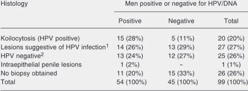
Table 2. Number and percentage (in parentheses) of men positive or negative for human papillomavirus (HPV) DNA and the histological type of their lesions when examined by biopsy.
Histology Men positive or negative for HPV/DNA
Positive Negative Total
Koilocytosis (HPV positive) 15 (28%) 5 (11%) 20 (20%)
Lesions suggestive of HPV infection1 14 (26%) 13 (29%) 27 (27%)
HPV negative2 13 (24%) 12 (27%) 25 (26%)
Intraepithelial penile lesions 1 (2%) - 1 (1%)
No biopsy obtained 11 (20%) 15 (33%) 26 (26%)
Total 54 (100%) 45 (100%) 99 (100%)
1Papillomatosis, hyperplasia, keratinization, or hyperkeratosis. 2Normal, or chronic
non-specific inflammatory process.
evident clinical lesions and 42.2% isolated lesions consistent with subclinical infection (Table 1). The following areas were found to suffer HPV attack: the frenular delta (31.4%), the preputial mucosa (29.6%), the glans-preputial sulcus (20.4%), the glans-penis (9.3%), the penile shaft (7.4%), and the ure-thral canal (1.9%).
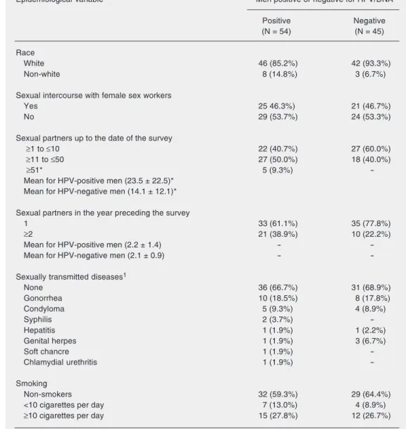
Table 3. Epidemiological variables investigated in the present study.
Epidemiological variable Men positive or negative for HPV/DNA
Positive Negative
(N = 54) (N = 45)
Age
≤19 5 (9.3%) 2 (4.4%)
≥20 to ≤29 23 (42.6%) 19 (42.2%)
≥30 to ≤39 16 (29.6%) 9 (20.0%)
≥40 to ≤49 6 (11.1%) 14 (31.1%)
≥50 to ≤59 4 (7.4%) 1 (2.2%)
Mean age for HPV-positive men (30.7 ± 10.3 years) -
-Mean age for HPV-negative men (33.0 ± 9.5 years) -
-Age at first intercourse
≤16 43 (79.6%) 26 (57.8%)
≥17 to ≤20 10 (18.5%) 17 (37.8%)
≥21 to ≤30 1 (1.9%) 2 (4.4%)
Mean age for HPV-positive men (15.5 ± 2.8 years) -
-Mean age for HPV-negative men (16.2 ± 2.6 years) -
-Circumcision
Circumcised 7 (13.0%) 3 (6.7%)
Uncircumcised 47 (87.0%) 42 (93.3%)
Condom use with female sex workers
Never 4 (16.0%) 6 (28.6%)
Sometimes 5 (20.0%) 7 (33.3%)
Frequently 5 (20.0%) 2 (9.5%)
Always 11 (44.0%) 6 (28.6%)
Condom use with sexual partner
Never 31 (57.4%) 28 (62.2%)
Sometimes 15 (27.8%) 15 (33.3%)
Frequently 4 (7.4%) 1 (2.2%)
Always 4 (7.4%) 1 (2.2%)
Educational level
Illiterate 1 (1.9%) 1 (2.2%)
Primary school (complete or incomplete) 32 (59.2%) 1 (2.2%)
Secondary school(complete or incomplete) 18 (33.4%) 13 (28.9%)
University (complete or incomplete) 3 (5.6%) 5 (11.1%)
Marital status
Single 9 (16.7%) 8 (17.8%)
Married 28 (51.9%) 27 (60%)
Cohabiting 17 (31.5%) 10 (22.2%)
Marital stability (number of years married)
≥1 to ≤2 23 (42.6%) 16 (35.6%)
≥3 to ≤5 10 (18.5%) 7 (15.6%)
≥6 22 (40.7%) 22 (48.9%)
Mean for HPV-positive men (6.7 ± 7.4 years) -
-Mean for HPV-negative men (9.1 ± 8.2 years) -
traumas during sexual activity.
The collection of material followed by peniscopy was justified by the possibility of working with minimum amounts of viral DNA in the penile samples obtained through scrapings, which might imply false-negative results for DNA/HPV, although the collec-tion covers the areas that are most affected
by HPV (6,24,25). The study cannot infer the percentage of false-negative results for DNA/HPV, but false-negative results could be explained by 1) the limitations of brush use to obtain the sample (coverage of scraped areas, pressure applied to perform the scrap-ing, and the limitation of the brush itself in obtaining the sample); 2) by the limited
Table 3 continued.
Epidemiological variable Men positive or negative for HPV/DNA
Positive Negative
(N = 54) (N = 45)
Race
White 46 (85.2%) 42 (93.3%)
Non-white 8 (14.8%) 3 (6.7%)
Sexual intercourse with female sex workers
Yes 25 46.3%) 21 (46.7%)
No 29 (53.7%) 24 (53.3%)
Sexual partners up to the date of the survey
≥1 to ≤10 22 (40.7%) 27 (60.0%)
≥11 to ≤50 27 (50.0%) 18 (40.0%)
≥51* 5 (9.3%)
-Mean for HPV-positive men (23.5 ± 22.5)* Mean for HPV-negative men (14.1 ± 12.1)*
Sexual partners in the year preceding the survey
1 33 (61.1%) 35 (77.8%)
≥2 21 (38.9%) 10 (22.2%)
Mean for HPV-positive men (2.2 ± 1.4) -
-Mean for HPV-negative men (2.1 ± 0.9) -
-Sexually transmitted diseases1
None 36 (66.7%) 31 (68.9%)
Gonorrhea 10 (18.5%) 8 (17.8%)
Condyloma 5 (9.3%) 4 (8.9%)
Syphilis 2 (3.7%)
-Hepatitis 1 (1.9%) 1 (2.2%)
Genital herpes 1 (1.9%) 3 (6.7%)
Soft chancre 1 (1.9%)
-Chlamydial urethritis 1 (1.9%)
-Smoking
Non-smokers 32 (59.3%) 29 (64.4%)
<10 cigarettes per day 7 (13.0%) 4 (8.9%)
≥10 cigarettes per day 15 (27.8%) 12 (26.7%)
Data are reported as number and percentage (in parentheses) of men positive or negative for human papillomavirus (HPV). 1The same individual sometimes gave more than one response to this question.
amount of human cells contained in the sample; 3) by the amplification technique used for the b-globin, and 4) the presence of inhibitors or the technique used to extract DNA. Studies comparing different sampling methods must be performed and the method used to extract human and viral genetic ma-terial should be improved.
Koilocytosis was the most frequent his-tological finding (27.8%) in the men who were positive for HPV DNA (Table 2). Hippeläinen et al. (24) found that 53.7% of a group of HPV DNA-positive men exhibited koilocytosis, while Nicolau et al. (26) using the same methodology, found a 50% fre-quency of koilocytosis. The results of our study did not allow us to infer that koilocyto-sis is a pathognomonic characteristic of HPV infection of the male urinogenital area be-cause there were as many false-positives as false-negatives, a fact which may lead to false information being given to the patient. The mean age of our group of men who showed HPV DNA was 30.7 years (Table 3), as compared to the mean of 38 years in the study by Bleeker et al. (27) and 26.5 years in the study by Baken et al. (28), although the case-controlled study carried out in different countries by Franceschi et al. (29) showed no association between age and HPV infec-tion.
With respect to educational level (Table 3), our sample may not have been represen-tative of all HPV-contaminated males in the city of Caxias do Sul and it is quite possible that men with completed secondary or higher education would have better access to more reliable information on the disease they were infected with and were also able to attend private clinics for treatment. All the men in the group positive for HPV DNA had only basic or secondary education and belonged to the segment of society that has least ac-cess to information and whose only acac-cess to medical assistance is via the public health service. These data suggest that health edu-cation is very precarious and that an
epide-miological survey should be conducted to ascertain what level of sex education this group of men receives in school and at home. There was no association between mari-tal status of the men and the presence of HPV DNA (Table 3). The study of the asso-ciation between marital stability ≥6 years and marital status revealed the presence of HPV DNA in 12.5% of single men, in 23.5% of cohabiting men and in 60.7% of married men. There was a statistically significant (P = 0.025) association between the presence of HPV DNA in married males and in males with marital stability ≥6 years.
The age at first sexual encounter (Table 3) showed that 79.6% of HPV DNA-positive men had initiated sexual activity when they were ≤16 years old, but this was not statisti-cally significant. In a 2002 study of men in the US, Castellsagué et al. (30) found that 22.9% of uncircumcised men had their first sexual experience when they were less than 16 years old while only 4.2% of circumcised men had their first sexual experience at this age. Colombian and Spanish males were the subjects of a study published in 1997 by Castellsagué et al. (31), who found that 20.1% of males had their first sexual encounter when they were less than 15 years old, while Franceschi et al. (29) found that 16% of males had their first sexual encounter at ≤16 (P = 0.67). In our study, men in the HPV DNA-positive group had their first sexual encounter at a mean age of 15.5 years but there was no statistically significant associa-tion between the presence of HPV DNA and age at first sexual encounter. Hippeläinen et al. (25) published an epidemiological study which showed that Finnish males had their first sexual encounter when they were 16.2 ± 1.5 years old. Rotola et al. (5) showed that the mean age at which Italian males in the city of Bologna had their first sexual en-counter was 18 years.
re-lated to the total number of sexual partners up to the date of the survey, with men who had the highest number of sexual partners also having the highest risk (P = 0.038) of being positive for HPV DNA (Table 3). Castellsagué et al. (30) studied uncircum-cised men who had had less than five sexual partners up to the time of the study and found that 12.5% of them were positive for HPV DNA, while among men who had had more than five sexual partners up to the time of the study the percentage of HPV DNA-positive subjects increased to 44.7%. Fran-ceschi et al. (29) found a highly significant association (P < 0.01) between the presence of HPV DNA and the number of sexual partners up to the date of the study, with 21.1% of men having less than 10 sexual partners being positive for HPV DNA, as opposed to 43.3% of men having more than 10 sexual partners.
The number of sexual partners in the year preceding our study (Table 3) was a highly significant (P = 0.021) variable concerning the presence of HPV DNA; however, this variable has not been previously evaluated by researchers in this area. The results ob-tained show us that the higher number of sexual partners is correlated to higher risks of being HPV DNA positive.
We found that for the shortest period of marital stability (≥1 to ≤2 years) 42.6% of subjects were positive for HPV DNA and for the highest period of marital stability (≥6 years) 40.7% of the subjects were positive. Both the minimum and maximum marital stability showed the highest percentages of HPV DNA-positive men (Table 3). In their 1993 study, Hippeläinen et al. (25) found that 68.2% of men who had had stable rela-tionships for up to 2 years were positive for HPV DNA, while only 31.7% of men who had had stable relationships for more than two years were HPV DNA positive. These results were also not statistically significant. The analysis showed that 59.3% of HPV DNA-positive males smoked tobacco (Table
3). However, as previously observed by Rotola et al. (5), Hippeläinen et al. (24,25), Franceschi et al. (29), and Wikstrom et al. (32), no significant correlation was observed between smoking and HPV positiveness in males. The absence of correlation between smoking and HPV infection in males, and the positive correlation observed in females (33) can be considered as an indicative that tobacco smoking, local immunity and the different structure of epithelial tissue of the female genital tract favor infection by HPV in females and the genesis of precursor and invasive lesions in the neck of the uterus.
Of the men in our HPV DNA group, 13% were circumcised, but there was no statisti-cally significant association between circum-cision and HPV DNA infection (Table 3). Castellsagué et al. (30) studied 292 circum-cised and 847 uncircumcircum-cised men in differ-ent countries and found that 5.5% of circum-cised and 19.6% of uncircumcircum-cised men were infected with HPV, and found an association between male circumcision and a reduced risk of genital infection with HPV. There is a reduced risk of cervical cancer in women with circumcised sexual partners, but with a high risk of HPV infection for the women.
female sex workers mentioned that their con-tact was casual and in the past. It is known that female sex workers have a higher risk of acquiring and disseminating sexually trans-mitted diseases because they have a greater number of sexual partners. To clarify this question, there is the need for a more elabo-rate epidemiological study in which this vari-able and other aspects of sexual behavior are surveyed in more detail.
About 84% of the HPV DNA-positive group used condoms (sometimes, frequently or always) during sexual activity with fe-male sex workers but this was not statisti-cally significant (Table 3). Franceschi et al. (29), in a similar Brazilian study, found that condom use was 86% and that 37% of the men studied in different countries regularly used condoms during sexual intercourse with female sex workers and demonstrated that the HPV-negative group made better use of condoms in sexual relationships with female sex workers. Although our study did not confirm the important protective role of condoms, this factor in itself indicates the possible existence of other unidentified epi-demiological or behavioral risk factors in the male population infected with HPV and that new epidemiological studies on condom use are needed to better understand our re-sults. In our study, 57% of the HPV DNA-positive men never used condoms with their regular female (non-sex worker) partner while 62.2% of HPV DNA-negative men never used a condom under the same cir-cumstances (Table 3). Hippeläinen et al. (25) pointed out the importance of condoms as a protective factor against HPV infection. We found that a previous history of sexu-ally transmitted disease in the HPV DNA-positive group (33.3%)was not statistically significant (Table 3). Wikstrom et al. (32) found that 23% (not significant) of males studied had previously had a sexually trans-mitted disease, while Hippeläinen et al. (24) reported an incidence of 15.8%. In our study the sexually transmitted diseases most
fre-quently reported by both groups were (in decreasing order) gonorrhea, acuminated condyloma, syphilis, and genital herpes but there was no statistically significant associa-tion between a history of sexually transmit-ted disease and the presence or absence of HPV DNA. Hippeläinen et al. (24) detected
Chlamydia infection and gonorrhea in their study.
Our study demonstrated for the first time that there is an association between the mean number of female sexual partners in the year preceding the study and the presence of HPV DNA, strengthening the belief that the higher the number of sexual partners the greater the chance of acquiring and transmit-ting HPV.
73% of the viruses encountered were types 6 and 11.
The results of the present study are con-sistent with the epidemiological data avail-able in the literature, except for our data on the number of female sexual partners up to the time of the study (‘lifetime sexual ners’) or the number of female sexual part-ners in the year preceding the study and HPV infection. HPV infection was frequent in the male sexual partners of women with CIN, indicating that a systematic study of this population is needed so that these couples can be better informed.
Our results, taken together with literature data, show that there is little agreement be-tween the prevalence of infection with HPV in men at high risk of HPV infection and
women carrying high and low grade lesions of the neck of the uterus. This may be ex-plained by the different levels of biological activity and differences in local immunity and organization of the genital epithelia of each sex. These factors can favor viral trans-mission and multiplication and predispose both sexes towards infection with one or more viruses. The different levels of biologi-cal activity and differences in lobiologi-cal immuni-ty and organization of the genital epithelia of each sex indicate that a prospective study is needed using samples of genital epithelia collected from a reasonable number of sexual partners. Thus, a more detailed epidemio-logical study can be carried out to gather more information on the biological behavior of infection by human papillomavirus.
References
1. Howley PM & Lowy DR (2001). Papillomaviruses and their replica-tion. In: Knipe DM & Howley PM (Editors), Fields Virology. 4th edn. Lippincott Williams & Wilkins Publishers, New York, 2197-2229. 2. WHO (1995). IARC Working Group on the evaluation of
carcino-genic risks to humans. Human papillomaviruses (HPV). Summary of data reported and evaluation, 64. WHO, Basel, Switzerland. 3. Menzo S, Monachetti A, Trozzi C et al. (2001). Identification of six
putative novel human papillomaviruses (HPV) and characterization of candidate HPV type 87. JournalofVirology, 75: 11913-11919. 4. de Villiers E, Fauquet C, Broker T et al. (2004). Classification of
papillomaviruses. JournalofVirology, 324: 17-27.
5. Rotola A, Costa S, Monini P et al. (1994). Impact of sexual habits on the clinical evaluation of male HPV infection. EuropeanJournalof
Epidemiology, 10: 373-380.
6. Siegel JF & Mellinger BC (1992). Human papillomavirus in the male patient. UrologicClinicsofNorthAmerica, 19: 83-91.
7. Brown T, Yen-Moore A & Tyring S (1999). An overview of sexually transmitted diseases. Part II. Journalof theAmericanAcademyof
Dermatology, 41 (Part 1): 661-677; quiz 678-680.
8. Kotoulas IG, Cardamakis E, Relakis K et al. (1996). Penoscopic diagnosis of flat condyloma and penile intraepithelial neoplasia. IV. Urethral reservoir. GynecologicandObstetricInvestigation, 41: 55-60.
9. Wikstrom A, Hedblad M, Johansson B et al. (1992). The acetic acid test in evaluation of subclinical genital papillomavirus infection: a comparative study on penoscopy, histopathology, virology and scanning electron microscopy findings. GenitourinaryMedicine, 68: 90-99.
10. Law C, Qassim M, Thompson C et al. (1991). Factors associated with clinical and sub-clinical anal human papillomavirus infection in homosexual men. GenitourinaryMedicine, 67: 92-98.
11. Mandal D, Haye K, Ray T et al. (1991). Prevalence of occult human papillomavirus infection, determined by cytology and DNA hybrid-ization, in heterosexual men attending a genitourinary medicine clinic. InternationalJournalofSTDandAIDS, 2: 351-355. 12. Nuovo GJ, Hochman HA, Eliezri YD et al. (1990). Detection of
human papillomavirus DNA in penile lesions histologically negative for condylomata. Analysis by in situ hybridization and the polymer-ase chain reaction. American Journal of Surgical Pathology, 14: 829-836.
13. Omar R, Choudhury M, Fischer J et al. (1991). A “Pap” test for men? Male urethral smears as screening tool for detecting subclinical human papillomavirus infection. JournalofUrology, 37: 110-115. 14. Wikstrom A, Lidbrink P, Johansson B et al. (1991). Penile human
papillomavirus carriage among men attending Swedish STD clinics.
InternationalJournal of STD and AIDS, 2: 105-109.
15. Ji H, Yliskoski M, Vayrynen M et al. (1991). Colposcopic analysis of genital human papillomavirus infections during an 8-year prospec-tive follow-up. InternationalJournalof Gynecologyand Obstetrics, 36: 291-300.
16. Levine R, Crum C, Herman E et al. (1984). Cervical papillomavirus infection and intraepithelial neoplasia: a study of male sexual part-ners. InternationalJournalof Gynecology andObstetrics, 64: 16-20. 17. Sand P, Bowen L, Blischke S et al. (1986). Evaluation of male consorts of women with genital human papilloma virus infection.
InternationalJournal ofGynecology andObstetrics, 68: 679-681.
18. Schneider A, Kirchmayr R, De Villiers E et al. (1988). Subclinical human papillomavirus infections in male sexual partners of female carriers. JournalofUrology, 140: 1431-1434.
265: 472-477.
20. Hildesheim A, Schiffman M, Gravitt P et al. (1994). Persistence of type-specific human papillomavirus infection among cytologically normal women. JournalofInfectiousDiseases, 169: 235-240. 21. Saiki R, Gelfand D, Stoffel S et al. (1988). Primer-directed enzymatic
amplification of DNA with a thermostable DNA polymerase. Sci-ence, 239: 487-491.
22. Bernard H, Chan S, Manos M et al. (1994). Identification and as-sessment of known and novel human papillomaviruses by polymer-ase chain reaction amplification, restriction fragment length poly-morphisms, nucleotide sequence, and phylogenetic algorithms.
JournalofInfectiousDiseases, 170: 1077-1085.
23. Tabrizi SN, Tan J, Quinn M et al. (1992). Detection of genital human papillomavirus (HPV) DNA by PCR and other conventional hybridisation techniques in male partners of women with abnormal Papanicolaou smears. GenitourinaryMedicine, 68: 370-373. 24. Hippeläinen M, Yliskoski M, Saarikoski S et al. (1991). Genital
human papillomavirus lesions of the male sexual partners: the diag-nostic accuracy of peniscopy. GenitourinaryMedicine, 67: 291-296. 25. Hippeläinen M, Syrjanen S, Koskela H et al. (1993). Prevalence and risk factors of genital human papillomavirus (HPV) infections in healthy males: a study on Finnish conscripts. SexuallyTransmitted
Diseases, 20: 321-328.
26. Nicolau SM, Martins NV, Ferraz PE et al. (1997). Importance of peniscopy, oncologic cytology and histopathology in the diagnosis of penile infection by human papillomavirus. Revista Paulista de
Medicina, 115: 1330-1335.
27. Bleeker MC, Hogewoning CJ, Van Den Brule AJ et al. (2002). Penile lesions and human papillomavirus in male sexual partners of women
with cervical intraepithelial neoplasia. Journalof theAmerican
Acad-emy ofDermatology, 47: 351-357.
28. Baken LA, Koutsky LA, Kuypers J et al. (1995). Genital human papillomavirus infection among male and female sex partners: prevalence and type-specific concordance. Journal of Infectious
Diseases, 171: 429-432.
29. Franceschi S, Castellsague X, Dal Maso L et al. (2002). Prevalence and determinants of human papillomavirus genital infection in men.
BritishJournalofCancer, 86: 705-711.
30. Castellsagué X, Bosch FX, Munoz N et al. (2002). Male circumci-sion, penile human papillomavirus infection, and cervical cancer in female partners. NewEngland Journal of Medicine, 346: 1105-1112.
31. Castellsagué X, Ghaffari A, Daniel RW et al. (1997). Prevalence of penile human papillomavirus DNA in husbands of women with and without cervical neoplasia: a study in Spain and Colombia. Journal
ofInfectiousDiseases, 176: 353-361.
32. Wikstrom A, Popescu C & Forslund O (2000). Asymptomatic penile HPV infection: a prospective study. InternationalJournal ofSTD and AIDS, 11: 80-84.
33. Villa LL (1997). Human papillomaviruses and cervical cancer.
Ad-vancesinCancerResearch, 71: 321-341.
34. Paesi SO, Serafini EP & Madi SRC (2003). Determinação e tipagem do papillomavirus humano (HPV) em amostras de população feminina atendida no Ambulatório de Patologia Cervical do Ambulatório Central da Universidade de Caxias do Sul. Revista de Ciências Médicas da Universidade de Caxias do Sul e da