Received: January 7, 2012 Accepted: March 21, 2012
Conflict of Interests: The authors state that there are no financial and personal conflicts of interest that could have inappropriately influenced their work.
Copyright: © 2011 Oliveira et al.; licensee EDIPUCRS. This is an Open Access article distributed under the terms of the Creative Commons Attribution-Noncommercial-No Derivative Works 3.0 Unported License.
Evaluation of a strategic practice demonstration
method applied to endodontic laboratory classes
Avaliação de um método estratégico de demonstração prática
aplicado nas aulas de endodontia laboratorial
Adriana Pachêco de Oliveira a
Erica dos Santos Carvalho a
José Luiz Lage-Marques b
Vanessa Cavalli b
Sandra Márcia Habitante b
Denise Pontes Raldi b
a Post-Graduate Program in Dental Sciences, Division of Endodontics, Department of Dentistry, University of Taubate, Taubate, SP, Brazil b Division of Endodontics, Department of Dentistry, University of Taubate, Taubate, SP, Brazil
Correspondence: Erica dos Santos Carvalho
Rua Expedicionário Ernesto Pereira, 110 Taubate, SP – Brazil
12020-340
E-mail: [email protected] Abstract
Purpose: To evaluate a strategic method of real time visual demonstration of the operatory procedures using an intraoral camera system and monitor videos, comparing them to those of conventional laboratory classes.
Methods: Fifty-two structured, multiple choice questionnaires were applied to undergraduate students of the 4th year (G1) and 3rd year (G2) submitted to the traditional and strategic teaching
methods, respectively. These tests were also able to detect the main problems faced by the students during the training of this operatory phase.
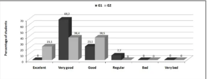
Results: Students of both groups (G1- 30.8% and G2- 34.6%) considered the access cavity to be one of the most difficult phase of endodontic treatment. The results of the evaluation among the 3rd year students demonstrated that 23.1% graded the new method as excellent,
38.4% as very good and 38.5 % as good, whereas none of the students (0%) considered the method to be regular, bad or very bad. A minor accident occurrence (P<0.05) was reported by the 3rd year students (G1- 50% and G2- 34.6%). Conclusion: The new strategy was found
to favor learning, reduce the incidence of errors and was appraised as efficient by the students. Key words: Dental education; Endodontics; teaching
Resumo
Objetivo: Avaliar um método estratégico nas aulas práticas, utilizando micro câmeras e monitores de vídeo para transmitir demonstrações da técnica operatória, comparando-o com o método de ensino aprendizagem clássico.
Metodologia: Foram aplicados 52 questionários direcionados aos alunos do 4º ano (grupo 1) submetidos a esta nova prática e aos do 3º ano (grupo 2) submetidos a metodologia de ensino tradicional. Este questionário também possibilitou detectar os principais problemas enfrentados pelos discentes durante o aprendizado desta etapa operatória.
Resultados: A fase de cirurgia de acesso foi considerada de difícil execução por ambos os grupos (G1- 30,8 % e G2- 34,6%), sendo a manobra de localização dos orifícios de entrada dos canais a etapa mais complicada. Quanto à avaliação da metodologia empregada no 3º ano, 23,1% dos alunos a consideraram excelente, 38,4% ótima, e 38,5% boa, ao passo que nenhum dos estudantes (0%) consideraram o método como regular, ruim ou péssimo. Uma menor ocorrência de erros (P<0,05), foi relatada pelos alunos do 3º ano (G1- 50% e G2- 34.6%).
Conclusão: A nova estratégia de ensino favoreceu a aprendizagem, diminuiu a incidência de erros e foi considerada eficaz pelos alunos.
Introduction
Teaching Endodontics to undergraduate students includes scientiic concepts and laboratory skill practice. Laboratory practice provides one of the best opportunities for active learning (1)and the adequate performance of endodontic techniques relies on speciic knowledge regarding anatomic details of the teeth to be treated (2). However, in practice, perception of these details is limited since direct visualization of the pulp chamber is minimal during the operatory act (3). The endodontic cavity preparation is the initial phase of endodontic treatment and allows visibility and access to each canal oriice, facilitating instrumentation (4,5). Laboratory practical classes to teach students to access the endodontic cavity take place at the beginning of the undergraduate’s preclinical training and these classes constitute one of the most dificult phases in training since numerous errors are performed by the students at this stage related to their lack of experience (6).
Such details make the learning process a dificult task for dental students, who become anxious and insecure about performing endodontic procedures. Thus, the search for strategic methods that improve learning skills becomes a constant challenge for the educators (7,8). Students who have never performed a technique must irst be guided through the process; thus, a visual demonstration of laboratory procedures is a key element in teaching pedagogy (9).
Current technology advancements now play an important role in the teaching process. The use of interactive learning methods are now a pedagogical tool to aid learning process (10-12).This new technology offers new and fascinating tools (computers, video imaging, digitized radiology and electronic data transmission), which are considered to be good resources for the learning process (13,14).
Due to these considerations and the difficulties experienced by students during pre-clinical training, adjustments in the conventional teaching/learning process are necessary. The aim of this study was to evaluate an
alternative method applied during laboratory classes on the endodontic cavity preparation and compare this to conventional methodology, according to the students’ own perception of their learning.
Methods
This study was analyzed and approved by the Ethical Research Committee (protocol # 348/2008) and the subjects of the study (undergraduate students) signed a term of informed consent. Fifty-two questionnaires were divided equally into two groups (n=26). In Group 1, the questionnaires were answered by students of the 4th year,
who received the traditional learning process (theoretical classes and lectures followed by laboratory practice with extracted teeth ixed in a mannequin). In Group 2, the questionnaires were answered by undergraduate students of the 3rd year, who had the same theoretical classes and
lectures as Group 1 and who, before the laboratory class, were submitted to an alternative teaching method. Real-time practical demonstrations to show the access cavity technique were performed on extracted teeth in the laboratory using an intraoral camera system (MICAM ci-SHARP, Curitiba Dentistry, Curitiba, PR, Brasil). The images were captured by a CCD micro camera (Curitiba Dentistry, Curitiba, PR, Brasil); sent through image processing electronics and the sequences of the procedures were transmitted by a cable to a 42-inch HD LCD television monitor (JVC- LT-42WX70, Kowloon, Hong Kong, Japan).
The questionnaire was prepared using the Sphinx Léxica
V. 5 software (Sphinx Brasil, Canoas, RS, Brasil) and the
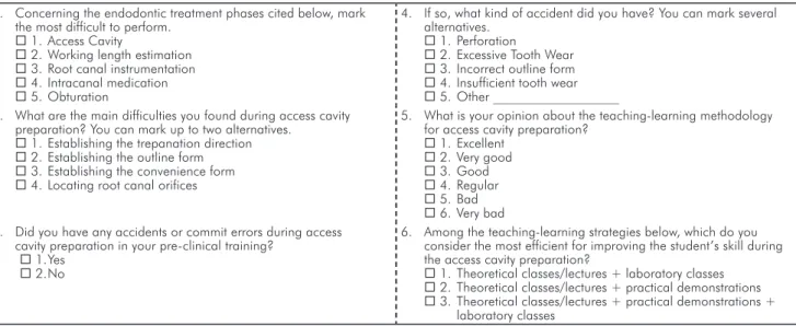
questions were elaborated in order to show which endodontic treatment phase was more complex in the students’ opinion, which were the primary problems faced during the access cavity phase, as well as the most common errors/accidents made at this stage. Furthermore, the students’ opinions and concerns about the teaching strategies were also attached to the questionnaire (Table 1).
Table 1. Questionnaire applied to the undergraduate students of the third and fourth year of UNITAU
1. Concerning the endodontic treatment phases cited below, mark the most difficult to perform.
1. Access Cavity
2. Working length estimation 3. Root canal instrumentation 4. Intracanal medication 5. Obturation
4. If so, what kind of accident did you have? You can mark several alternatives.
1. Perforation
2. Excessive Tooth Wear 3. Incorrect outline form 4. Insufficient tooth wear 5. Other ___________________ 2. What are the main difficulties you found during access cavity
preparation? You can mark up to two alternatives. 1. Establishing the trepanation direction 2. Establishing the outline form 3. Establishing the convenience form 4. Locating root canal orifices
5. What is your opinion about the teaching-learning methodology for access cavity preparation?
1. Excellent 2. Very good 3. Good 4. Regular 5. Bad 6. Very bad 3. Did you have any accidents or commit errors during access
cavity preparation in your pre-clinical training? 1. Yes
2. No
6. Among the teaching-learning strategies below, which do you consider the most efficient for improving the student’s skill during the access cavity preparation?
1. Theoretical classes/lectures + laboratory classes 2. Theoretical classes/lectures + practical demonstrations 3. Theoretical classes/lectures + practical demonstrations +
A previous pilot evaluation of the questionnaire was performed and handed out to twenty-six undergraduate students of the 4th year (students not submitted to the
experiment itself). This initial evaluation aimed to correct and adapt the questionnaire to be applied. The undergraduate students of this pilot evaluation were only subjected to the traditional method of teaching.
Results
The results of the survey (%) are represented in Table 2. Undergraduates of the 4th and 3rd years(G1 and G2) revealed
that access cavity preparation is the most complex endodontic treatment stage (34.6% and 30.8%, respectively). Both groups indicated the locating the root oriices as the most dificulty procedure during endodontic cavity preparation (Table 3).
Table 2. Technical difficulties in endodontic treatment stages
(%), according to undergraduate students of the 4th (G1) and
3rd (G2) years
Technical Difficulty G1 (%) G2 (%)
Access Cavity 30.8 34.6
Working length estimation 19.2 33.5
Instrumentation 26.9 15.4
Intracanal Medication 3.8 3.8
Obturation 23.1 7.7
Table 3. Main difficulties during the access cavity preparation
(%), reported by undergraduate students of the 4th (G1) and 3rd
(G2) years
Main difficulties G1 (%) G2 (%)
Perforation 26.9 24.0
Outline Form 21.2 44.0
Convenience Form 25.0 32.0
Locating root canal orifices 65.4 48.0
Fig. 3. Evaluation of teaching methodologies reported by
undergraduate students of the 4th (G1) and 3rd (G2) year.
The results of the remaining questions of this study are represented in Figures 1 to 3. The data obtained for the occurrence of error/accidents were submitted to statistical analysis by the Chi-square test. The results indicated that undergraduate students of the 3rd year presented a lower
percentage of error/accidents compared to 4th year students
(P< 0.05).
All the 3rd year (100%) and 92.3% of the 4th year students
elected the association of theoretical classes/lectures with practical demonstrations and laboratory classes as the best teaching-learning method. A minority of the 4th year
undergraduates (7.7%) considered the traditional teaching-learning (theoretical classes/lectures + practical classes) to be the most eficient strategy for improving the student’s skill endodontic cavity preparation.
Fig. 1. Occurrence of error/accidents during access cavity
preparation by the undergraduate of the 4th (G1) and 3rd (G2) year.
Fig. 2. Types of error/accidents most commonly performed during access cavity preparation by the undergraduate of the
4th (G1) and 3rd (G2) year.
Discussion
Endodontic access cavity preparation is often considered to be one of the most dificult phases of endodontic treatment and is frequently associated with unsuccessful endodontic procedures (4,5).This occurrence could be observed in the present study in both groups of undergraduates evaluated (G1-34.6% and G2-30.8 %). The reason for these errors is possibly due to the no experience of the students once the access cavity preparation constitutes the initial phase of endodontic treatment and is also the irst contact of the student with endodontic practice.
occurrence of errors and accidents during the endodontic cavity preparation, carried out by the students of 3rd year
(G2), were signiicantly lower when compared with the students of the 4th year (G1). This observation reinforces
the effectiveness of the strategy applied.
Aragon et al. (7)related that their dental students regularly complained about how dificult it was for them to visually appreciate the live demonstrations taught, since the mannequin’s oral cavity is small. In addition, students have to take sequential turns during these demonstrations to be able to get close enough to their row instructor while a particular task is performed. The method tested in this study with the use of the intraoral micro camera, connected directly to video monitors, allows the ampliication of the images and facilitates the visualization of the surgical act with all the details of the procedures. This method corroborates the fundamental role of the technology in the current context of the teaching models, increasing and facilitating the levels of learning (8,10,12).
Brüllman et al. (3)has also showed in an in vitro study the practical application of computer-aided techniques for detecting root canal oriices through the access cavity using a video camera mounted on a microscope. A total of 165 extracted human teeth (molars and premolars) were used as test data to collect 8,250 images via screenshots for the evaluation of the detection quality. The software provided a detection sensitivity of 90.1%, with only 11.9% of the images as false-positive detections. The study shows that
computer-aided recognition of root canal oriices with video cameras is possible.
Another important goal of this study was to reveal the impact of this method with regard to the improvement in the quality of the learning process, based on the students’ point of view. Students’ responses indicated that this strategy was appreciated by all (100%) the students who experienced it (G2). The results of this study also suggest that the demonstration of the surgical act in real time may be used in other phases during preclinical training in Endodontics, as an adjunct to conventional teaching, to improve and facilitate students’ learning. In addition, the video output can be recorded. The recorded images can then be edited using video-editing software. This procedure can be especially beneicial if students have the opportunity to refer back to the material repeatedly (16,18).
Educators must break paradigms, modify ineffective education processes and seek adequate teaching methods in order to fulill the students’ real needs. Additionally, teachers must constantly evaluate the results of these changes, to adequate and increasingly improve them.
Conclusions
The alternative teaching method strategy favored learning and reduced the incidence of errors. Additionally, the related methodology was considered good and interesting in the students’ opinion.
1. Maldarelli GA, Hartmann EM, Cummings PJ, Horner RD, Obom KM, Shingles R, et al. Virtual lab demonstrations improve students’ mastery of basic biology laboratory techniques. JMBE 2009; 10:51-7.
2. Pécora JD, Sousa Neto MD, Saquy PC, Woelfel JB. In vitro study of root canal anatomy of maxillary second premolars. Braz Dent J 1993;3:81-5.
3. Brüllmann DD, Alvarez P, Willershausen B. Recognition of root canal orifices in video sequences as a future support system during endodontic treatment. J Endod 2009;35: 1400-3.
4. Patel S, Rhodes J. A practical guide to endodontic access cavity preparation in molar teeth. Br Dent J 2007;203:133-40.
5. Johnson BR. Endodontic access. Gen Dent 2009;57:570-7.
6. Gulabivala K. Setting up an MSc programme in endodontics. Int Endod J 1991;24:216-21. 7. Aragon CE, Zibrowski EM. Does exposure to a procedural video enhance preclinical dental
student performance in fixed prosthodontics? J Dent Educ 2008;72:67-71.
8. Al-Jewair TS, Qutub AF, Malkhassian G, Dempster LJ. A systematic review of computer-assisted learning in endodontics education. J Dent Educ 2010;74:601-11.
9. Janik JM. Access cavity preparation. Dent Clin North Am 1984;28:809-18.
10. Tracey A, Bridget D. Virtual labs in the online biology course: student perceptions of effectiveness and usability. J Online Learn Teach 2007;3:105-11.
11. Nance ET, Lanning SK, Gunsolley JC. Dental anatomy carving computer-assisted instruction program: an assessment of student performance and perceptions. J Dent Educ 2009;73:972-9.
12. Handal B, Groenlund C, Gerzina T. Academic perceptions amongst educators towards eLearning tools in dental education. Int Dent J 2011;61:70-5.
13. Rosenberg H, Grad HA, Matear DW. The effectiveness of computer-aided, self-instructional programs in dental education: a systematic review of the literature. J Dent Educ 2003;67:524-32.
14. Marras I, Nikolaidis N, Mikrogeorgis G, Lyroudia K, Pitas I. A virtual system for cavity preparation in Endodontics. J Dent Educ 2008;72:494-502.
15. Bogacki RE, Best A, Abbey LM. Equivalence study of a dental anatomy computer-assisted learning program. J Dent Educ 2004;68:867-71.
16. Wright EF, Hendricson WD. Evaluation of a 3-D interactive tooth atlas by dental students in dental anatomy and endodontics courses. J Dent Educ 2010;74:110-22.
17. Handelsman J, Ebert-May D, Beichner R, Bruns P, Chang A, DeHaan R et al. Education. Scientific teaching. Science 2004;304:521-2.