152
Rev Odonto Cienc 2012;27(2):152-155Received: September 20, 2011 Accepted: June 29, 2012
Conflict of Interests: The authors state that there are no financial and personal conflicts of interest that could have inappropriately influenced their work.
Copyright: © 2011 Böttcher et al.; licensee EDIPUCRS. This is an Open Access article distributed under the terms of the Creative Commons Attribution-Noncommercial-No Derivative Works 3.0 Unported License.
Original Article
Calcium hydroxide removal: Effectiveness of
ultrasonic and manual techniques
Remoção de hidróxido de cálcio: Eficácia das técnicas
ultrassônica e manual
Daiana Elisabeth Böttcher a
Nicole de Mello Rahde b
Fabiana Soares Grecca a
a School of Dentistry, Federal University of Rio Grande do Sul (UFRGS), Porto Alegre, RS, Brazil b School of Dentistry, Pontifical Catholic University of Rio Grande do Sul (PUCRS), Porto Alegre, RS, Brazil
Correspondence:
Fabiana Soares Grecca Avenida Ramiro Barcelos, 2492 Porto Alegre, RS – Brazil 90035-003
E-mail: [email protected]
Abstract
Purpose: Different techniques have been proposed to improve the removal of calcium hydroxide. The purpose of this in vitro study was to compare the effectiveness of ultrasound and manual techniques in the removal of calcium hydroxide from root canals.
Methods: Thirty-eight single-rooted teeth were divided into two groups of 19 teeth each. The teeth were prepared using the crown-down technique with an apical master file #50. After shaping, the canals were radiographed and dressed with calcium hydroxide. After 14 days, the calcium hydroxide was removed with the ultrasound technique in group I and through manual filing in group II. The teeth were then radiographed again to evaluate the removal of the paste. To assess the calcium hydroxide removal, the radiographs were scanned and analyzed based on the gray levels. The independent samples and Student’s t-tests (a=0.05) were used for each group to compare the efficacy of calcium hydroxide removal between the groups and among thirds (cervical, middle and apical).
Results: There was no difference between ultrasound and manual techniques in the removal of calcium hydroxide from root canals. There were no statistical differences among the thirds analyzed.
Conclusion: Neither manual nor ultrasonic techniques completely removed calcium hydroxide from the root canal.
Key words: Endodontics; calcium hydroxide
Resumo
Objetivo: Diferentes técnicas são propostas para melhorar a remoção da pasta de hidróxido de cálcio. O objetivo do presente estudo in vitro foi comparar a efetividade das técnicas ultrassônica e manual para a remoção do hidróxido de cálcio do canal radicular.
Metodologia: Trinta e oito dentes monorradiculares foram divididos em dois grupos de 19 dentes cada. Os dentes foram preparados através da técnica de coroa-ápice até o instrumento memória #50. Após o preparo, os canais foram radiografados e, então, preenchidos com hidróxido de cálcio. A remoção da pasta no grupo I foi feita com o uso do ultrassom enquanto que no grupo II, a pasta foi removida através da técnica manual. Os dentes foram radiografados novamente para avaliar a remoção do hidróxido de cálcio. As radiografias foram digitalizadas e os terços avaliados de acordo com os níveis de cinza para quantificar a remoção do hidróxido de cálcio. Os testes para amostras independentes e o t de Student (a=0.05) foram aplicados para comparar a remoção da pasta entre os grupos e entre os terços do canal.
Resultados: Não houve diferença entre as técnicas ultrassônica e manual para a remoção do hidróxido de cálcio do canal radicular. Não houve diferenças estatísticas na comparação ente os terços avaliados.
Conclusão: Nem a técnica manual nem a ultrassônica removeram completamente o hidróxido de cálcio do canal radicular.
Rev Odonto Cienc 2012;27(2):152-155
153
Böttcher et al.
Introduction
In endodontics, the use of intracanal dressing is important
between treatment sessions for periapical periodontitis.
Calcium hydroxide (CH) has been used to inactivate
bacterial LPS (1), to induce the formation of mineralized
tissue (2) and to change the pH in the tubules through the
diffusion of hydroxyl ions (3), thereby contributing to the
success of endodontic therapy.
In order to maximize the properties of CH, the pulp
space should be illed with the medication (4-6). However,
the removal of CH may be dificult, which raises questions
regarding the appropriate method to use for the effective
removal of the paste and the consequences of keeping
the medication in the root canal illing (7-11). Different
irrigating solutions and hand iles have been widely used
for this purpose (8,10,12).
Several studies (13-15) have demonstrated the ability
of ultrasound to promote greater root canal cleansing by
removing the smear layer. However, its use in the removal
of CH has shown different results depending on the paste
vehicle or the irrigating solution used (16-18).
Thus, the aim of this
in vitro
study was to compare
the effectiveness of ultrasound and manual techniques in
removing CH from root canals.
Methods
This study (protocol number 17525) was approved
by the Institutional Review Board and by the Research
Ethics Committee of the School of Dentistry at the Federal
University of Rio Grande do Sul (Brazil).
Thirty-eight human single-rooted teeth with fully formed
apices and straight root canals were selected for this study.
The teeth were stored in saline solution until they were used.
Roots with cracks, caries, open apices, or resorptive defects
were excluded from the study.
The coronal portions of the teeth were removed with
diamond disks, standardizing the root length to 14 mm. A
diamond bur (KG Sorensen, Barueri, SP, Brazil) was used to
gain straight-line access to the root canal. Using a syringe and
a 27-gauge needle (Ultradent Products Inc., Indaiatuba, SP,
Brazil), the canal was irrigated with 2 mL of a 2.5% sodium
hypochlorite solution (NaOCl) (Biodinâmica Química e
Farmacêutica Ltda, Ibiporã, PR, Brasil) between each iling.
A #10 K-Flexoile (Dentsply Maillefer, Tulsa, OK, USA)
was used to remove the pulp tissue and introduced further
into the root canal until just the tip was visible at the apical
foramen. The working length was determined by subtracting
1 mm from this length. The canal was instrumented using
the crown-down technique from the working length up to a
#50 master apical ile.
A inal rinse with 3 mL of 17% EDTA
(Biodinâ-mica, Ibiporã, PR, Brazil) and 2 mL of 2.5% NaOCl was
performed.
Radiographs (R1) were taken after the root canal
preparation using Pró-70 (Prodental Equipamentos
Odon-tológicos LTDA, Ribeirão Preto, SP, Brazil), which was
operated at 70 kVp and 8 mA with a focus-ilm distance
of 40 cm and 0.4 s of exposure. Ultraspeed periapical
ilm (Eastman Kodak Corp., Rochester, NY, USA) was
used. The radiographs were processed during the same
session, using an automatic method (DentX 9000
®, DentX,
Elmsford, NY, USA) with new solutions and a cycle of
4.5 minutes. The root apices were sealed with sticky
wax. The samples were dried and illed with CH (Calen, S.S.
White Artigos Dentários Ltda., Rio de Janeiro, RJ, Brazil;
Composition: 2.5 g calcium hydroxide, 0.5 g zinc oxide,
0.05 g colophony and 1.75 mL polyethylene glycol 400).
The CH paste was placed using a threaded ML endodontic
syringe (S. S. White Artigos Dentários Ltda., Rio de
Janeiro, RJ, Brazil) with a 27-gauge needle (Terumo,
Tokyo City, Tokyo, Japan). Radiographs were taken
to evaluate the quality of the CH illing as previously
described.
The access openings were sealed with a temporary illing
material (Vigodent S/A Indústria e Comércio, Rio de Janeiro,
RJ, Brazil). Teeth were stored in an incubator for 14 days at
37°C and 100% relative humidity. After this period, samples
were randomly assigned to two groups.
For the utrasonic group (group 1, n=19), CH paste
was removed using 2 mL of a 2.5% NaOCl solution and
ultrasonic activation was delivered for 1 minute using the
Enac (Osada Electric Co, Tokyo City, Tokyo, Japan). The
frequency of the oscilatting instrument (#30 Flexoile) was
ixed at 25 kHz. The activation was followed by a rinse with
3 mL of a 17% EDTA solution for 3 minutes. Canals were
then dried with paper points.
For the manual group (group 2, n=19), CH paste was
removed using 2 mL 2.5% NaOCl with a #30 Flexoile,
which was placed through the working length and moved up
for 1 minute. Manual removal was followed by 3 mL 17%
EDTA for 3 minutes. Canals were then dried with paper
points.
As previously described, radiographs were taken to
evaluate the empty canals (R2).
The digitization of the radiographs was carried out using
an Epson Perfection 2450
®scanner (Epson, Long Beach, CA,
USA) with a transparency adaptor. The images were captured
in their original size, with 300 dpi, 8 bit mode, providing 256
grey levels. All radiographs were simultaneously captured.
The images were exported to the Adobe Photoshop program
(Adobe Systems Inc., San Jose, CA, USA). The canals were
divided into cervical, middle and apical thirds and, using
the histogram tool, the optical density of each third was
recorded based on a scale of 256 possible shades of gray,
with black representing zero and white representing 255. The
evaluation was performed by a calibrated examiner, and the
Intraclass Correlation Coeficient (ICC) test revealed a good
level of agreement (0.99).
154
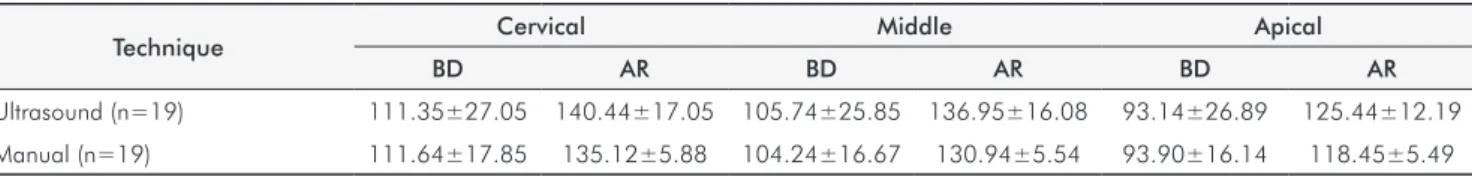
Rev Odonto Cienc 2012;27(2):152-155 Calcium hydroxide removalTable 1. Comparison of means and standard deviations of the gray levels obtained with ultrasound and manual techniques in the cervical, middle and apical thirds before dressing (BD) and after removal (AR) of calcium hydroxide
Technique Cervical Middle Apical
BD AR BD AR BD AR
Ultrasound (n=19) 111.35±27.05 140.44±17.05 105.74±25.85 136.95±16.08 93.14±26.89 125.44±12.19
Manual (n=19) 111.64±17.85 135.12±5.88 104.24±16.67 130.94±5.54 93.90±16.14 118.45±5.49
Technique Thirds
Cervical Middle Apical
Ultrasound (n=19) 29.09±22.21 23.48±17.22 31.22±19.17
Manual (n=19) 26.69±15.15 32.30±20.31 24.55±15.59
P .065 .101 .202
Table 2. Comparison of mean differences and standard deviations of the gray levels obtained with ultrasound and manual techniques in the cervical, middle and apical thirds, before dressing and after removal of calcium hydroxide
Results
The gray values before and after CH dressing for each
group are shown in Table 1. The results showed that after
removing the CH from the root canal, the gray levels were
statistically different (
P
=0.05) from those prior to illing,
i.e.
,
the canals did not return to their initial empty state prior to
illing with calcium hydroxide. This difference occurred in
all thirds, regardless of the technique applied.
To compare the ultrasound and manual techniques, the
differences in gray levels after CH removal and before
dressing was evaluated for each third. There was no
statistically signiicant difference (
P
>0.05) between the
techniques (Table 2).
Discussion
The results of this study showed that neither one of
the techniques removed CH completely. In all thirds, the
gray scale values obtained using optical density after the
removal of CH were different from the values obtained
after preparation of the empty root canal. These results are
similar to the indings of previous studies, which showed
considerable amounts of CH remnants independent of the
removal technique used (9,12,16-18).
Despite the excellent antimicrobial characteristics, these
remnants of CH paste can reduce the canal permeability
by promoting the formation of calcium carbonate particles
and interfering with the sealing ability of endodontic
sealers (7-9,11,19). Therefore, different techniques have
been proposed to improve the removal of CH (16,17,20,21).
An irrigant solution in conjunction with ultrasonic
vibration was directly associated with the removal of organic
and inorganic debris from the root canal walls (12,16).
Thus, the effectiveness of irrigation depends on both the
mechanical lushing action and the chemical ability to
dissolve tissue (7,8,21,22).
The effect of ultrasonic agitation of the irrigants has
been evaluated with contradictory results. In our study,
the use of ultrasound did not improve CH removal from
the root canal when compared to the manual technique. In
contrast, van der Sluis
et al
.
(16) reported that the use of
ultrasound was more eficient than conventional irrigation in
removing CH. Wiseman
et al.
(18) also found better results
when using passive ultrasonic irrigation for CH removal
compared to sonic activation. However, it is noteworthy that
both authors have reported the persistence of remnants in the
canals.
Perhaps the different methods used to assess eficacy
may be the reason that some authors report better results
with the use of ultrasound. Various methods were applied to
evaluate the presence of remnants (10,18,23,24), making it
dificult to compare studies regarding evaluation
methodo-logies.
Besides the use of ultrasound, simultaneous factors
such as the type of vehicle (oily vehicles are more dificult
to remove) (21) and the use of chelating agents (8,10) are
important for improving the eficiency of CH removal.
Nevertheless, canal irregularities may be inaccessible for
conventional irrigation procedures, and CH may remain in
these extensions (16,24).
Conclusion
Rev Odonto Cienc 2012;27(2):152-155
155
Böttcher et al.
1. Safavi KE, Nichols FC. Effect of calcium hydroxide on bacterial lipopolysaccharide. J Endod 1993;19:76-8.
2. Holland R, de Mello W, Nery MJ, Bernabe PF, de Souza V. Reaction of human periapical tissue to pulp extirpation and immediate root canal filling with calcium hydroxide. J Endod 1977;3:63-7.
3. Çalt S, Serper A, Ozcelik B, Dalat MD. pH changes and calcium ion diffusion from calcium hydroxide dressing materials through root dentin. J Endod 1999;25:329-31.
4. Rahde N. de M, Figueiredo JA, Oliveira EP. Influence of calcium hydroxide points on the quality of intracanal dressing filling. J Appl Oral Sci 2006;14:219-23.
5. Simcock RM, Hicks ML. Delivery of calcium hydroxide: comparison of four filling techniques. J Endod 2006;32:680-2.
6. Torres CP, Apicella MJ, Yancich PP, Parker MH. Intracanal placement of calcium hydroxide: a comparison of techniques, revisited. J Endod 2004;30:225-7.
7. Barbizam JV, Trope M, Teixeira EC, Tanomaru-Filho M, Teixeira FB. Effect of calcium hydroxide intracanal dressing on the bond strength of a resin-based endodontic sealer. Braz Dent J 2008;19:224-7.
8. Çalt S, Serper A. Dentinal tubule penetration of root canal sealers after root canal dressing with calcium hydroxide. J Endod 1999;25:431-3.
9. Margelos J, Eliades G, Verdelis C, Palaghias G. Interaction of calcium hydroxide with zinc oxide-eugenol type sealers: a potential clinical problem. J Endod 1997;23:43-8. 10. Salgado RJ, Moura-Netto C, Yamazaki AK, Cardoso LN, de Moura AA, Prokopowitsch I.
Comparison of different irrigants on calcium hydroxide medication removal: microscopic cleanliness evaluation. Oral Surg Oral Med Oral Pathol Oral Radiol Endod 2009;107:580-4.
11. Böttcher DE, Hirai VHG, Da Silva Neto UX, Grecca FS. Effect of calcium hydroxide dressing on the long-term sealing ability of two different endodontic sealers: An in vitro study. Oral Surg Oral Med Oral Pathol Oral Radiol Endod 2010;110:386-9.
12. Kenee DM, Allemang JD, Johnson JD, Hellstein J, Nichol BK. A quantitative assessment of efficacy of various calcium hydroxide removal techniques. J Endod 2006;32:563-5. 13. Alaçam T, Demirtola N, Misirligil A, Ayhan N, Gokay O. In vivo comparison of antimicrobial
effectiveness of conventional and ultrasound activated irrigation techniques in root canal therapy. Bull Tokyo Dent Coll 1987;28:19-22.
14. Guerisoli DM, Marchesan MA, Walmsley AD, Lumley PJ, Pecora JD. Evaluation of smear layer removal by EDTAC and sodium hypochlorite with ultrasonic agitation. Int Endod J 2002;35:418-21.
15. Serafino C, Gallina G, Cumbo E, Monticelli F, Goracci C, Ferrari M. Ultrasound effects after post space preparation: An SEM study. J Endod 2006;32:549-52.
16. van der Sluis LW, Wu MK, Wesselink PR. The evaluation of removal of calcium hydroxide paste from an artificial standardized groove in the apical root canal using different irrigation methodologies. Int Endod J 2007;40:52-7.
17. Balvedi RP, Versiani MA, Manna FF, Biffi JC. A comparison of two techniques for the removal of calcium hydroxide from root canals. Int Endod J 2010;43:763-8.
18. Wiseman A, Cox TC, Paranjpe A, Flake NM, Cohenca N, Johnson JD. Efficacy of sonic and ultrasonic activation for removal of calcium hydroxide from mesial canals of mandibular molars: a microtomographic study. J Endod 2011;37:235-8.
19. Ricucci D, Langeland K. Incomplete calcium hydroxide removal from the root canal: a case report. Int Endod J 1997;30:418-21.
20. Naaman A, Kaloustian H, Abboud NN, Ounsi HF, Ricci C, Medioni E. Influence of calcium hydroxide intracanal medication on the sealing ability of warm gutta-percha. Gen Dent 2008;56:348-52.
21. Nandini S, Velmurugan N, Kandaswamy D. Removal efficiency of calcium hydroxide intracanal medicament with two calcium chelators: volumetric analysis using spiral CT, an in vitro study. J Endod 2006;32:1097-101.
22. Lee SJ, Wu MK, Wesselink PR. The efficacy of ultrasonic irrigation to remove artificially placed dentine debris from different-sized simulated plastic root canals. Int Endod J 2004;37:607–12.
23. Balvedi RPA, Versiani MA, Manna FF, Biffi JCG. A comparison of two techniques for the removal of calcium hydroxide from root canals. Int Endod J 2010;43:763-8.
24. Rödig T, Hirschleb M, Zapf A, Hülsmann M. Comparison of ultrasonic irrigation and RinsEndo for the removal of calcium hydroxide and Ledermix paste from root canals. Int Endod J 2011;44:1155-61.