Quim. Nova, Vol. 35, No. 4, 771-774, 2012
Artigo
*e-mail: [email protected]
AN ANTIMICROBIAL ALKALOID AND OTHER METABOLITES PRODUCED BY Penicillium sp. AN ENDOPHYTIC FUNGUS ISOLATED FROM Mauritiaflexuosa L. f.
Hector Henrique Ferreira Koolen*, Elzalina Ribeiro Soares, Felipe Moura Araújo da Silva, Richardson Alves de Almeida e Afonso Duarte Leão de Souza
Departamento de Química, Universidade Federal do Amazonas, Av. Gal. Rodrigo Otavio, 3000, 69077-000 Manaus - AM, Brasil Lívia Soman de Medeiros e Edson Rodrigues Filho
Departamento de Química, Universidade Federal de São Carlos, Rod. Washington Luiz, km 235, 13565-905 São Carlos - SP, Brasil Antonia Queiroz Lima de Souza
Escola Superior de Ciências da Saúde, Universidade do Estado do Amazonas, Av. Carvalho Leal, 1777, 69065-001 Manaus - AM, Brasil
Recebido em 20/7/11; aceito em 4/11/11; publicado na web em 20/1/12
The alkaloid glandicoline B (1) and six other compounds: ergosterol (2), brassicasterol (3), ergosterol peroxide (4), cerevisterol (5), mannitol (6) and 1-O-α-D-glucopyranoside (7) were isolated from Penicillium sp. strain PBR.2.2.2, a fungus from Mauritia flexuosa
roots. The structures of the isolated metabolites were established by spectral analysis. MeOH extract of the fungal mycelium at 500 µg mL-1 exhibited antimicrobial activity against Staphylococcus aureus and the compound 1 at 100 µg mL-1 was active against S. aureus, Micrococcus luteus and Escherichia coli. The relationship between the bioactive properties of the fungus PBR.2.2.2 and those achieved for glandicoline B, as well the potential of this substance as bactericide is discussed.
Keywords: Penicillium; Mauritia flexuosa; glandicoline B.
INTRODUCTION
Plant-associated microorganisms are well known as great pro-ducers of natural products, being rich sources for biologically active metabolites with a wide-ranging application.1 Their ability to use different solid substrates for their own metabolism, including vegetal sources, makes them a very interesting biochemical-chemical agent in nature, specially interfering in the host-plant development.2 The host Mauritia flexuosa L. f.is a big palm tree from the Arecaceae family widespread by all South America.3 Its main constituents are flavonoids and triterpenoids.4 Plants of this species are used in the Amazonian folk-medicine, for burns and used as vermifuge.3 The fruits of this palm trees are also used to produce biofuels, cosmetics and as food by the local population.5
The Penicillium genus comprises more than 200 species, distrib-uted through the entire world, being found in the most uncommon environments, such as the permafrost soil of the Artic,6 some waste water from mines,7 and deep ocean sediments.8 Fungi from this genus are known as great producers of bioactive metabolites such as antimicrobial,9 immunosuppressants and cholesterol-lowering agents,10 anti-HIV11 and antitumor drugs.8 Endophytes are a poorly investigated group of microorganisms that represent an abundant and dependable source of bioactive and chemically novel compounds with potential for exploitation in a wide variety of medical, agricultural, and industrial arenas.12 In this context, the present work describes the isolation of some compounds from the Penicillium strain PBR.2.2.2 associated with the Amazonian palm tree M. flexuosa and their an-tibacterial activities.
RESULTS AND DISCUSSION
The EtOAc extract from the mycelial mass of the Penicillium sp. PBR 2.2.2 was chromatographed on silica gel columns and preparative
HPLC to give seven compounds (1-7) (Figure 1). The compounds 2-7 were identified as ergosterol,13 brassicasterol,14 ergosterol peroxide,15 cerevisterol,16 mannitol,17 and 1-O-α-D-glucopyranoside.18
Compound 1 was obtained as a yellow gum. Its 13C NMR spec-trum exhibited 22 signals which were assigned, by DEPT 135 and HSQC experiments to two methyls, one methylene, nine methines and ten quaternary carbons. Its APCI-MS spectrum contains an ion peak of [M+H]+ at m/z 420, consistent with the molecular formula C22H21N5O4 which also was in accordance with the NMR data. The
Figure 1. Structures from the isolated compounds 1-7 N
N
HN
O HO
N NH O OH
E
∆7
1
2
3
7,8 dihydro
HO
O O
HO OH
OH
OH HO
OH
OH OH
OH
O OH
HO
HO
OH O
4
5
6
7
HO 4
5 6
7 3a
7a 24
25
23 22 21 8 9
10 11
12 13 14
15 16 17
18 1
19 20 2
3
S R
Koolen et al.
772 Quim. Nova
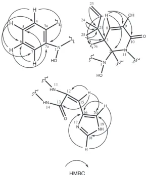
identification of 1 as the indole alkaloid glandicolin B (Figures 2-3) was made sure as follow. Its 1H NMR spectrum showed two duplets--like signals at δH 7.57 (H-4) and 6.96 (H-7) along with two triplets at δH 7.24 (H-6) and 7.01 (H-5), all signals with coupling constant of J = 7.7 Hz, characteristics of an orto bi-substituted aromatic ring, which was confirmed by the COSY correlations of H-5 with H-4 and H-6, together with the correlation of H-6 and H-7. Besides, the HMBC correlations of these hydrogen atoms were useful to determine the six aromatic carbons and the C-3, at δc 54.3, which was detected in correlation with the H-4. This aromatic ring was achieved as part of the indole skeleton of 1 principally by the HMBC correlations among the signal at δH 5.37 (1H, s, H-8) with the C-3 as well with the signals at δc 103.5 (C-2), 127.8 (C-3a), 144.4 (C-9) and 161.0 (C-10). The isoprenyl unit could be verified by the HMBC correlations of hydrogen signals at δH 1.32 (3H, s, H-24), 1.29 (3H, s, H-25), 5.10 (1H, dd, J = 10.8, 16.0, H-23 cis) and 5.03 (1H, dd, J = 16.0, 17.2, H-23 trans) with the carbon signal at δC 43.6 (C-21) as well by the COSY correlations of both H-23 one each other and with the me-thine hydrogen at δH 6.22 (1H, dd, J = 10.8, 17.2, H-22). The H-24 and H-25 also have HMBC correlation with the C-3 ratifying this carbon as the connection point of the isoprenyl group of 1. Finally the histidine group was indicated by the deshielded signals at δH 8.26 (1H, s, H-15), 7.90 (1H, s, H-18) and 7.37 (1H, s, H-20) and their HMBC correlations: H-15 with carbon signals at δc 167.2 (C-13), 131.4 (C-20) and 127.4 (C-12), H-18 with a signal at δc 128.8 (C-16) and the C-20, and H-20 with a signal at δc 137.6 (C-18) and the C-16. While no correlation could be observed between the histidine moiety and the remainder of 1, the chemical shifts of C-12, C-13 and C-2, all non-hydrogenated carbons, are in coherence with the known structure of glandicolin B. Since this substance is the direct precursor to meleagrin which is the O-methylated version of the glandicoline B, its absolute configurations on the double bond between C-12 and C-15 and on the C-2 and C-3 were deduced as E, S and R, respec-tively, as established by crystallography for meleagrin.19 All NMR data obtained for 1 are in the Table 1.
The CID spectra of the product ions of the peak at m/z 420 at 15 eV also confirmed 1, mainly by the fragments at m/z 351 and 334 corresponding to the losses of the isoprenyl and hydroxyl groups respectively, the ion 289 corresponds to a formamide loss from m/z 334. The ions at m/z 261 and 233 are derived from CO losses from the ion at m/z 289.
Isolation of Glandicoline B has been published only once previ-ously, from a soil strain of Penicillium glandicola,20 and its identifica-tion was done by comparison between its 1H NMR (acetone-d6), IR, UV and MS (EI) spectra and meleagrin data. All NMR (methanol-d4) and MS (APCI) data of 1 are published for the first time for glandicoline B. This substance is an important intermediate in the
Figure 2.1H-1H NOE and COSY correlations for the compound 1 N
N
HN
O HO
N NH O OH
4
5
6
7 3a
7a 24
25 23
22
21 8 9
10
11 12
13 14
15
16
17
18 1
19 20 2
3
NOE COSY
Figure 3.1H-13C HMBC correlations for the compound 1
N
HO 4 5
6
7 3a
7a 1
N N
HO
O OH 24
25 23
21 8 9
10
11
1 2 3
HN
HN
O
N NH 11
12
13 14
15
16
17
18 19 20
H
H
H
H
3a H
H
H H
HMBC
Table 1. Spectral data of compound 1, δ in ppm (1H, 500 MHz; 13C, 125 MHz)
δ
c δH, mult, (J in Hz) COSY HMBC NOE
1
-2 103.5 3 54.3 3a 127.8
4 125.8 7.57, d, (7.7) H5 C3, C6, C7a H24 5 123.7 7.01, t, (7.7) H4, H6 C3a, C7
6 129.4 7.24, t, (7.7) H5, H7 C4, C7a 7 113.1 6.96, d, (7.7) H6 C3a, C5 7a 149.8
8 110.8 5.37, s C2, C3, C3a, C9, C10 H23, H24 9 144.4
10 161.0 11 -12 127.4 13 167.4 14
-15 108.2 8.26, s C12, C13, C20 H20 16 128.8
17
-18 137.6 7.90, s C16, C20
19
-20 131.4 7.37, s C16, C18 H15
21 43.6
22 146.7 6.22, dd, (10.8; 17.2)
23a 113.7 5.10, dd, (10.8; 16) C21 23b 113.7 5.03, dd, (17.2; 16) C21
24 n.d. 1.32, s C3, C21 H4, H8
An antimicrobial alkaloid and other metabolites produced by Penicillium sp. 773
Vol. 35, No. 4
biosynthesis of the mycotoxins meleagrin and oxalin,21 which are pro-duced by some Penicillium species, as well the N-hydroxylated form of the glandicoline A, which is derived from some rearrangements reactions of the diketopiperazine alkaloid roquefortine C, another mycotoxin with neurotropic and antibiotic activities, commonly found in Penicillium spp. cultivation.22
Biological assay
The mycelial extract in methanol of the Penicillium sp. strain PBR.2.2.2 exhibited antimicrobial activity at 500 µg mL-1 against Staphylococcus aureus and compound 1 was active at 100 µg mL-1 against S. aureus, Micrococcus luteus and Escherichia coli. The other six compounds did not showed activity against those bacteria. The glandicoline B exhibited a low bactericide activity if compared with the positive controls norflaxacin and tetracycline. While other compounds of the roquefortine/oxaline biosynthesis22 have been showed important biological activities, e. g. the cytotoxicity of some diterpenoid derivates of meleagrin8 and the bactericide activity of the roquefortines,23 there is no previous report about biological activities for glandicoline B.
EXPERIMENTAL
Instrumental and chromatographic materials
The optical rotation was measured in MeOH using a Perkin Elmer® 241 polarimeter with a sodium lamp at 598 nm at 33 °C. 1D and 2D NMR experiments were obtained in CD3OD and CDCl3 (Cambridge Isotope®) using a Varian® INOVA 500 spectrometer op-erating at 500 MHz for 1H and 125 MHz for 13C. TMS was used as internal standard. Spectra of APCI-IT-MS were obtained in an LCQ fleet equipment (Thermo®) operating in positive mode. The analysis of product ions until MS4 was performed by collision-induced dis-sociation (CID) with energy of 15 eV. TLC was performed using silica gel 60 PF254 (Merck®) and spots were visualized under UV light and sprayed vanillin-H2SO4 reagent, followed by heating at 100 °C. The fractionation was performed in CC using normal phase silica 230-400 mesh (Merck®). Anlytical HLPC was performed on a Shimadzu SPD-M10AVP diode array detector, LC-10AD pumps, SIL-10ADVp auto sampler and UV-vis SPD-6A detector. PreparativeHPLC: Shimadzu UV detector LC-6AD pump, UV SPD-10AV detector and manual injector. Analytical HPLC column: Phenomenex, Luna, Phenyl-Hexyl (5 µm, 250 mm x 4.60 mm); preparative HPLC column: Phenomenex, Luna, Phenyl-Hexyl (5 µm, 250 mm x 21.20 mm).
Isolation and identification of Penicillium sp. PBR.2.2.2
Roots of M. flexuosa were collected in June 2008 in the campus of the Universidade Federal do Amazonas (UFAM) in the city of Manaus, Brasil, and authenticated by the Herbarium of the UFAM where a voucher specimen (no. 7282) is deposited. The plant material was washed with detergent and sterile water for external cleaning, then fragments were immersed in 70% alcohol, later in 3% hypochlorite solution and finally in sterile water.12 After this process, plant frag-ments were inoculated in Petri dishes containing ISP2-agar medium and incubated up to thirty days. At 24-h intervals, after the third day, fragments of agar containing hyphae of the newly born endophytic fungi were removed and transferred to test tubes with the same medium and incubated at 26 °C for 30 days. After this period the mitosporic fungi were purified by the Tween technique.12 The iden-tification of the isolated endophytic fungus Penicillium sp. PBR.2.2.2 was conducted by microculture of it in different solid media: PDA (potato, dextrose and agar), ISP2 (international Streptomyces project
number 2) and Czapek-agar. The vegetative and reproductive micro-morphological structures observed in the slides of the culture with 24 and 48 h in optical microscope were compared with the literature.24 A voucher was deposited in the fungal collection of the GEMMA group of the UFAM.
Cultivation and extraction
Penicillium sp. strain PBR.2.2.2 was grown on PDA culture me-dium at 26 °C for 8 days. After this period a suspension of its cells was prepared, quantified using McFarlandscaleand inoculated (50 µL) in 60 flasks of 1 L containing 300 mL of ISP2 liquid medium (10 g corn starch, 4 g yeast extract, 10 g malt, 4 g dextrose for every 1 L of distilled water). The cultivation was made at 26 °C in static mode for 21 days. The mycelial mass (1.7 kg) was ground and extracted with methanol (3 x 1 L) providing 10.5 g of crude extract.
Isolation of the chemical constituents
The methanolic extract was submitted to liquid vacuum chroma-tography on silica gel CC (70-230 mesh). A gradient system elution was applied (hexane:EtOAc 9:1 and 3:7, EtOAc 100% and MeOH 100%) resulting in 4 fractions which were monitored by silica gel TLC. The both hexane:EtOAc fractions were rechromatographed on a low-pressure silica gel CC (230-400 mesh) (h x Ø = 40 x 2 cm) eluted with dichloromethane, ethyl acetate and methanol gradients affording ergosterol 2 (DCM 100%, 13.4 mg), brassicasterol 3 (DCM:EtOAc 95:5, 16.2 mg), ergosterol peroxide 4 (DCM:EtOAc 85:15, 34.7 mg), cerevisterol 5 (DCM:EtOAc 1:1, 4.8 mg). The EtOAc 100% fraction was analysed in analytical HPLC. Isocratic elution with 50% MeOH at a flow rate of 0.7 mL min-1 gave the best chromatographyc resolu-tion. According to the scaling up from analytical HPLC conditions, the EtOAc 100% fraction was submitted to reverse phase prepara-tive HPLC, with 50% MeOH at 21.3 mL min-1 and UV detection at 245 nm, to afford 1 (23.5 mg). The MeOH 100% fraction when rechromatographed in a low-pressure silica gel CC (230-400 mesh) (h x Ø = 30 x 1.5 cm) eluted with dichloromethane, ethyl acetate and methanol gradient afforded mannitol 6 (12.9 mg) and 1-O-α-D -glucopyranoside 7 (4.2 mg).
Glandicoline B (1): yellow gum; [α]D + 23.8 (c 0.14 in MeOH); 1H-NMR (500 MHz, CD
3OD), 13C (125 MHz), COSY and HMBC: Table 1; APCI-MS daughter ions (15 eV): m/z 420 (5) ([M+H]+, 403 (5), 374 (2), 351 (10), 334 (20), 316 (10), 289 (100).
Antimicrobial assay
Koolen et al.
774 Quim. Nova
CONCLUSIONS
Penicillium sp. strain PBR.2.2.2 is the first reported endophytic fungus from the palm tree M. flexuosa. From its bioactive mycelium extract were isolated seven compounds, including the rare indole alka-loid glandicoline B, which exhibited moderate antimicrobial activities against S. aureus, M. luteus and E. coli. This alkaloid, an intermediate in the biosynthesis of the roquefortine/oxaline pathway,22 was isolated previously only once, from Penicillium glandicola,a species found in Russian permafrost soil,20 a totally different environment from that from where obtained the Penicillium sp. strain PBR.2.2.2. All NMR and MS data of glandicoline B, new in the literature, confirm the structure previously described.20 The bioactive properties of the fungus PBR.2.2.2 can be attributed, at least in part, to glandicoline B, the major component of its mycelium and a potential agent for bactericide application.
SUPLEMENTARY MATERIAL
The NMR 1H, HMBC, HSQC, COSY, NOE, NMR 13C and MS spectra of 1 are available with free access in a pdf file, at http:// quimicanova.sbq.org.br.
ACKNOWLEDGEMENTS
The authors are grateful to Fundação de Amparo à Pesquisa do Estado do Amazonas (FAPEAM) and Conselho Nacional de Desenvolvimento Científico e Tecnológico (CNPq) for the financial support.
REFERENCES
1. Gunatilaka, A. A.; J. Nat. Prod. 2006, 69, 526. 2. Strobel, G. A.; Microbes Infect. 2003, 5, 544. 3. Rull, V.; Rev. Paeobot. Palynol. 1998, 100, 122.
4. Williams, C. A.; Harborne, J. B.; Phytochemistry1973, 12, 2430. 5. Passos, M. A. B.; Mendonça, M. S.; Acta Amazonica2006, 36, 436.
6. Sonjak, S.; Frisvad, J. C.; Gunde-Cimerman, N.; Microbiol. Ecol. 2006,
52, 207.
7. Stierle, D. B.; Stierle, A. A.; Patacini, B.; J. Nat. Prod.2007, 70, 1820. 8. Du, L.; Li, D.; Zhu, T.; Cai, S.; Wang, F.; Xiao, X.; Gu, Q.; Tetrahedron
2009, 65, 1033.
9. Gao, S. S.; Li, X. M.; Li, C. S.; Proksch, P.; Wang, B. G.; Biorg. Med. Chem. Lett. 2008, 21, 2897.
10. Kwon, O. E.; Rho, M. C.; Song, H. Y.; Lee, S. W.; Chung, M. Y.; Lee, J. H.; Kim, Y. H.; Lee, H. S.; Kim, Y. K.; J. Antibiot. 2002, 55, 1008. 11. Singh, S. B.; Zink, D. L.; Guan, Z.; Collado, J.; Pelaez, F.; Felock, P. J.;
Hazuda, D. J.; Helv. Chim. Acta2003, 86, 3380.
12. Souza, A. Q. L.; Souza, A. D. L.; Astolffi-Filho, S.; Belém-Pinheiro, M. L.; Sarquis, M. L.; Moura, M. I.; Pereira, J. O.; Acta Amazonica 2004,
34, 195.
13. Shirane, N.; Takenaka, H.; Ueda, K.; Hashimoto, Y.; Katoh, K.; Ishii, H.;
Phytochemistry 1996, 41, 1308.
14. Bruun, T.; Phytochemistry1976, 15, 1180.
15. Yue, J. M.; Chen, J.; Lin, Z.; Sun, H. D.; Phytochemistry2001, 56, 806. 16. Kawagishi, H.; Katsumi, R.; Sazawa, T.; Mizuno, T.; Hagiwara, T.;
Nakamura, T.; Phytochemistry 1988, 27, 2779.
17. Branco, A.; Santos, J. D.; Pimentel, M. A. M.; Osuna, J. A. T.; Lima, L. S.; David, J. M.; Ind. Crop. Prod. 2010, 32, 510.
18. Agrawal, P. K.; Phytochemistry1992, 31, 3307.
19. Kawai, K.; Nozawa, K.; Nakajima, S.; Iitaka, Y.; Chem. Pharm. Bull.
1984, 32, 94.
20. Kozloviskii, A. G.; Vinokurova, N. G.; Reshetilova, A. T.; Sakhavoskii, V. G.; Baskunov, B. P.; Seleznev, S. G.; Appl. Biochem. Microbiol. 1994,
30, 414.
21. Reshetilova, T. A.; Vinokurova, N. G.; Khmelenina, V. N.; Kozlovsky, A. G.; Microbiology1995, 64, 29.
22. Overy, D. P.; Nielsen, K. F.; Smedsgaard, J.; J. Chem. Ecol.2005, 31, 2373.
23. Aninat, C.; Hayashi, H.; André, F.; Delaforge, M.; Chem. Res. Toxicol.
2001, 14, 1259.
24. Kiffer, E.; Morelet, M.; The Deuteromycetes – Mitosporic Fungi – Classification and Generic Keys, 2nd ed., Science Publishers: New York,