327
Hypoalgesic effect of Bernard’s diadynamic currents on healthy
individuals*
Efeito hipoalgésico das correntes diadinâmicas de Bernard em indivíduos saudáveis
Bruna Fortunato Camargo
1, Mariana Mendonça dos Santos
1, Richard Eloin Liebano
2* Received from the City of São Paulo University (UNICID). São Paulo, SP.
1. Graduating Student, Physical Therapy Course, City of São Paulo University (UNICID). São Paulo, SP, Brazil.
2. Professor of the Graduation and Master Physical Therapy Course, City of São Paulo University (UNICID). São Paulo, SP, Brazil.
Correspondence to:
Prof. Richard Eloin Liebano, M.D. Rua Cesário Galeno, 448/475 - Tatuapé 03071-000 São Paulo, SP.
E-mail: [email protected]
SUMMARY
BACKGROUND AND OBJECTIVES: Diadynamic currents are alternate currents rectiied in complete waves or half waves and were developed by Pierre Bernard. These currents are used in the clinical practice for anal-gesia and soft tissue healing; however, without scientiic evidences. This study aimed at investigating the hypoal-gesic effect of Bernard’s diadynamic currents in healthy individuals and the sensory discomfort of each current.
METHOD: Participated in this study 75 healthy volun-teers, being 35 males and 40 females aged from 18 to 60 years. Volunteers were randomly distributed in ive study groups (15 participants per group), as follows: ixed diphase (DF), ixed monophase (MF), short pe -riods (CP), long pe-riods (LP) and control group (CG). Diadynamic currents were applied for 15 minutes to the non-dominant forearm and pressure pain thresholds were measured on hand and forearm before, during and 15 minutes after currents application.
RESULTS: There has been no statistically signiicant
difference among groups on hand pressure pain thresh-old in the 5th minute (p = 0.490), 10th minute (p = 0.590), 15th minute (p = 0.996) and 30th minute (p = 0.489). There
has also been no signiicant differences among groups on forearm in the 5th minute (p = 0.767), 10th minute (p
= 0.439), 15th minute (p = 0.395) and 30th minute (p =
0.915). There has been no statistically signiicant dif
-ference in discomfort evaluated in the 5th minute (p =
0.087) and 10th minute (p = 0.055). However, in the 15th
minute, CP current has shown a lower discomfort index as compared to MF (p = 0.021).
CONCLUSION:There has been no difference in pres-sure pain threshold among studied groups.
Keywords: Electrical stimulation therapy, Pain, Pain threshold.
RESUMO
JUSTIFICATIVA E OBJETIVOS: As correntes di-adinâmicas são correntes alternadas retiicadas em on -das completas ou semion-das e foram desenvolvi-das por Pierre Bernard. Essas correntes são utilizadas na prática clínica para analgesia e reparação de lesões de tecidos moles; entretanto, sem evidências cientíicas. O objetivo deste estudo foi investigar o efeito hipoalgésico das cor-rentes diadinâmicas de Bernard em indivíduos saudáveis e o desconforto sensorial de cada corrente.
MÉTODO: Foram recrutados 75 voluntários saudáveis,
sendo 35 homens e 40 mulheres na faixa etária de 18 a 60 anos. Os voluntários foram distribuídos aleatoriamente em cinco grupos de estudo (15 participantes por grupo), a saber: difásica ixa (DF), monofásica ixa (MF), curtos períodos (CP), longos períodos (LP) e grupo controle (CG). As correntes diadinâmicas foram aplicadas du-rante 15 minutos no antebraço não dominante e medidas de limiar de dor por pressão foram realizadas na mão e no antebraço antes, durante e 15 minutos após a apli-cação das correntes.
diferen-ça estatisticamente signiicante no desconforto avaliado no 5° minuto (p = 0,087) e 10° minuto (p = 0,055). No entanto, no 15° minuto a corrente CP apresentou menor índice de desconforto quando comparado à corrente MF (p = 0,021).
CONCLUSÃO: Não houve diferença no limiar de dor por pressão entre os grupos de estudo.
Descritores: Dor, Limiar da dor, Terapia por estimu-lação elétrica.
INTRODUCTION
Diadynamic currents were developed in France by den -tist Pierre Bernard in the early 1950s. These are alternate currents rectiied in complete or half waves, with fre
-quency of 50 and 100 Hz1. According to Pierre Bernard,
these currents have a broad analgesic effect on soft tis-sue injuries and systemic disorders.
Diadynamic currents are classiied in ive types and each one has different physiological and therapeutic effects. So, the choice of the current to be used depends on the proposed therapeutic objective2,3.
Fixed double-phase current induces fast and temporary analgesia by masking central nervous system, in addi-tion to having spasmolytic effect. Fixed monophase cur -rent is indicated for muscle electrical stimulation and improvement of local circulation. Shorts periods current has more intense circulatory effects.
Long period’s current is characterized by persistent an-algesic effect. Its syncopated rhythm aims at muscle contractions. However, the effects of each current seem to be based on practical experience of professionals us -ing diadynamic currents rather than on results of con-trolled experimental studies. In addition, there are no scientiic evidences conirming that such currents have analgesic effects.
So, our study aimed at evaluating the effect of diady-namic currents on pressure pain threshold and sensory discomfort of healthy individuals.
METHOD
Participated in this study 75 healthy volunteers, being 35 males and 40 females, aged from 18 to 60 years, who were interviewed by investigator 1 to check the presence of contraindications to procedures, including upper limbs nervous injury, pain, pregnancy, chronic diseases, pacemaker, epilepsy, allergy to electrodes, pain killers, skin injuries or lack of sensitivity where electrodes were to be positioned.
After signing the free and informed consent term,
partic-ipants were randomly allocated to one of the ive study groups, with 15 participants per group: ixed diphase (DF), ixed monophase (MF), long periods (LP), short periods (CP) and control group (CG). Patients were ran -domized by means of the sequentially numbered opaque and sealed envelopes method. Volunteers were divided by gender to assure the same number of males and fe-males in each group.
This study was approved by the Ethics Committee, City of São Paulo University, under protocol 13526064/2010.
Volunteers’ preparation
Upper limb was cleaned with soap and water before marking electrode sites and pressure algometry area. These areas were marked with adequate pen, with vol -unteers sitting in front of a table and the forearm in the supine position.
Two pressure pain threshold measurement areas were marked on the non-dominant arm: 1. 3.5 cm distally from the anatomic snuffbox toward the midline of the dorsal interosseous muscle; 2. On anterior forearm, 8.5 cm proximally to the ist distal fold4-6.
Aluminum electrodes used had the standard size of 4.3 cm x 11.3 cm, covered by a wet sponge and positioned as follows: a) on distal ist fold; b) on lateral forearm, 10 cm proximally to the distal ist fold. These electrodes stimu -lated the supericial radial nerve and the median nerve. A demo of the treatment was made on the dominant fore -arm before PPT reading. Participants of groups DF, MF, CP and LP were not informed to which group they be -longed; they were only informed that would feel a sensa -tion of paresthesia.
All groups were submitted to the same procedures, since electrodes placement until current application, however in the control group individuals were informed that they would receive no current during the 15 minutes.
Current amplitude adjustment was standardized for groups DF, MF, CP and LP, increasing 1 mA of current every second until participants would report strong, however comfortable paresthesia. As from this moment, application time started to be measured. Participants of all groups were asked every 5 minutes whether they were comfortable, and have illed an evaluation form during this period regarding their discomfort with the treatment, except CG.
Pressure pain threshold measures
Wagner FDX pressure algometer gauged according to manufacturer’s instructions. Electrodes were placed on the forearm of all participants and the electrical stimula-tion panel was covered during PPT measurement so that the investigator would not know which type of current was being applied.
During PPT measurement, algometer’s circular probe with area of 1 cm2 was placed perpendicular to the skin
and pressed at a constant speed of approximately 5 New
-tons per second (N/s)4. Participants were asked to close
their eyes and to say “stop” when pressure or discomfort became pain sensation. Three Newton measures were collected from each area every time and mean was used for data analysis. Pressure in kPa (kilo Pascal) was cal -culated by the following formula P [Pa} = F [N]/A [m2],
where P is pressure, F is applied force and A is the al
-gometer tip area4. Treatment (BDC) was not interrupted
for PPT measurement.
In the two areas, hand and forearm (Figure 1), PPT was measured before starting current application (0 min), at 5 min, 10 min and 15 min of application, and 15 min after current application. During the study, PPT readings of both areas were randomly taken. PPT measuring or -der randomization was also made by opaque and sealed envelopes. All participants had two demos of PPT col -lection on their dominant arm to assure that they under-stood the PPT measure concept before starting the study.
to quantify current discomfort detected by the participant during 5, 10 and 15 minutes of application, except for the control group who did not receive electrical current.
Data analysis
A mean of three PPT measures was used for analysis. Variation of baseline values (pretreatment) was evalu -ated by the Analysis of Variance (ANOVA) of one in -tergroup pathway. When ANOVA detected statistically signiicant difference Tukey’s post hoc was used. Sig -niicance level was established as p < 0.05.
RESULTS
Participated in this study 75 individuals with mean age of 28.16 ± 10.2 years (SD=10.2) body mass index (BMI) of 24.07 ± 4.0 kg/m². One MF group volunteer was ex -cluded because at 5 minutes of current application he presented severe urticarial reaction on forearm.
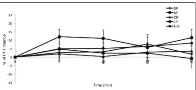
Percentages of changes in pressure pain threshold on hand and forearm are shown in graphs 1 and 2, respectively.
Figure 1 – Position of electrodes on forearm and PPT measuring ar -eas on forearm (A) and hand (B).
Sensory discomfort analysis
To evaluate the level of discomfort during electrical stim-ulation, the 10-cm visual analog scale (VAS) was used where the left edge means “very comfortable” and the
right edge means “very uncomfortable”7. VAS was used
Graph 1 – Percentages of changes in pressure pain threshold on hand of experimental groups.
DF = ixed diphase; MF = ixed monophase; CP = short periods; CP = long periods; CG = control group.
%
o
f
PPT
ch
a
n
g
e
Time (min)
Graph 2 – Percentage of changes in forearm pressure pain threshold in experimental groups.
DF = ixed diphase; MF = ixed monophase; CP = short periods; CP = long periods; CG = control group.
Time (min)
%
o
f
PPT
ch
a
n
g
Hand pressure pain threshold statistical analysis has not shown signii cant differences among groups at 5 min-utes (p = 0.490), 10 minmin-utes (p = 0.590), 15 minmin-utes (p = 0.966) and 30 minutes (p = 0.489).
Forearm pressure pain threshold statistical analysis has also not shown signii cant differences among groups at 5 minutes (p = 0.767), 10 minutes (p = 0.489), 15 minutes (p = 0.395) and 30 minutes (p = 0.915).
With regard to sensory discomfort, there has been no sta-tistically signii cant difference at 5 minutes (p = 0.087) and 10 minutes (p = 0.055). However, there has been less discomfort in CP group at 15 minutes, as compared to the MF group (p = 0.021) (Graph 3).
been low back pain relief in disc disease patients with the use of diadynamic currents1.
In our study, LP group current had a trend to hand hypoalgesia, since pain threshold has increased in the beginning of the application, however there has been no statistically signii cant difference among groups. MF group had a trend to hyperalgesia because pain threshold has decreased along time, however also without statistically signii cant difference.
Our results have shown that DF current does not induce fast analgesia as advocated by Pierre Bernard. On fore-arm, all currents had hypoalgesic trend as compared to the control group, however also without statistically sig-nii cant differences. Our results coni rm the i ndings of other authors who have also not found signii cant dif-ferences with the use of DF current in an experimental ischemic pain model in healthy subjects2. It is possible
that patients with pathophysiological tissue changes and pain will respond more favorably to diadynamic currents than healthy individuals submitted to induced pain1,13. According to sensory discomfort scale results, CP cur-rent induced the least discomfort among curcur-rents, being more comfortable as compared to MF. So, our data sug-gest that MF current should not be used for analgesia be-cause in addition to being more uncomfortable for peo-ple, it does not induce further hypoalgesia as compared to other currents.
New studies are needed, with a higher number of par-ticipants, as well as with patients with pain and/or pathophysiological tissue changes to coni rm or not our results.
CONCLUSION
There has been no pressure pain threshold difference among groups, that is, no signii cant hypoalgesic effect was obtained among applied currents and there have been no signii cant differences among groups receiv-ing currents and the CG. CP current has induced less
sensory discomfort in the 15th minute of stimulations as
compared to the MF current.
REFERENCES
1. Ratajczak B,Hawrylak A,Demidas A,et al. Effective-ness of diadynamic currents and transcutaneous elec-trical nerve stimulation in disc disease lumbar part of spine. J Back Musculoskelet Rehabil. 2011;24(3):155-9. 2. Hämäläinen O, Kemppainen P. Experimentally in-duced ischemic pain and so-called diaphase i x current. Scand J Rehab Med. 1990;22(1):25-7.
Graph 3 – Sensory discomfort during diadynamic currents application.
*CP current group had less discomfort as compared to MF group at 15 minutes of application (p = 0.021)
DF = i xed diphase; MF = i xed monophase; CP = short periods; LP = long periods.
DISCUSSION
Electrotherapeutic resources to control pain in different painful syndromes is becoming more popular in the clin-ical practice and provides improved functionality and
quality of life of individuals11,12. Diadynamic currents
are often used in the clinical practice to control pain and heal biological tissues, in spite of the few studies avail-able in databases such as LILACS, Scielo and Medline, to show the efi cacy of such currents, in addition to their characteristics12,13. Pressure algometer has been used as
measurement tool, which is currently an effective tool to identify pain threshold4-6,8-10.
A study has used diadynamic currents (DF and LP) with and without 1% hydrocortisone iontophoresis to treat chronic low back pain. Each current was applied for 5 minutes and both groups had statistically signii cant pain
improvement after seven consecutive sessions13. Similar
3. Bertolini GRF, Breda D. Uso das correntes diadinâmi -cas de Bernard (DF e CP) no tratamento de hiperidrose – Avaliação de 10 casos. Fisioter Brasil. 2002;3(4):231-6. 4. Pantaleão MA, Laurino MF, Gallego NL, et al. Ad -justing pulse amplitude during TENS application pro -duces greater hypoalgesia. J Pain. 2011;12(5):581-90. 5. Cowan S, Mckenna J, McCrum-Gardner E, et al. An investigation of the hypoalgesic effects of TENS deliv-ered by a glove electrode. J Pain. 2009;10(7):694-701. 6. Moran F, Leonard T, Hawthorne S, et al. Hypoalgesia in response to transcutaneous electrical nerve stimula-tion (TENS) depends on stimulastimula-tion intensity. J Pain. 2011;12(8):929-35.
7. Barr JO, Wenssenbuehler SA, Cleary CK. Effective -ness and comfort of transcutaneous electrical nerve stimulation for older persons with chronic pain. J Geriatr Phys Ther. 2004;27(3):93-9.
8. Liebano RE, Rakel B, Vance CG, et al. An investiga-tion of the development of analgesic tolerance to TENS in humans. Pain. 2011;152(2):335-42.
9. Tong KC, Lo SK, Cheing GL. Alternating frequen
-cies of transcutaneous electric nerve stimulation: does it produce greater analgesic effects on mechanical and thermal pain thresholds? Arch Phys Med Rehabil. 2007;88(10):1344-9.
10. Rakel B, Cooper N, Adams HJ, et al. A new tran -sient sham TENS device allows for investigator blind -ing while deliver-ing a true placebo treatment. J Pain. 2010;11(3):230-8.
11. Ferreira LL, Cavenaghi S, Marino LHC. Recursos eletroterapêuticos no tratamento da dor oncológica. Rev Dor. 2010;11(4):339-42.
12. Guerra TEC, Bertolini GRF. Efeitos da variação da rampa de entrega do delta F sobre a acomodação da cor -rente interferencial em mulheres saudáveis. Rev Dor. 2012;13(1):25-9.
13. Carvalho AR, Fungueto EM, Canzi IM, et al. Cor -rentes diadinâmicas de Bernard e iontoforese no trata-mento da dor lombar. Fisioter Mov. 2005;18(4):11-9.
Submitted in May 14, 2012.