ABSTRACT
BACKROUND AND OBJECTIVES:Most widely used
treat-ment modality for temporomandibular disorders is the occlusal splint. Low-level lasertherapy has been used as therapeutic agent, however as isolated treatment. So, this study aimed at evaluating the efect of the association of low-level lasertherapy and occlusal splint to treat temporomandibular disorders.
METHODS: Participated in the study 25 selected patients ac-cording to the Research Diagnostic Criteria for Temporoman-dibular Disorders protocol. Control group (CG) was made up of 12 asymptomatic volunteers. Two groups were randomly formed: “splint-laser” (SLG), being treated with occlusal splint and associated low-level lasertherapy; “splint” (SG), treated with occlusal splint only. Jaw movements, pain at palpation and self-perception of signs and symptoms were investigated before and after treatment.
RESULTS: here has been signiicant decrease in pain at palpa-tion and reported pain according to self-perceppalpa-tion of signs and symptoms for both groups, however more signiicant for SLG. here has been increased amplitude of jaw movements with sig-niicant diference after treatment for both groups.
CONCLUSION: he association of low-level lasertherapy and occlusal splint to treat temporomandibular disorders has pro-moted more marked pain decrease as compared to occlusal splint alone. Placebo efect should not be discarded and should be test-ed in future studies.
Keywords: Low-level lasertherapy, Occlusal splints, Temporo-mandibular joint disorders.
Low-level lasertherapy associated to occlusal splint to treat
temporomandibular disorder: controlled clinical trial
Laserterapia de baixa intensidade associada ao uso de placa oclusal no tratamento de
disfunção temporomandibular: estudo clínico controlado
Melissa de Oliveira Melchior1, Ana Paula Zanetti Brochini2, Marco Antonio Moreira Rodrigues da Silva2
1. Universidade de São Paulo, Faculdade de Odontologia de Ribeirão Preto, Departamento de Odontologia Restauradora, Ribeirão Preto, SP, Brasil.
2. Universidade de São Paulo Faculdade de Odontologia de Ribeirão Preto, Ribeirão Preto, SP, Brasil.
Submitted in September 05, 2016.
Accepted for publication in February 01, 2017.
Conlict of interests: none – Sponsoring sources: Coordenação de Aperfeiçoamento de Pes-soal de Nível Superior.
Correspondence to: Av. do Café s/nº, Monte Alegre 14040-904 Ribeirão Preto, SP, Brasil. E-mail: [email protected]
© Sociedade Brasileira para o Estudo da Dor
RESUMO
JUSTIFICATIVA E OBJETIVOS: A modalidade de tratamento mais empregada para disfunção temporomandibular é a placa oclusal. A laserterapia de baixa intensidade tem sido empregada como agente terapêutico, porém como tratamento isolado. As-sim, o objetivo deste estudo foi analisar o efeito da associação da laserterapia de baixa intensidade ao uso da placa oclusal como tratamento para disfunção temporomandibular.
MÉTODOS: Participaram do estudo 25 pacientes seleciona-dos de acordo com o protocolo Research Diagnostic Criteria for Temporomandibular Disorders. O grupo controle (GC) foi for-mado por 12 voluntários assintomáticos. Dois grupos foram formados por sorteio: “placa-laser” (GPL), que recebeu trata-mento com placa oclusal e laserterapia de baixa intensidade associada; “placa” (GP), que recebeu tratamento apenas com placa oclusal. Os movimentos mandibulares, a dor à palpação e autopercepção dos sinais e sintomas, foram investigados antes e após os tratamentos.
RESULTADOS: Houve diminuição signiicativa da dor à palpa-ção e da dor relatada de acordo com a autoperceppalpa-ção dos sinais e sintomas para ambos os grupos tratados, porém de forma mais acentuada para o GPL. Houve aumento da amplitude dos movi-mentos mandibulares com diferença signiicativa após os trata-mentos para ambos os grupos.
CONCLUSÃO: A associação da laserterapia de baixa intensi-dade ao tratamento da disfunção temporomandibular com placa oclusal promoveu diminuição mais acentuada do sintoma do-loroso dolorosa quando comparado ao tratamento apenas com placa oclusal. O efeito placebo não deve ser descartado e deverá ser testado em estudos futuros
Descritores: Placas oclusais, Terapia a laser de baixa intensidade, Transtornos da articulação temporomandibular.
INTRODUCTION
Both acute and chronic pain are still a major reason for look-ing for medical and dental treatment and are a major chal-lenge for professionals dealing with orofacial pain (OFP)1,2.
Temporomandibular disorders (TMD) are among most common OFP. TMD may be understood as a set of clini-cal changes involving the stomatognathic system, where pain is the primary reason for looking for treatment. It is classified as musculoskeletal pain, OFP subtype especially characterized by spontaneous pain in orofacial muscles and/
or temporomandibular joints (TMJ) which worsens during stomatognathic functions3-5. Currently, its etiology involves
predisposing, perpetuating and worsening factors which should be taken into consideration in the diagnosis to es-tablish a treatment approach which is in general multidisci-plinary, according to the needs of each case1,5,6.
Occlusal splint is the most common modality to treat TMD, with positive results widely shown in the literature, both for aspects related to painful sensitivity and those related to biomechanics and neuromuscular system7,8.
Low-level laser (LLL) has been used as alternative therapy for pain relief in muscle and joint TMD presentations for inducing analgesic, anti-inflammatory and biomodulator ef-fect of physiologic cell functions6,9-11.
Studies have shown that LLL is efficient as therapeutic agent for decreasing pain and increasing jaw movement ampli-tude6,9-12. In light of the above, this study aimed at
evalu-ating the effect of the association of low-level laserthera-py with the use of Functional Anatomic Research Center (FARC) occlusal splint, on pain perceived by TMD patients, as compared to the use of occlusal splint alone.
METHODS
This study was developed in the Faculdade de Odontolo-gia de Ribeirão Preto, Universidade de São Paulo, and vol-unteers have signed the Free and Informed Consent Term (FICT).
Thirty subjects were selected in a tertiary clinic for TMD patients, of whom 25 have participated in the study till the end, in a total convenience sample of 20 females and 5 males, for having been carried out in compliance with the demand of assistance of the above-mentioned service. Inclusion criteria were TMD diagnosis according to the Research Diagnostic Criteria for Temporomandibular Dis-orders (RDC/TMD)3. Subjects lacking teeth preventing
the installation of the occlusal splint, those with central or peripheral neurologic disorders, history of head and neck tumors or trauma, presence of systemic inflammatory dis-eases and use of analgesics in the last month, and submitted to TMD treatment or others related to the stomatognathic system up to one year before were excluded. Systemic in-flammatory diseases and use of analgesics for less than one month were controlled.
Control group (CG) was made up of 12 asymptomatic vol-unteers paired by age and gender to TMD subjects.
To every subject diagnosed with TMD, one of the following treatments was consecutively directed, forming two groups: 1) Splint group (SG): 15 subjects (12 females and 3males) being treated with occlusal splint alone manufactured and adjusted by a dentist;
2) Splint-laser group (LSG): 10 subjects (8 females and 2 males) being treated with low level lasertherapy together with occlusal splint manufactured and adjusted by a dentist. This group has lost patients before treatment completion who were not included in results analyses: 2 by withdrawal
and 3 for being unable to come twice a week to comply with the laser application protocol.
Subjects were evaluated sitting on dental chair, in a room with adequate lighting, by a dentist (different from the professional in charge of the treatments), before (A1) and after (A2) treatments. Major complaint and the presence of oral parafunctional habits were investigated. Evaluation was based on RDC/TMD Axis I3. Jaw movement amplitude
was measured with digital caliper rule (Mitutoyo, Co., Ltd., Suzhou, China). Pain at palpation was investigated based on the same protocol, adding trapezius (upper portion) and sternocleidomastoid (medial portion) muscles, routinely in-vestigated in this service, and pain intensity was indicated by subjects in a numerical scale from zero to 10, where zero is no pain and 10 the worst imaginable pain. The choice of pain at palpation rather than pain at pressure threshold (PPT) was done due to its relation with pain intensity per-ception which we tried to investigate8,9,13.
To investigate subjects’ perception of their signs and symp-toms, they have answered the “Protocol to determine TMD signs and symptoms for Multiprofessional centers (ProTM-DMulti)13. The first part is made up of questions admitting
just positive and negative answers. The second part indicates how much each sign or symptom is severe in different daily situations, such as at emergence, chewing, speaking and at rest, using a numeric scale from zero to 10 where zero is total lack of sign or symptom and 10 most possible severity. Sum of scores attributed to each sign/symptom in the four investigated situations may vary from zero to 40, indicating higher severity as sum increases.
Occlusal splint: groups SG and SLG received occlusion splint model FARC, developed by the University of Milan, fol-lowing the biomechanical model proposed by Ferrario & Sforza7 (acrylic resin splint with 2 mm thickness and
con-tacts of second premolar to second permanent molar, with-out anterior static or dynamic contacts). Usage orientation has followed the protocol of the University of Milan: daily and nightly in the first two weeks and then nightly for three more weeks, with previously proven positive results8.
Low-level lasertherapy (LLL): SLG patients were treated with LLL three times a week during the five weeks of treatment with the occlusal splint (total of 10 sessions). Equipment was THERA LASER (DMC, LTDA - São Carlos, São Paulo - Brazil), which emits radiation obtained as from stimu-lation of a semiconductor diode formed by Gallium-Alu-minum Arsenide (AsGaAI) with wavelength of 830nm, in continuous emission. Protocol was the same as previously tested12: infrared laser, with wavelength of 780 nm, fixed
power of 70 mW and doses of 105J/cm2. Exposure time was
60 seconds per painful point.
crossed by the auriculotemporal nerve; masseter muscle (3 most painful points identified by digital palpation being one at the origin, one at the body and one at muscle insertion); anterior temporal muscle (one most painful point, identi-fied by digital palpation). Application modality on muscles and joint region was punctual and with direct contact of radiation emission tip with skin to prevent reflection phe-nomenon9-12,14.
Biosafety: used laser belongs to Class 3b according to ANSI classification, needing preventive care during its applica-tion, with the use of goggles for dentists and patients, and the compliance with official safety standards of the Inter-national Standard CEI IEC 825-1. Application sites were cleaned with 70oGL alcohol.
This study was approved by the Ethics Committee for Re-search with human beings (CAAE 0080.0.138.000-10).
Statistical analysis
Initial evaluation data (A1) and evaluation after five weeks of treatment with occlusal splint (A2) were considered for data analysis, both for SG and SLG. Control group was evaluated only once. For measurement interval data, of reason or ordinals presenting normal distribution, such as jaw movement data, ProTMDMulti and pain at palpa-tion parametric tests were used. ANOVA test was used to compare among groups (CG x SG x SLG). T test for inde-pendent samples was used to compare differences between evaluations (A1-A2) of experimental groups (SG x SLG). This analysis was carried out to know the real gain of each group. For intragroup data analysis (A1 x A2), t test for paired samples was used.
RESULTS
Only one subject had isolated muscle TMD. Others had as-sociation with joint dysfunctions. When asked about major complaints leading them to look for treatment, the following reports were given: headaches (60%), facial pain (52%), TMJ pain (20%) and noises (16%), dental wear (12%), earache (8%) and neck ache (4%). Noxious oral habits were report-ed by all subjects, with more frequency by TMD subjects. Among reported habits, there were teeth clenching (vigil bruxism) (76%), sleep bruxism (64%), use of chewing gum (64%) and nail biting (56%). he same habits were reported
by asymptomatic subjects in the following ratio: 0%, 25%, 33.3% and 16.6%, respectively.
With regard to jaw movements, the comparison among groups (ANOVA) has shown that experimental groups were diferent initially (A1) for mouth opening, laterality and protrusion evaluations (p<0.05). After treatment (A2) there has been no statistical diference between SLG and SG in all movements (p>0.05); comparison of experimental groups with CG has shown diference for opening (CG x SG, p<0.05; CG x SLG, p<0.01) and right laterality (CG x SLG, p<0.05) in A1; in A2 there has been mouth opening diference only between SG and CG (p<0.05).
In comparing A1 and A2 (intragroups) (Student t test for paired data) there has been signiicant diference between both experimental groups (p<0.01). To better visualize jaw movement amplitude evolution between both proposed treat-ments, comparative analyses of “A1 – A2” subtraction be-tween experimental groups (Student t – independent data) were carried out. Results have shown no diference (p>0.05) between groups with regard to jaw movement amplitude evo-lution, that is, both proposed treatments provided positive and satisfactory results for this item. Mean and standard de-viation of jaw movements are shown in table 1.
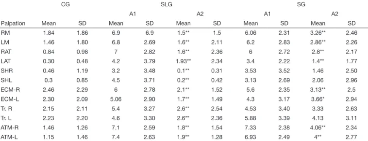
For pain at palpation, comparison between groups (ANOVA) has shown signiicant diference in A1 between CG and SLG for TMJ and masseter, anterior temporal, sternocleidomas-toid (medial portion) to the right (p<0.01), supra-hyoid to the left and trapezius (upper portion) to the right muscles (<0.05); between CG and SG for TMJ and masseter, anterior temporal, sternocleidomastoid (medial portion) (p<0.01), su-pra-hyoid and trapezius (upper portion) to the left (p<0.05). here has been no diference between SLG and SG in this phase.
However, scores attributed to pain at palpation after treat-ment (A2) by SLG was not diferent from that attributed by CG (p>0.05), even in muscles not submitted to lasertherapy; but were diferent in some sites as compared to SG (left mas-seter, right anterior temporal, TMJ – p<0.05). his latter has also shown diferences in pain at palpation scores in speciic sites, similarly to SLG, as compared to CG (left masseter, right anterior temporal – p<0.05, TMJ – p<0.01).
When comparing A1 and A2 (Student t – paired data) there has been pain at palpation improvement according to scores attributed by subjects, with signiicant diference in SLG for
Table 1. Mean and standard deviation of jaw opening, right laterality, left laterality and protrusion movements for control group and splint and splint-laser groups, both with temporomandibular disorders, before and after proposed treatments
CG SLG SG
A1 A2 A1 A2
Mean SD Mean SD Mean SD Mean SD Mean SD
Opening 56.38 5.65 43.59 6.43 53.17 6.17 47.13 6.16 50.13 6.16
Right laterality 8.35 1.75 6.03 2.22 9.84 1.67 7.76 1.91 8.35 1.75
Left laterality 8.55 1.17 6.62 2.52 10.85 1.55 8.13 2.76 9.34 2.76
Protrusion (mm) 7.76 1.63 7.29 1.16 9.98 1.68 6.17 2.49 8.1 2.49
masseter, anterior temporal, supra-hyoid, sternocleidomas-toid (medial portion), trapezius (upper portion) and TMJ (p<0.01); and for SG there has been diference for masseter, anterior temporal, TMJ and right (p<0.01) and left (p<0.05) sternocleidomastoid muscles (medial portion).
Comparative analyses of “A1-A2” subtraction (Student t – independent data) between experimental groups have shown diference only in right masseter palpation, with lower scores attributed by SLG. Table 2 shows mean values and standard deviation of scores attributed by subjects to pain at palpation. According to ProTMDMulti part I questionnaire data, abso-lute frequency of TMD signs and symptoms for each group in initial and inal evaluation were obtained, and decreased number of reports were observed in the inal phase for both groups. hese data are shown in table 3.
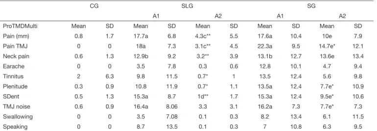
According to ProTMDMulti part II questionnaire data, se-verity of each sign or symptom was determined by the sum of scores attributed to the four questioned situations (emer-gence, chewing, speaking, at rest). Scores varied from zero to 40, being that the higher the value the more severe the TMD. Table 4 shows mean scores attributed to signs and symptoms evaluated by ProTMDMulti in each group, in the two evalu-ation moments (A1 and A2).
Comparison between groups (ANOVA) has shown that in the initial evaluation there has been signiicant diference only between experimental groups and control group for muscle pain, TMJ pain and noise, dental sensitivity (p<0.01) and neck pain (p<0.05). SG was diferent from CG also in aural plenitude (p<0.05). SLG and SG were not diferent at ex-periment onset (p>0.05), however at inal evaluation SLG was not diferent from CG (p>0.05), but was diferent from SG for muscle pain, TMJ pain (p<0.01), neck pain and dental sensitivity (p<0.05) and this group was diferent from CG with regard to the same initial symptom (p<0.05).
In comparing A1 and A2 (Student t – paired data), SLG had signiicant diference for seven evaluated symptoms with Pro-TMDMulti: muscle pain, TMJ pain and noises, neck pain, dental sensitivity (p<0.01), tinnitus and aural plenitude (p<0.05). For SG there has been signiicant improvement in four reported symptoms: TMJ pain and noise (p<0.01), den-tal sensitivity and aural plenitude (p<0.05).
Comparative analyses of “A1 – A2” subtraction (Student t – independent data) have shown diference between experimen-tal groups (p<0.05) for muscle pain, TMJ pain, neck pain,
Table 2. Mean, standard deviation and comparison (Student t for paired data) of scores attributed by subjects to pain at palpation, for control group, splint-laser group and splint group, before and after proposed treatments
CG SLG SG
A1 A2 A1 A2
Palpation Mean SD Mean SD Mean SD Mean SD Mean SD
RM 1.84 1.86 6.9 6.9 1.5** 1.5 6.06 2.31 3.26** 2.46
LM 1.46 1.80 6.8 2.69 1.6** 2.11 6.2 2.83 2.86** 2.26
RAT 0.84 0.98 7 2.82 1.6** 2.36 6 2.72 2.8** 2.17
LAT 0.30 0.48 4.2 3.79 1.93** 2.34 3.4 2.22 1.4** 1.77
SHR 0.46 1.19 3.2 3.48 0.1** 0.31 3.53 3.52 1.46 2.50
SHL 0.3 0.85 4.5 3.71 0.2** 0.42 3.13 2.69 2.06 2.96
ECM-R 2.46 2.29 6 2.78 2.1** 1.52 5.6 2.35 3.13** 2.5
ECM-L 2.30 2.09 5.06 2.90 1.7** 1.49 4.3 3.17 3.66* 2.94
Tr. R 2.15 2.11 5.4 3.27 2.6** 2.54 4.53 3.40 3.33 2.63
Tr. L 2.23 2.20 4.6 3.30 2.6** 2.36 5.88 3.39 4.13 3.11
ATM-R 1.46 1.26 7.1 2.59 1.8** 1.54 7.33 2.38 4.06** 2.34
ATM-L 1.15 1.46 7.4 2.63 1.9** 1.28 6.93 2.49 4** 2.77
CG = control group; SLG = splint-laser; SG = splint group; A1 = before treatment; A2 = after treatment; RM = right masseter; LM = left masseter; RAT = right anterior temporal; LAT = left anterior temporal; SHR = supra-hyoid to the right; SHL: supra-hyoid to the left; ECM-R = right sternocleidomastoid; ECM-L = left sternocleido-mastoid; Tr. R = right trapezius; Tr. L = left trapezius; ATM-R = right temporomandibular joint; ATM-L = left temporomandibular joint. *signiicant difference (p<0.05); **signiicant difference (p<0.01).
Table 3. Absolute frequency of signs and symptoms in the three stu-died groups, according to answers to ProTMDMulti part I protocol, before and after proposed treatments
Signs and symptoms A1 A2
CG SG SLG SG SLG
Muscle pain 0 15 10 8 2
Muscle fatigue 0 12 9 7 2
TMJ pain 0 15 7 9 2
TMJ noises 0 15 8 8 4
headache 3 15 9 7 2
Earache 0 9 5 4 0
Tinnitus 1 9 6 6 2
Aural plenitude 1 12 8 6 1
Dificulty
Mouth opening 0 10 8 5 1
Mouth closing 0 5 4 2 0
Chewing 0 11 7 5 3
Yawning 0 9 9 5 6
Swallowing 0 6 3 2 0
Speaking 0 7 3 4 0
dental sensitivity and diiculty to swallow, that is, positive evolution of these symptoms was better evaluated by SLG subjects being that remaining symptoms had positive evolu-tion according to percepevolu-tion of both groups, without signii-cant diference (p>0.05).
DISCUSSION
TMD is a term used for musculoskeletal facial pain conditions involving several signs and symptoms, being pain the primary motivator for looking for treatment3-5,15. his way, this study
has based its analyses on painful perception of daily situa-tions and on intensity of pain at palpation8,9,13. Methodology
for sample structuring (by convenience) and its size (n) was similar to previous studies which have evaluated the efects of TMD therapies8,9,13, being the irst study on the association of
LLL to concomitant use of occlusal splint, performed during the clinical routine of a tertiary service to TMD patients. Major complaints reported by investigated subjects were sim-ilar to previous studies3,13, being that head and face pain were
more frequent (60 and 52%, respectively), suggesting comor-bidity between them. he presence of TMD seems to cause excitatory impact in some types of headaches, and vice-versa, especially in patients more susceptible to central sensitization phenomenon, as it is the case with chronic orofacial pain15.
Parafunctional habits are risk factors for TMD and OFP, be-cause they may overload teeth and masticatory system dur-ing maintained contractions16. Grinding teeth at sleep (sleep
bruxism) was reported by 64% of studied sample and teeth tightening (vigil bruxism) was reported by 76% of cases. Rel-evance of parafunctional oral habits on TMD pathophysiol-ogy is variable according to individuals, but they have been associated to painful TMD in a previous study16. In this
study, proposed method has not considered a correlation
analysis allowing predicting the inluence of such habits on TMD symptoms of the studied sample, which may represent a limitation of the study. Clinically, it is up to the professional to analyze this relationship in each case to consider it during diagnosis, treatment plan and prognosis, as factor contribut-ing to the presentation16,17.
Jaw mobility restriction is considered a major clinical TMD sign3,5. Although subjects before treatment had no limitations
according to normality patterns, at the end there has been signiicant increase in movement amplitude for both treated groups, which has also been observed in previous study8,
be-ing SLG values higher that SG values. his has allowed the relection that individual amplitude may be larger than the normality pattern and mask an individual movement restric-tion. And although a signiicant diference in mouth opening movement between CG and SG after treatment, there has been approximation between values found for treated groups and control group. his indicates the eiciency of both proposed treatments, where further painless jaw movements freedom is needed to recover stomatognathic system functionality8,9,13.
Biomodulator LLL efect might have favored muscle lexibil-ity and pain remission, when ofering efects which occlusal splint alone is unable to produce, complementing conven-tional treatment. Results suggest that the association of LLL to conventional treatment may more eiciently contribute to the handling of cases with jaw mobility diiculties, because its light promotes analgesia and has anti-inlammatory efect on muscles and joints6,14, that is, its action mechanisms are
diferent from those of the occlusal splint, however comple-menting them. his hypothesis however would have been bet-ter tested with the presence of an additional group treated with occlusal splint and laser-placebo (just guide-light) which was not possible due to characteristics of the equipment used. It is known that expectation added to treatment experience
Table 4. Mean and standard deviation of scores attributed by subjects to signs and symptoms investigated with ProTMDMulti protocol, for control group, splint-laser and splint groups before (A1) and after (A2) proposed treatments. ANOVA for analysis between groups; Student t for paired data for intragroup analysis
CG SLG SG
A1 A2 A1 A2
ProTMDMulti Mean SD Mean SD Mean SD Mean SD Mean SD
Pain (mm) 0.8 1.7 17.7a 6.8 4.3c** 5.5 17.6a 10.4 10e 7.9
Pain TMJ 0 0 18a 7.3 3.1c** 4.5 22.3a 9.5 14.7e* 12.1
Neck pain 0.6 1.3 12.9b 9.2 3.2** 3.9 13.1b 12.7 13.6e 13.4
Earache 0 0 3.5 7.8 0.3 0.6 12.8 10.1 4.7 9.4
Tinnitus 2 6.3 9.8 11.5 0.7* 1 13.5 12.4 5.6 9.8
Plenitude 0.3 0.9 10.8 11.9 0.7* 1.1 13.5a 12.4 7.7e* 10.9
SDent 0.5 1.3 15.3a 8.7 1d** 1.7 15.3a 12.4 9.5e* 10.6
TMJ noise 0.6 0.9 16.4a 8.06 3.3 3.1 16.2a 7.3 7.7e* 7.3
Swallowing 0 0 3.5 7.08 0.1 0.3 8.2 13.4 6.1 11.5
Speaking 0 0 8.7 13.5 0.1 0.3 7 10.8 6.3 9.5
induces placebo efect18, which could have been the case with
this study, because such efect was shown with LLL in previ-ous studies10,11.
Due to the subjectivity of pain, its diagnosis, mostly done by its description, is in general not accurate with regard to diferent variables, such as individual threshold, perception, emotional aspects and individual discomfort, that is, each in-dividual learns to attribute the term “pain” to their sensations by means of their personal experiences4,15. “ProTMDMulti”
protocol was developed, tested and validated to investigate people’s perception of the presentation of their primary com-plaint13.
According to this protocol, it was possible to observe that subjects treated with LLL associated to splint had relief in 7 out of 10 investigated signs and symptoms, versus four in subjects conventionally treated with splint alone. In addition, comparison of subtraction of values found in the two evalu-ation moments of this study (A1-A2) has shown signiicant diference (p<0.05) between groups (SG x SLG) for muscle pain, TMJ pain, neck pain, dental sensitivity and diiculty to swallow, being these better evaluated by SLG subjects after treatment. It has also to be considered that subjects’ percep-tion could have been inluenced by the placebo efect, not tested in this study, induced by more marked pain decrease expectation in face of a more complete treatment with more frequent professional-patient contact, stimulating brain areas of pain modulating neurotransmitters release18.
Palpation of orofacial and cervical muscles was used as di-agnostic method for muscle sensitivity changes, as well as to evaluate the efects of proposed treatments. Cervical region evaluation was suggested for often presenting TMD-related disorders19,20. As with the evaluation of signs and symptoms
perception, pain at palpation after treatments has also im-proved for both TMD groups, but more markedly for SLG, especially in sites submitted to LLL. his might be the result of LLL analgesic and anti-inlammatory efects6,10,14, in
addi-tion to its placebo efect, thus potentiating the efect of the occlusal splint treatment. Not directly treated cervical mus-cles (sternocleidomastoid – medial portion – and trapezius – upper portion) had also signiicant decrease in sensitivity to palpation (Table 2), possibly due to the inluence of the orofacial region with which they have relation, or due to the placebo efect6,10,11,14,18-20.
Finally, the association of therapies for TMD, involving oc-clusal splint and LLL, has shown better efect in decreasing pain and increasing jaw movement amplitude as compared to occlusal splint alone, conirming that it is an easy to apply method, accessible to the clinician and of low cost to patients. However, the necessary availability of time twice a week was one limitation of this study, considering the number of sub-jects not concluding the treatment. Future studies involving the association of these therapies with larger samples will be necessary to conirm statistical results, which should be considered with care in this study. In addition, controlling aspects which could have inluenced results, such as parafunc-tional habits, LLL placebo efect, speciic joint and muscle
TMD diagnosis, as well as their randomized distribution in diferent groups shall help the reliable understanding of the tested association of treatments.
CONCLUSION
he protocol of therapies association proposed in this study has shown more positive results as compared to isolated con-ventional treatment, suggesting that complementary therapy with low-level laser potentiates its efects when simultane-ously applied.
REFERENCES
1. Zakrzewska JM. Multi-dimensionality of chronic pain of the oral cavity and face. J Headache Pain. 201325;14(1):37.
2. Awamleh L, Pun H, Lee JC, Avivi-Arber L. Decreased face primary motor cortex (face-M1) excitability induced by noxious stimulation of the rat molar tooth pulp is dependent on the functional integrity of face-M1 astrocytes. Exp Brain Res. 2015;233(4):1261-72.
3. Dworkin SF, LeResche L. Research diagnostic criteria for temporomandibular disor-ders: review, criteria, examinations and speciications, critique. J Craniomandib Di-sord. 1992;6(4):301-55.
4. Progiante PS, Pattussi MP, Lawrence HP, Goya S, Grossi PK, Grossi ML. Prevalence of temporomandibular disorders in an adult Brazilian Community Population using the Research Diagnostic Criteria (Axes I and II) for temporomandibular disorders (he Maringá Study). Int J Prosthodont. 2015;28(6):600-9.
5. Maixner W, Diatchenko L, Dubner R, Fillingim RB, Greenspan JD, Knott C, et al. Orofacial pain prospective evaluation and risk assessment study-the OPPERA study. J Pain. 2011;12(11 Suppl):T4-11.e1- 2.
6. Herpich CM, Amaral AP, Leal-Junior EC, Tosato J de P, Gomes CA, Arruda E, et al. Analysis of laser therapy and assessment methods in the rehabilitation of tem-poromandibular disorder: a systematic review of the literature. J Phys her Sci. 2015;27(1):295-301.
7. Ferrario VF, Sforza C. Biomechanical model of the human mandible in unilateral clench: distribution of temporomandibular joint reaction forces between working and balancing sides. J Prosthet Dent. 1994;72(2):169-76.
8. Vieira e Silva CA, da Silva MA, Melchior M de O, de Felício CM, Sforza C, Tartaglia GM. Treatment for TMD with occlusal splint and electromyographic control: appli-cation of the FARC protocol in a Brazilian population. Cranio. 2012;30(3):218-26. 9. Melchior MO, Venezian GC, Machado BC, Borges RF, Mazzetto MO. Does low
intensity laser therapy reduce pain and change orofacial myofunctional conditions? Cranio. 2013;31(2):133-9.
10. Moraes Maia ML, Ribeiro MA, Maia LG, Stuginski-Barbosa J, Costa YM, Porporatti AL, et al. Evaluation of low-level laser therapy efectiveness on the pain and mastica-tory performance of patients with myofascial pain. Lasers Med Sci. 2014;29(1):29-35. 11. Chen J, Huang Z, Ge M, Gao M. Eicacy of low-level laser therapy in the treat-ment of TMDs: a meta-analysis of 14 randomised controlled trials. J Oral Rehabil. 2015;42(4):291–9.
12. da Silva MA, Botelho AL, Turim CV, da Silva AM. Low level laser therapy as an adjunctive technique in the management of temporomandibular disorders. Cranio. 2012;30(4):264-71.
13. de Felicio CM, Melchior Mde O, Da Silva MA. Clinical validity of the protocol for multi-professional centers for the determination of signs and symptoms of temporo-mandibular disorders. Part II. Cranio. 2009;27(1):62-7.
14. Sancakli E, Gökçen-Röhlig B, Balik A, Öngül D, Kipirdi S, Keskin H. Early results of low-level laser application for masticatory muscle pain: a double-blind randomized clinical study. BMC Oral Health. 2015;15(1):131-6.
15. Speciali JG, Dach F. Temporomandibular dysfunction and headache disorder. Heada-che. 2015; 55(Suppl 1):72-83.
16. Fernandes G, Franco-Micheloni AL, Siqueira JT, Gonçalves DA, Camparis CM. Para-functional habits are associated cumulatively to painful temporomandibular disorders in adolescents. Braz Oral Res. 2016;30(1): e15.
17. Takeuchi T, Arima T, Ernberg M, Yamaguchi T, Ohata N, Svensson P. Symptoms and physiological responses to prolonged, repeated, low-level tooth clenching in humans. Headache. 2015;55(3):381-94.
18. Reicherts P, Gerdes AB, Pauli P, Wieser MJ. Psychological placebo and nocebo efects on pain rely on expectation and previous experience. J Pain. 2016;17(2):203-14. 19. Silveira A, Gadotti IC, Armijo-Olivo S, Biasotto-Gonzalez DA, Magee D. Jaw
dys-function is associated with neck disability and muscle tenderness in subjects with and without chronic temporomandibular disorders. Biomed Res Int. 2015;2015:512792. 20. von Piekartz H, Pudelko A, Danzeisen M, Hall T, Ballenberger N. Do subjects with