Corresponding Author: www.eerp.usp.br/rlae
Corresponding Author: Tânia Couto Machado Chianca
Universidade Federal de Minas Gerais. Escola de Enfermagem Av. Alfredo Balena, 190
CEP: 30130-100, Belo Horizonte, MG, Brasil E-mail: [email protected]
Corneal injuries: incidence and risk factors in the
Intensive Care Unit
1Andreza Werli-Alvarenga
2Flávia Falci Ercole
3Fernando Antônio Botoni
4José Aloísio Dias Massote Mourão Oliveira
5Tânia Couto Machado Chianca
6Patients hospitalized in the Intensive Care Unit (ICU) may present risk for corneal injury due to
sedation or coma. This study aimed to estimate the incidence of corneal injuries; to identify the
risk factors and to propose a risk prediction model for the development of corneal injury, in adult
patients, in an intensive care unit of a public hospital. This is a one year, prospective cohort study
with 254 patients. The data were analyzed using descriptive statistics, univariate and logistic
regression. Of the 254 patients, 59.4% had corneal injuries and the mean time to onset was 8.9
days. The independent variables that predispose to risk for punctate type corneal injury were:
duration of hospitalization, other ventilatory support device, presence of edema and blinking less
than five times a minute. The Glasgow Coma Scale and exposure of the ocular globe were the
variables related to corneal ulcer type corneal injury. The injury frequencies were punctate type
(55.1%) and corneal ulcers (11.8%). Risk prediction models for the development of punctate
and corneal ulcer type corneal injury were established.
Descriptors: Corneal Diseases; Corneal Ulcer; Risk Factors; Diagnosis of Nursing; Intensive Care
Units; Nursing.
1 Supported by Conselho Nacional de Desenvolvimento Científico e Tecnológico (CNPq) Brazil, process # 477867/2008-1 and by Fundação de Amparo à Pesquisa do Estado de Minas Gerais (FAPEMIG), Brazil, process # CDS-APQ-00726-08.
2 RN, Doctoral Student in Nursing, Escola de Enfermagem, Universidade Federal de Minas Gerais, Brazil. Hospital Universitário Risoleta Tolentino Neves, Belo Horizonte, MG, Brazil. E-mail: [email protected].
3 RN, Ph.D. in Epidemiology, Adjunct Professor, Escola de Enfermagem, Universidade Federal de Minas Gerais, Brazil. E-mail: [email protected].
4 Physician, Ph.D. in Infectology, Adjunct Professor, Faculdade de Medicina, Universidade Federal de Minas Gerais, Brasil. Hospital Risoleta Tolentino Neves, Belo Horizonte, MG, Brazil. E-mail:[email protected].
5 Physician, Hospital São Geraldo, Belo Horizonte, MG, Brazil. Hospital das Clínicas, Universidade Federal de Minas Gerais, MG, Brazil. E-mail: [email protected].
Lesões na córnea: incidência e fatores de risco em Unidade de Terapia Intensiva
Pacientes internados em unidade de terapia intensiva (UTI) podem apresentar risco
para lesão na córnea devido à sedação ou coma. Este estudo teve por objetivo estimar
a incidência das lesões na córnea, identiicar os fatores de risco e propor modelo de
predição de risco para o desenvolvimento de lesão na córnea, em pacientes adultos,
em unidade de terapia intensiva, de um hospital público. É estudo de coorte prospectiva
de um ano, com 254 pacientes. Os dados foram analisados por estatística descritiva,
univariada e de regressão logística. Dos 254 pacientes, 59,4% tiveram lesão na córnea e
o tempo médio para o seu aparecimento foi de 8,9 dias. As variáveis independentes que
predispõem ao risco para lesão na córnea, tipo puntacta, foram: tempo de internação,
outro dispositivo de assistência ventilatoria, presença de edema e piscar de olhos menor
que cinco vezes por minuto. Escala de coma de Glasgow e exposição de globo ocular
foram as variáveis relacionadas à lesão na córnea do tipo úlcera de córnea. As lesões
foram do tipo puntacta (55,1%) e úlceras de córnea (11,8%). Modelos de predição de
risco para lesões na córnea do tipo puntacta e úlcera foram estabelecidos.
Descritores: Doenças da Córnea; Úlcera da Córnea; Fatores de Risco; Diagnóstico de
Enfermagem; Unidades de Terapia Intensiva; Enfermagem.
Lesiones en la córnea: incidencia y factores de riesgo en Unidad de Terapia Intensiva
Pacientes internados en Unidad de Terapia Intensiva (UTI) pueden presentar riesgo de
lesión en la córnea debido a la sedación o al coma. Este estudio tuvo por objetivo estimar
la incidencia de las lesiones en la córnea; identiicar los factores de riesgo y proponer un
modelo de predicción de riesgo para el desarrollo de lesión en la córnea, en pacientes
adultos, en Unidad de Terapia Intensiva, de un hospital público. Estudio de cohorte
prospectivo de un año con 254 pacientes. Los datos fueron analizados por estadística
descriptiva, univariada y de regresión logística. De los 254 pacientes, 59,4% tuvieron
lesión en la córnea y el tiempo promedio para su aparición fue de 8,9 días. Las variables
independientes que predisponen al riesgo de lesión en la córnea tipo punteada fueron:
tiempo de internación, otro dispositivo de asistencia ventilatoria, presencia de edema y
parpadeo de ojos menor que cinco veces por minuto. La escala de coma de Glasgow y la
exposición del globo ocular fueron las variables relacionadas a la lesión en la córnea del
tipo úlcera de córnea. Las lesiones fueron del tipo punteada (55,1%) y úlceras de córnea
(11,8%). Modelos de predicción de riesgo para lesiones en la córnea del tipo punteada
y úlcera fueron establecidos.
Descriptores: Enfermedades de la Córnea; Úlceras Corneales; Factores de Riesgo;
Diagnóstico de Enfermería; Unidades de Cuidados Intensivos; Enfermería.
Introduction
In the Intensive Care Unit (ICU) severely ill patients
are hospitalized that are usually dependent on technology
and the use of medications for the maintenance of life.
These patients are at risk for developing corneal injury
due to multiple factors, among these the most prominent
cause is ocular exposure(1). The role of the intensive care
nurse in prevention and monitoring to identify changes
injury is an infectious or inlammatory lesion in the
corneal tissue that can affect the surface or deep layers,
being classiied as traumatic, supericial, infectious,
degenerative, keratoconus or miscellaneous(5-7). In
the adult ICU, the most frequent injuries are of the
traumatic, supericial and infectious types(1,8-14). The
most common traumatic injury in hospitalized patients
in the ICU is corneal abrasion. This is a supericial
injury in the epithelium, subject to implementation of
care for its regression(1-14). Supericial injuries refer to supericial punctate keratitis and to exposure keratitis.
They can be caused by ocular exposure, with ineffective
palpebral closure and inadequate lacrimal luid
quality(1-4). Among the most common infectious lesions
is infectious ulcerative keratitis or bacterial corneal
ulcers(1-14). These injuries can be adequately prevented
or treated. Conversely, they can lead to temporary or
permanent visual impairment, depending on the degree
of tissue involvement. In this respect, in ICU patients,
the mechanisms responsible for the lubrication and
protection of the eye may be compromised. The palpebra
is preserved in sedated/comatose patients, as long as its
integrity is maintained(5).
Normally, the eyes are kept closed during the period
of sleep due to the contraction of the orbicularis oculi
muscle. In the states of sedation or coma, orbicularis
muscle relaxation occurs reducing its contraction,
which makes passive complete eye closure dificult.
Additionally, sedation and coma may compromise the
random eye movements, cause loss of the blinking
relex and compromise the lacrimal ilm. Other factors involved in the formation of the lacrimal ilm include the
administration of drugs such as atropine, antihistamines
and tricyclic antidepressants. These factors seriously
compromise the corneal and conjunctival defenses(13) and may result in supericial keratopathy and inlammatory
diseases of the cornea, leading to ulceration and
perforation and thus, to permanent damage. These
exposures can occur in ICU patients, on average, from
48 hours to one week after hospitalization(9,13).
In randomized controlled studies(1,8,12), the rate of
occurrence of corneal injury in ICU patients remained
between 3.33% and 22%. Another study found that 60%
of the ICU patients who receive sedation for more than
48 hours developed corneal abrasion, detected in 42%
of the cases within the irst week of hospitalization. In
turn, abrasion leads to an increased risk of infections and
ulcerations(13). A study to estimate the incidence and risk
factors for corneal injury in ICU patients is necessary
since the problem is current and relevant. Therefore, this
study aimed to estimate the incidence of corneal injury,
to identify the risk factors and to propose a model for
predicting risk of corneal injury. It is considered important
that nursing diagnoses are identiied in critically ill patients,
initially in studies of incidence, such as in the case of
the proposed study. It is believed that the identiication
of nursing diagnoses fosters better care planning and
better communication between the nurses and the team,
in addition to the recognition of phenomena considered
important for investigation and description.
Methods
This is a prospective cohort study, conducted in an
adult ICU of a public hospital in Belo Horizonte - MG.
This is a large general hospital, with 30 ICU beds for
the hospitalization of patients in the clinical and surgical
specialties. It is a reference for trauma and
non-trauma emergencies and part of the Brazilian National
Health System (SUS). The sample size calculation was
based on an overall incidence of the events of interest
(corneal injury) of 57.1% in a pilot study previously
conducted. Considering a margin of error of 10%, a
signiicance level of 5% (type I error) and a power of
90% (1-type II error), a systematic random sample was
estimated of approximately 254 subjects. The criteria
for patient inclusion were: to be older than 18 years;
to not present corneal injury on admission; to remain
hospitalized in the ICU for a period exceeding 24 hours;
and to consent to participate in the research or to have
their participation authorized by the person responsible
through the Terms of Free Prior Informed Consent. To
collect data an instrument, constructed and tested in the
pilot study, was used, containing the sociodemographic
and clinical variables and the risk factors for developing
corneal injury identiied in the literature(1,8-9,11-14).
The dependent variables for the determination of
the incidence were corneal injury and type of corneal
injury - punctate and corneal ulcer. The independent
variables (risk factors for injury to the cornea), selected
from the literature(1,8-9,11-14) were: ICU; degree of
contamination from the surgery; ventilatory method;
age; gender; origin; duration of hospitalization in the
UTI; duration of hospitalization until the appearance
of the corneal injury; Acute Physiology and Chronic
Health Evaluation II - APACHE II severity-of-disease
classiication system; Therapeutic Interventions Scoring
System (TISS 28); type of patient; American Society
of Anesthesiologists (ASA); medical diagnosis; duration
of height; presence of edema; localization of edema;
sedation; Glasgow Coma Scale (GCS); Ramsay Sedation
Scale; intubation; tracheostomy (TQT); other ventilatory
support device; mechanical ventilation (MV); duration
of MV; non-invasive ventilation (NIV); duration of NIV;
inspired oxygen fraction (FiO2); positive end-expiratory
pressure (PEEP); eyes blinks per minute; exposition
of the ocular globe; degree of exposition of the ocular
globe; conjunctival hemorrhage; ocular colonization/
infection; secretion of the right and of the left eye;
microorganism of the right and the left eye; pneumonia;
medication in use; nutritional status; Cumulative
Water Balance (CWB); degree of headboard elevation;
Endotracheal Tube (ETT) or TQT Fixation; temperature
of the unit; dosage of albumen; leukocyte count; total
protein dosage; serum sodium dosage.
Before starting the data collection an intensive
care nurse and physician were trained to evaluate the
cornea by an ophthalmologist. This training consisted
of theoretical and practical content regarding corneal
injury. The ophthalmologist was considered the “gold
standard” for the performance of the corneal evaluation.
A Kappa coeficient of 0.77 (substantial agreement)
was found between the intensive care physician and
the ophthalmologist and of 0.88 (almost perfect
agreement) between the intensive care nurse and the
ophthalmologist. The Cronbach’s alpha was calculated
to evaluate the internal consistency and reliability of the
evaluation. The value of 0.91 was found which shows
excellent reliability in the test conducted.
The pilot study had a duration of 30 days and allowed
the average time of data collection to be estimated,
the instrument to be tested and the calculation of the
sample size to be performed. After concordance was
reached among the examiners and the testing of the
instruments and calculation of the sample size, the
study was conducted from May 2008 to May 2009. Data
collection was performed by the intensive care nurse,
ive times a week. For the corneal evaluation a drop of luorescein was placed in each eye of the patient and
approximately three minutes allowed to pass. Then the
ophthalmoscope with a cobalt ilter for the evaluation
of the cornea was positioned, under very low ambient
light conditions for the best quality of examination.
The collection of the cultures of the secretion of the
conjunctiva with antibiogram was performed after 24
hours of hospitalization, and when the patients had signs
and symptoms of ocular infection. For the collection of
the material two plastic loops and two chocolate agar
culture medias were used. The data obtained were
transferred, treated and processed in the Statistical
Package for the Social Sciences (SPSS) version 16.0
and in Minitab 15.1. The double entry procedure was
used to avoid digitation errors. The descriptive analysis
was conducted using simple frequency distributions,
measures of central tendency (mean and median) and measures of variability (standard deviation). The
incidence of corneal injury and the identiication of the
risk factors for the injury were determined. Univariate
analysis was conducted and measures of association
between the dependent and independent variables were
calculated using the chi-square test (χ2) or the Fisher
exact test, in the case of qualitative variables. For the quantitative variables, the Student t test for comparing
two groups (association with punctate injury and corneal
ulcer) was performed.
In all tests, a signiicance level of 5% (a=0.05) was used. To estimate the strength of association between
the dependent and independent variables the odds
ratio (OR) was used, with a conidence interval of 95%
and a p-value=0.05. Multiple logistic regression was
performed to estimate risk for corneal injury prediction
models, punctate and corneal ulcer, through the Forward
method. The Hosmer and Lemeshow test was used
to evaluate the adequacy of the estimated models.
The variables used to estimate the models were the independent variables that, in the univariate analysis,
were statistically signiicant (p≤0.20). Some independent variables were excluded because they were correlated
with other variables already included in the model. The
study was approved by the Research Ethics Committee
of the Federal University of Minas Gerais, COEP/UFMG,
Protocol No. ETIC 008/08.
Results
Characteristics of the patients
Of the 254 subjects it was found that the majority
(66.1%) were male. Only 21.7% were surgical patients
and 63.4% came from the Emergency Unit beds for
critical patients. The average age was approximately
55.9 years (18-100). The mean of the APACHE II was
19 and 37.2 for the TISS 28. The mean duration to the
onset of corneal injury was 8.9 days. During the data
collection period there was a loss of 60 patients (23.6%)
due to death and 194 patients (76.4%) due to referral,
with 187 of these (96.4%) to inpatient units. In relation
to the ventilatory support devices, 199 (78.3%) were
intubated, 200 (78.7%) on mechanical ventilation and
device. Macronebulization was used with 114 (57.3%)
patients and non-invasive ventilation with only 16
(6.3%). Regarding conjunctival secretion, 48 patients (18.89%) were found to have ocular infections, 27
(10.6%) in the right eye and 21 (8.3%) in the left eye.
10 (3.94%) patients presented infection in both eyes.
In relation to the medical diagnosis on admission to the ICU, pulmonary diseases were the most frequent,
affecting 91 (35.8%) patients.
Incidence and risk factors for corneal injury for the patients included in the study
Of the 254 subjects 151 corneal injuries were
identiied. The overall incidence of this type of injury was
59.4% for the period of study. The incidence of punctate
type corneal injury was 55.1% and of the corneal ulcer type, 11.8%. The number of injuries of the corneal
ulcer type was calculated based on the number of ulcers
identiied in the irst evaluation plus the number of
punctate type injuries that evolved into corneal ulcers in the period stipulated for the study. There was regression
of the punctate type injuries in 14.3%.
Corneal injury risk prediction model
The qualitative variables that presented signiicant
association (p≤0.05) with corneal injury were: origin of
the patient; type of patient; presence of neurological
disease; intubation; mechanical ventilation or TQT;
use of another ventilatory support device; pneumonia;
result of the conjunctival secretion culture of the right
eye, result of the conjunctival secretion culture of the
left eye; presence of edema; localization of the edema;
patient sedated; eye blinks per minute; exposure of the
ocular globe; area of ocular globe exposure; presence
of conjunctival hemorrhage; use of vasoactive drugs;
use of antihypertensives; use of antibiotics (ATB); use
of diuretics; use of hypnotics/sedatives/anxiolytics; use
of antifungal medication; use of other non-categorized
drugs; use of vitamins; use of bronchodilators; use of
neuromuscular blockers; and ixation of the ETT/TQT/
macronebulization/nasal cannula.
The qualitative variables that presented signiicant
association (p≤0.05) with corneal injury were: duration
of hospitalization; duration of hospitalization until the
appearance/regression of the corneal injury; age;
APACHE II; TISS 28; PEEP; duration of MV in days;
GCS, and CWB. The probability that a patient develops
a corneal injury was estimated in the risk prediction
model with the logistic regression equation using the
Forward method. In the adjustment of the model, those
independent variables considered statistically signiicant
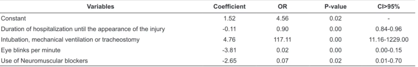
(p≤0.05) for corneal injury were used (Table 1).
Table 1 - Estimates of the Logistic Regression to deine the inal model in relation to the dependent variable corneal
injury. Belo Horizonte, MG, Brazil, 2008-2009
Variables Coefficient OR P-value CI>95%
Constant 1.52 4.56 0.02
-Duration of hospitalization until the appearance of the injury -0.11 0.90 0.00 0.84-0.96
Intubation, mechanical ventilation or tracheostomy 4.76 117.11 0.00 11.16-1229.00
Eye blinks per minute -3.81 0.02 0.00 0.00-0.15
Use of Neuromuscular blockers -2.65 0.07 0.02 0.01-0.70
χ2=12.583; p=0.127 R2=62.5%.
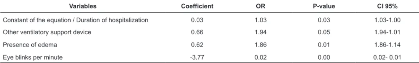
Punctate type corneal injury risk prediction model
The qualitative variables with signiicant association
(p≤0.05) for punctate type injury were: origin; type of
patient; kidney disease; neurological disease; intubation/
MV or TQT; other ventilatory support device; pneumonia;
result of the conjunctival discharge culture of the right
eye; presence of edema; patient sedated; eye blinks
per minute; exposure of the ocular globe; area of ocular
exposure; use of antihypertensives, use of vasoactive
drugs, use of diuretics; use of hypnotics/sedatives/
anxiolytics; use of vitamins; use of bronchodilators;
use of neuromuscular blockers; and ixation of ETT/
TQT/macronebulization/nasal cannula. The quantitative
variables with signiicant association (p≤0.05) for punctate type injury were: duration of hospitalization;
age; APACHE II; TISS 28; PEEP; duration of MV in days;
and GCS. The probability that a patient develops a
punctate type corneal injury was estimated in the risk
Table 2 - Independent variables considered in the inal model in relation to the dependent variable punctate type
corneal injury. Belo Horizonte, MG, Brazil, 2008-2009
Variables Coefficient OR P-value CI 95%
Constant of the equation / Duration of hospitalization 0.03 1.03 0.03 1.03-1.00
Other ventilatory support device 0.66 1.94 0.05 1.94-1.01
Presence of edema 0.62 1.86 0.01 1.86-1.14
Eye blinks per minute -3.77 0.02 0.00 0.02- 0.01
χ2=3.880; p=0.868; R2=56.4%.
Corneal ulcer type corneal injury risk prediction model
The variables in the prediction model for corneal
ulcer type corneal injury with statistical signiicance
were: duration of hospitalization; other ventilatory
assistance device; presence of edema; and less than
ive eye blinks per minute. The quantitative variables
that presented signiicant association (p≤0.05) with the corneal ulcer were: APACHE II, TISS 28, weight, duration of MV in days, and GCS. The probability that a patient develops a corneal ulcer type corneal injury was estimated in the risk prediction model using the Forward
method (Table 3).
Table 3 - Independent variables considered in the inal model in relation to the dependent variable corneal ulcer type
corneal injury. Belo Horizonte, MG, Brazil, 2008-2009
Variables Coefficient OR P-value CI 95%
Exposure of the ocular globe 0.99 2.70 0.00 1.39-5.27
Glasgow Coma Scale >7 -0.253 0.77 0.00 0.73-0.83
χ2=7.077; p=0.314. R2=74.2%.
Although it contains only two variables (exposure of
the ocular globe and the Glasgow coma scale with values
between 11 and 15), the value of R2 indicates that this
model is able to explain 74.2% of the variability of the
probability of corneal ulcer development. In addition,
it is possible to conclude, through the Hosmer and
Lemeshow test, that the model is adequate to estimate
the probability of corneal ulcers development (p=0.31).
The total percentage of accuracy in the prediction of
the response, according to the estimated equation, was
89.1% (95.5% accuracy in the negatives and 37% in
the positives). It is important to observe that the OR
value of -0.253 of the Glasgow coma scale indicated that
for each one-unit increase in the scale, the likelihood of
developing a corneal ulcer decreases by 0.253 times.
Discussion
Among the 254 ICU patients it was found that
151 presented punctate and corneal ulcer type corneal
injuries. The incidence of punctate type corneal injury
was 55.1% and of the corneal ulcer type, 11.8%. By
combining the two, this number is greater than 59.4%,
since 19 punctate type injuries evolved into corneal ulcers
during the study period. Studies(15-16) have estimated
an incidence of corneal injury from 20 to 40%, though
they do not describe the characteristics of the patients
studied. In turn, other studies(1,8-9,12) have estimated an
incidence between 3.33% and 42%. It is noteworthy
that the studies cited above were developed in countries
with different social contexts to Brazil. Nursing care
to prevent injury to the cornea should be established
at admission of the patient to the ICU and for those
at risk for injury, coming from other inpatient units,
given the high incidence found (59.4%) in this study.
Considering that the corneal ulcer is a type of injury that
can often leave leukoma as sequel(5), their prevention
is imperative, even when found at the punctate type
injury stage. There was regression of the punctate in
14.3% of the cases. This number was estimated only for
this type of injury, because it is the type of injury that
resolves without medical treatment. At this stage the
nurse can provide skilled care, through early detection
and nursing interventions, with a view to its regression
without medical treatment. The mean time to onset of
injury to the cornea was 8.9 days. This is the critical
period of hospitalization for the patient to developed a
corneal injury. During this phase, the nurse and medical
that preventive actions can be implemented. Another
study(12) presents the mean time of onset of injury as
between 48 hours and 7 days. There are few studies that
estimate the mean time to the onset of injury and the
implementation of corneal care for ICU patients in Brazil,
making it dificult to compare and analyze the data. The variables that presented signiicant association
(p≤0.05) with corneal injury were: origin of the patient;
type of patient; presence of neurological disease;
intubation; mechanical ventilation or TQT; use of another
ventilatory support device; pneumonia; result of the
conjunctival secretion culture of the right eye; result of
the culture conjunctival secretion of the left eye; presence
of edema; location do edema; patient sedated; eye blinks
per minute; exposure of the ocular globe; area of ocular
globe exposure; presence of conjunctival hemorrhage;
use of vasoactive drugs; use of antihypertensives; use
of antibiotics (ATB); use of diuretics; use of hypnotics/
sedatives/anxiolytics; use of antifungal medication; use
of other non-categorized drugs; use of vitamins; use of
bronchodilators; use of muscular blocking; ixation of
the ETT/TQT/macronebulization/nasal cannula; duration
of hospitalization; duration of hospitalization until the
onset of corneal injury; age; APACHE II; TISS 28; PEEP;
MV duration in days; GCS; and CWB. The variables in
the corneal injury predictive model were as follows:
duration of hospitalization until onset/regression of the
injury; presence of ETT/MV/TQT; eye blinks per minute;
and use of neuromuscular blockers. The literature
(1,8-14) indicates as possible risk factors for corneal injury:
intubation; mechanical ventilation or TQT; result of the
conjunctival secretion cultures; edema; sedation; eye
blinks per minute; exposure of the ocular globe; area
of ocular globe exposure; use of hypnotics/sedatives/
anxiolytics; APACHE II; TISS 28; PEEP; MV duration in
days; GCS Some of these variables were conirmed as
risk factors in the prediction models constructed in the
present study, such as intubation; mechanical ventilation
or TQT; result of the conjunctival secretions culture; eye
blinks per minute, presence of edema, among others.
From the risk prediction model it was observed
that for every one-unit increase in the duration of the
hospitalization, the probability of injury development
decreased by 0.11. This data could be explained by
improvements in the clinical status of the patient with
the increase in the duration of the hospitalization. The
patient would no longer be intubated, on mechanical
ventilation or tracheostomy and would present an
adequate blink relex. These variables were estimated by
the model as risk factors for corneal injury. Intubation,
MV or TQT increases the chance of a patient developing
a corneal injury by 117.11 times when compared to
those who are not using these devices, while keeping
the other variables constant. This inding is corroborated
by several studies(1,8-17).
The eye blink rate of less than ive times per
minute increased the chance of a patient developing a
corneal injury by 45.46 times compared to a blink rate
of more than ive times per minute, while keeping the other variables constant. This risk factor is conirmed in
another study(10) which proposes a protocol for ocular
care in accordance with the number of times the ICU
patient blinks the eyes per minute. This is an important
variable in relation to risk for corneal injury. In turn, it
can be concluded that patients who use neuromuscular
blockers are 14.085 times more likely to develop a
corneal injury, when compared to those in which it was
not administered. In these cases, the patient may often
present exposure of the ocular globe when being given
neuromuscular blockers(2).
No study was identiied in the literature that
estimates the risk factors for punctate type and corneal
ulcer type injuries, therefore it was not possible to
establish a comparison with the results found in the
present study. In the available studies(1,8,13) the risk
factors are established from the pathophysiology of
the injury. For punctate type injury, with each
one-unit increase in the duration of the hospitalization, the
probability of developing the injury increases by 0.03
times. This could be explained by the severe proile
of the patient. The more severe the clinical status of
the patient, the greater the duration of hospitalization
and, consequently, the greater the risk for developing a
corneal injury.
Another important inding in the present study is
that a risk factor for punctate type injury, presented by
the patients, was the use of another ventilatory support
device, such as macronebulization, Venturi mask and the
nasal cannula. Patients using any of these devices have
a 1.96 times higher chance of developing punctate type
injury when compared with those who are not using
any of them, keeping the other variables constant. This
could be explained by the corneal exposure to oxygen at
concentrations greater than 21%(17). It can be concluded
that patients who present edema have a 1.86 times
higher chance of developing punctate type injury when
compared with those who do not present this, if the other
variables are kept constant. This variable is identiied as a
risk factor for corneal injury by several authors(1,8-13). The
Received: Nov. 10th 2010 Accepted: June 22nd 2011
patient developing a corneal ulcer by 2.7 times when
compared to those who have no exposure, keeping the
other variables constant. This data is also consistent
with the literature(1,8-14), because with the exposure
of the iris the cornea also remains exposed. Without
lubrication mechanisms, injuries in the epithelium
can be presented. When the state of dehydration
is maintained, there may be endothelial injury and
consequently a corneal ulcer may develop.
Conclusions
The incidence of the punctate type injury
encountered in this study was higher than those of other
corneal injuries. The prevention of punctate type injury in
adult ICU patients is essential and must be implemented
by the intensive care nurse. In the inal model, the
variables that predispose to risk for developing corneal
injuries were: duration of hospitalization until onset/
regression of the injury; intubation; MV or TQT; eye
blinks per minute; and use of neuromuscular blockers.
The variables that predispose to risk for punctate type
corneal injuries were: duration of hospitalization; other
ventilatory assistance device; presence of edema; and
eye blinks per minute. The variables that predispose to
risk for corneal ulcer type corneal injury are exposure
of the ocular globe and Glasgow coma scale values
between 11 and 15. According to the study performed, a
nursing diagnosis that contemplates the risk for corneal
injury can be said to be of fundamental importance due
to the high incidence of this type of injury subject to
prevention by the nursing staff. The timely nature of
the study conducted with a group of patients in public
teaching hospitals, can be identiied as a limiting factor
requiring a multicentric study to legitimize the external
validity of the study.
References
1. Dawson D. Development of a new eye care guideline
for critically ill patients. Intensive Crit Care Nurs.
2005;21(2):119-2.
2. Cortese D, Capp L, McKinley S. Moisture Chamber
versus lubrification for the prevention corneal ephitelial
breakdown. Am J Critical Care. 1995;4(6):425-8.
3. Elias ACGP, Matsuo T, Cardoso LTQ, Grion CMC.
Aplicação do sistema de pontuação de intervenções
terapêuticas (TISS 28) em unidade de terapia intensiva
para avaliação da gravidade do paciente. Rev.
Latino-Am. Enfermagem. 2006;14(3):324-9.
4. Guimarães RCM, Rabelo ER, Moraes MA, Azzolin K.
Gravidade de pacientes em pós-operatório de cirurgia
cardíaca: uma análise evolutiva segundo o TISS-28. Rev.
Latino-Am. Enfermagem. 2010;18(1):[6 telas].[acesso
10 nov 2010]. Disponível em: http://www.scielo.br/
scielo.php?script=sci_arttext&pid=S0104-1169201000
0100010&lng=pt&nrm=iso&tlng=pt
5. Ezra DG, Lewis G, Healy M, Coombes A. Preventing
exposure keratopathy in the critically ill: a prospective
study comparing eye care regimes. Br J Ophthalmol.
2005;89(8):1068-9.
6. Hernandez EV, Mannis MJ. Superficial keratophaty
in intensive care unit patients. Am J Ophthalmol.
1997;2:212-6.
7. Hudak CM, Gallo BM. Cuidados Intensivos de
Enfermagem. 7ª ed. São Paulo: Guanabara Koogan;
2006. 1013 p.
8. Imanaka H, Taenaka N, Nakamura J, Aoyama K,
Hosotani H. Ocular surface disorders in the critical ill.
Anesthesia & Analgesia. 1997;85(2):343-6.
9. Knaus WA, Draper EA, Wagner DP, Zimmerman JE.
APACHE II: A severity of disease classification system.
Crit Care Med. 1985;13:818-29.
10. Korollof N, Boots R, Lipman J, Thomas P, Rickard C,
Coyer F. A randomised controlled study of the efficacy
of hypromellose and lacri-lub combination versus
polyethylene/cling wrap to prevent corneal epithelial
breakdown in the semiconscious intensive care patient.
Intensive Care Med. 2004;30:1122-6.
11. Mercieca F, Suresh P, Morton A, Tullo A. Ocular
surface disease in intensive care unit patients. Eye.
1999;13(2):231-6.
12. Nember J. Eye Care for Intensive Care Patients. Best
Practice – The Joanna Briggs Institute. 2002;6:1-5.
13. Nember J. Eye Care for Patients in the ICU. Best Practice
– The Joanna Briggs Institute. 2006;106:72A-72D.
14. Paschoal MAV. Manual de Oftalmologia. Rio de
Janeiro: Cultura Médica; 2008. 356 p.Sivasankar S,
Jasper S, Simon S, Jacob P, John G, Raju R. Eye Care in
ICU. Indian J Crit Care Med. 2006;10(1):11-4.
16. So HM, Lee CCH, Leung AKH et al. Comparing the
effectiveness of polyethylene covers (GladwrapTM) with
lanolin (Duratears1) eye ointment to prevent corneal
abrasions in critically ill patients: A randomized controlled
study. Int J Nurs Studies. 2008;45:1565-71.
17. Rosenberg JB, Lewis A, Eisen MD. Eye care in the
intensive care unit: Narrative review and meta-analysis.