Abstract
Objective: To determine the incidence and risk factors of accidental extubation (AE) in a tertiary neonatal intensive care unit.
Methods: A prospective cohort study was conducted to determine AE incidence density per 100 patient-days, during a 23-month period, in 222 newborns receiving assisted ventilation (AV). Logistic regression analysis was used to determine risk factors for AE. The presence of a cyclical pattern in extubation rates, according to the variables of interest, was investigated by Cosinor analysis.
Results: The mean AE rate was 5.34/100 patient-days ventilated. AE-associated predictive variables were: subsequent use of the oral and nasal routes during AV [relative risk (RR) = 4.73; 95% conidence interval (95%CI) 1.92-11.60], AV duration (per day, RR = 1.03; 95%CI 1.02-1.04), and number of patient-days ventilated (RR = 1.01; 95%CI 1.01-1.02). According to the adjusted multiple regression analysis, total AV time was the only independent predictor of AE in this sample (RR = 1.02; 95%CI 1.01-1.03). AV time of 10.5 days showed an accuracy of 0.79 (95%CI 0.71-0.87) for the occurrence of AE. Cosinor analysis showed signiicant periodicity in overall AE rate and in the number of patient-days ventilated. There was a signiicant correlation between the number of patient-days ventilated and AE frequency.
Conclusion: Mean AE density was 5.34/100 patient-days ventilated.AV duration was the only independent predictor of AE. The best accuracy for AE occurrence was achieved at 10.5 days of AV duration.
J Pediatr (Rio J). 2010;86(3):189-195: Mechanical ventilation, risk factors, prematurity, intubation, incidence.
ORiginAl ARtiCle
Copyright © 2010 by Sociedade Brasileira de Pediatria189 introduction
Assisted ventilation (AV) through an endotracheal tube is used in the treatment of airway obstruction or respiratory failure1 and has contributed to the increased survival of
newborns.2 AV-related adverse effects are common, and,
among them, accidental extubation (AE) has been reported as the most frequent event in adult patients.3 In neonatal
intensive care units in the United States, AE is the fourth most common adverse event.4
AE or unplanned extubation is deined as every
unexpected or nonelective extubation5 resulting from
patient restlessness or caregiver activity during procedures at the bedside. Some characteristics should be considered
incidence and risk factors of accidental extubation
in a neonatal intensive care unit
Fabiana l. Carvalho,1 Maria Aparecida Mezzacappa,2 Roseli Calil,3 Helymar da Costa Machado4
1. Mestre, Saúde da Criança e do Adolescente. Fisioterapeuta Supervisora, Curso de Especialização em Fisioterapia Neonatal, Centro de Atenção Integral à Saúde da Mulher (CAISM), UNICAMP, Campinas, SP, Brazil.
2. Doutora, Saúde da Criança e do Adolescente. Professora, Departamento de Pediatria, Faculdade de Ciências Médicas, Universidade Estadual de Campinas (FCM/UNICAMP), Campinas, SP, Brazil. Setor de Neonatologia, Centro de Atenção Integral à Saúde da Mulher (CAISM), UNICAMP, Campinas, SP, Brazil. 3. Doutora, Saúde da Criança e do Adolescente. Médica assistente, Setor de Neonatologia, Centro de Atenção Integral à Saúde da Mulher (CAISM), UNICAMP,
Campinas, SP, Brazil. Departamento de Pediatria, FCM/UNICAMP, Campinas, SP, Brazil. 4. Mestre, Estatística. Estatístico, Câmara de Pesquisa, FCM/UNICAMP, Campinas, SP, Brazil.
This study was conducted in the Department of Pediatrics, School of Medicine, Universidade Estadual de Campinas (FCM/UNICAMP), Campinas, SP, Brazil, and at the Center of Integral Attention to Women’s Health (CAISM), UNICAMP, Campinas, SP, Brazil.
No conflicts of interest declared concerning the publication of this article.
Suggested citation: Carvalho FL, Mezzacappa MA, Calil R, Machado HC. Incidence and risk factors of accidental extubation in a neonatal intensive care unit. J Pediatr (Rio J). 2010;86(3):189-195.
in the diagnosis of this event, such as: tube displacement, presence of vocalization, sudden and unexplained air escape, gastric distension, radiologic evidence of endotracheal tube malpositioning,5 sudden cyanosis or desaturation, and
absence of respiratory movements or breathing.2
AE may require urgent reintubation in less controlled situations, often with repeated attempts, increasing the
risk of laryngeal injury, and the consequent stridor,6 and
physiologic changes such as hypoxemia and increased blood and intracranial pressure.7 Data on the incidence of AE in neonatal intensive care units are scarce. In these units, incidence ranges from 0.72 to 4.8 AE per 100 patient-days
ventilated,2,8-11 values that are higher than those found
in pediatric intensive care units (0.11-2.7/100 patient-days).5,8,12-14
Given the importance of this potentially preventable adverse effect, AE rate has been used as an indicator in the assessment of the quality of medical care.15 Taking into
consideration AE-associated morbidity,2 it seems important
that neonatal units establish the incidence of this adverse event and be informed about associated risk factors in order to be capable of identifying the need to implement interventions and measure their results.10,11
The objective of this study was to establish AE density
per 100 patient-days ventilated in a neonatal intensive care unit, in addition to identifying the occurrence of periodic events and newborn-related risk factors for AE.
Patients and methods
A prospective cohort study was conducted between
September 2006 and July 2008 involving newborns with
tracheal intubation admitted to the neonatal intensive care unit of a tertiary teaching hospital.
All newborns receiving AV for at least 12 hours were included, regardless of the route of intubation. Exclusion criterion was death before 24 hours of life.
AE was deined as every episode of unplanned tube
removal or loss during intensive care, regardless of when
or how it occurred, being identiied by partial or total
displacement of the endotracheal tube and/or impaired breathing on chest auscultation, whether or not these findings were confirmed by laryngoscopy. Episodes
requiring tube replacement due to conirmed or suspected
obstruction were not included.
The researcher in charge visited the unit daily, including weekends, to verify, in person, the number of newborns on AV and the route of intubation used. All newborns
were followed up until AV was discontinued. Information
on the occurrence of AE was obtained from reports by the physician and nursing staff and double checked on medical records.
Nasal intubation was the routine procedure in the unit, and the oral route was an alternative to technical dificulties during nasal insertion. Tube ixation was performed by the
physician in charge of the procedure, and tube position was
conirmed by radiography, being considered appropriate
when the tip of the tube was placed at the level of T1.16 The staff nurses in charge performed routine tube ixation, with
no predetermined periodicity, based on necessity, due to loss
of ixation (adherence) and/or presence of secretion. Tube
aspiration was performed by the nursing staff or physical therapists whenever necessary.
Newborns on AV received analgesia with a continuous
infusion of 0.5-2.0 µg/kg/h fentanyl, and, whenever necessary, sedation with midazolam.
Since 2004, this unit has assessed monthly AE rates and, for 9 months, monitored the characteristics of AE episodes through reports by the nursing staff of circumstances involving each event. The multiprofessional team underwent training and sensitization to the topic, the medical team was warned about the correct positioning of the tube, and an intubation-related care protocol was established.
In addition to intubation route, other variables were
studied: birth weight, gender, gestational age (GA), use of sedation and analgesia during AV, and total ventilation time (and AV duration for each intubation route) per entire
day, considering the irst and last day on AV.
GA was determined according to the methods by Capurro17 or New Ballard18 or based on the estimates by
early echography or amenorrhea.
In addition to absolute monthly AE numbers, AE density
was calculated per each 100 patient-days ventilated. This rate was obtained by dividing the absolute number of accidental extubations occurred in a month by the total number of patient-days ventilated multiplied by 100. Each day on ventilation was considered as one patient-day.19
This study was approved by the Research Ethics Committee of the institution, and the children’s parents or guardians signed a written consent inform.
Sample size (n = 222) was calculated to establish the incidence of the main infectious and mechanical complications of AV; in the present study, we decided to describe only AE. The AE rate used in sample size calculation was 2.7 AE/100 patient-days,12 with an alpha of 0.05 and sampling
error of 2.5%. Weighted sampling error was obtained for
each complication so that the lower conidence limit did not
restrict incidence to values 50% greater than the reference values adopted.
Variables were compared between groups with and without AE using the chi-square test and the Mann-Whitney test. Values for overall AE density and each intubation route were calculated per month. To establish the presence of periodicity in monthly AE rates, we used Cosinor analysis20
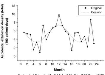
Figure 1 - Individual values and Cosinor curve for accidental extubation density per 100 patient-days ventilated during a 23-month study period
mesor (M), amplitude (A), acrophase (Phi), and period (Tau). Cosinor analysis20 was also used to compare the inluence
of variables on temporal series of AE density. The variables analyzed were: GA, birth weight, gender, use of analgesia/ sedation, intubation route, and total number of patient-days ventilated/month. Analysis of the cross-correlation function was used to establish the correlation between the number of patient-days ventilated and AE frequency per month.21 The
Kruskal-Wallis test was used to compare weight, GA and AV duration according to intubation route. The Wilcoxon test for related samples was used in the analysis of AE density according to intubation route.
Risk factors for AE were determined by univariate and
multivariate (stepwise) logistic regression analysis. In the
regression analysis, the number of patient-days ventilated was determined using mean values from the months in which the newborn remained on AV.
Receiver operating characteristic (ROC) curves were constructed for AV duration and overall AE occurrence and
for each intubation route. The signiicance level was set
at p < 0.05. The statistical software Statistical Analysis
System for Windows, version 9.1.3 (SAS Institute Inc, 2002-2003, Cary, NC, USA) was used for data analysis.
Results
During the 23-month study period, 222 newborns required AV, totaling 2,563 patient-days ventilated.
Fifteen subjects did not meet the inclusion criteria: AV
duration was less than 12 hours (n = 1), and parents or guardians were not found to sign the written consent form (n = 14). Six newborns were excluded due to death before 24 hours of life.
Of the total study subjects, 119 were included in AE
rates in more than one month, because AV duration was
greater than 30 days, totaling 341 assessments. Newborns’ distribution according to study year was 42 (18.9%) in 2006, 112 (50.5%) in 2007, and 68 (30.6%) in 2008. A total of 62 subjects had AE episodes (27.9%). Of these 62 subjects, 36 (58%) had a single AE episode. Recurrence of extubation occurred in 26 subjects, 13 had two episodes
and the remaining 13 were extubated in three or more
occasions. Seventeen subjects (27.4%) did not require reintubation within 48 hours after the AE episode.
The frequency of subjects using only one intubation route, either the nasal or oral route, was: 45.4 and 21.8%; 58.7 and 23.0%; 63.8 and 27.5%, respectively for weight
< 1,000 g, 1,000-2,500 g, and > 2,500 g. Forty-three
subjects were intubated using both the nasal and oral routes
and at any sequence.
Subjects with and without AE were signiicantly different
in all variables, except for distribution according to gender
and presence of analgesia/sedation (Table 1). Subjects
using both routes showed lower birth weight (p = 0.004), lower GA (p = 0.003), and longer AV duration (p < 0.001) when compared to those using a single route.
Monthly AE rate ranged from 0.92 to 9.77/100 patient-days, with a mean of 5.34 (Figure 1). Mean extubation rates
per year were 4.38±1.94, 5.36±2.59 and 4.73±2.16 in 2006, 2007 and 2008, respectively. AE rates according to
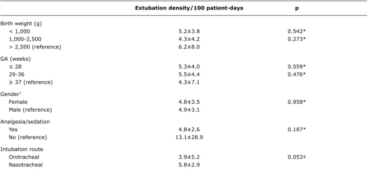
the variables analyzed are described in Table 2.
The occurrence of signiicant periodicity was veriied in
overall AE density of the period (Figure 1) and also for oral route (p = 0.009), nasal route (p = 0.031), and both routes
(p = 0.037). Signiicant periodicity was identiied in AE
density according to the number of patient-days ventilated
(p = 0.014). No cyclical pattern was observed in overall AE
rates when the other variables were analyzed.
There was a signiicant positive correlation (correlation coeficient = 0.723; p < 0.05) between the number
of patient-days ventilated per month and absolute AE
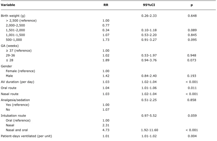
numbers. Univariate logistic regression analysis identiied
the following risk factors for AE: total AV time, AV duration per each intubation route, naso and orotracheal intubation routes, and number of patient-days ventilated (Table 3). Multivariate analysis showed that AV duration was the only independent predictor of AE. Every day on ventilation increased AE risk in 3% [relative risk (RR) = 1.03; 95%
conidence interval (95%CI) 1.02-1.04; p < 0.01] in the non-adjusted model and 2% (RR = 1.02; 95%CI 1.01-1.03; p < 0.001) in the model adjusted for weight
and GA covariables.
Without Ae (n = 160) With Ae (n = 62) p
Birth weight (g) 1,881.3±947.0 1,615.3±1,007.9 0.014*
Median 1,740.0 1,082.5
Minimum-maximum 540-4,670 545-4,500
< 1,000 31 24 0.009†
1,000-2,500 86 23
> 2,500 43 15
GA (weeks) 32.8±4.3 31.2±4.6 0.011*
Median 32.5 30.0
Minimum-maximum 25-41 24-39
≤ 28 27 21 0.022†
29-36 90 28
≥ 37 43 13
Gender (male/female)‡ 87/72 41/21 0.123†
AV duration (days) 6.7±6.5 25.6±25.3 < 0.001*
Median 5 16
Minimum-maximum 1-46 1-110
Use of analgesia/sedation§ 141 54 0.833†
Intubation route
Nasal 93 33 < 0.001†
Oral 47 6
Nasal and oral 20 23
extubation density/100 patient-days p
Birth weight (g)
< 1,000 5.2±3.8 0.542*
1,000-2,500 4.3±4.2 0.273*
> 2,500 (reference) 6.2±8.0
GA (weeks)
≤ 28 5.3±4.0 0.559*
29-36 5.5±4.4 0.476*
≥ 37 (reference) 4.3±7.1
Gender†
Female 4.8±3.5 0.958*
Male (reference) 4.9±3.1
Analgesia/sedation
Yes 4.8±2.6 0.187*
No (reference) 13.1±28.9
Intubation route
Orotracheal 3.9±5.2 0.053‡
Nasotracheal 5.8±2.9
table 1 - Characteristics of the study sample according to the presence of accidental extubation (n = 222)
AE = accidental extubation; AV = assisted ventilation; GA = gestational age. * Mann-Whitney test.
† Chi-square test.
‡ One case with undetermined gender. § 27 cases did not use sedation/analgesia.
Values expressed as mean ± standard deviation and absolute numbers.
table 2 - Mean values ± standard deviation of accidental extubation density according to neonatal variables and intubation route
GA = gestational age. * Cosinor analysis.
Variable RR 95%Ci p
Birth weight (g) 0.26-2.33 0.648
> 2,500 (reference) 1.00
2,000-2,500 0.77
1,501-2,000 0.34 0.10-1.18 0.089
1,001-1,500 1.07 0.53-2.20 0.845
500-1,000 1.73 0.91-3.27 0.095
GA (weeks)
≥ 37 (reference) 1.00
29-36 1.02 0.53-1.97 0.948
≤ 28 1.89 0.94-3.76 0.073
Gender
Female (reference) 1.00
Male 1.42 0.84-2.40 0.193
AV duration (per day) 1.03 1.02-1.04 < 0.001
Oral route 1.04 1.01-1.06 0.011
Nasal route 1.03 1.02-1.04 < 0.001
Analgesia/sedation 0.51-2.25 0.858
Yes (reference) 1.00
No 1.07
Intubation route 0.97-5.52 0.059
Oral (reference) 1.00
Nasal 2.31
Nasal and oral 4.73 1.92-11.60 < 0.001
Patient-days ventilated (per unit) 1.01 1.01-1.02 0.004
table 3 - Univariate logistic regression analysis for accidental extubation (n = 222)
95%CI = 95% confidence interval; AV = assisted ventilation; GA = gestational age; RR = relative risk.
AV duration with the best accuracy for total AE occurrence and for nasal and oral routes was achieved, respectively, at 10.5 days, 7.5 days, and 5.5 days. The
respective areas under the curve and 95%CI were: 0.79, 0.71-0.87, p < 0.001; 0.74, 0.62-0.87, p < 0.001; 0.69,
0.42-0.96, p = 0.12.
Discussion
In a cohort of 222 newborns, we obtained a mean AE
rate of 5.34/100 patient-days ventilated in a neonatal intensive care unit of a tertiary teaching public hospital. AV duration was the independent predictor of these events, and AV time of 10.5 days showed the best accuracy for the
occurrence of AE. A signiicant periodicity of AE incidence
rate was observed. A cyclical pattern was also observed in the number of patient-days ventilated with a positive correlation between this variable and AE numbers.
AE has been the object of several studies in adult and
pediatric intensive care units.3,5,12,13,22-25 However, we identiied only a few publications analyzing, in the neonatal
age group, incidence and circumstances accompanying AE,2,8-10 total AV time,2 length of hospital stay and mortality
in extubated patients,2 association between types of ixation
and AE,10 and one intervention study to reduce AE.11
The extubation density obtained in this study is higher than that reported in previous studies,2,8-11 and,
in some months, unacceptably high rates were observed, revealing the need for the implementation of an effective intervention.
In 2004, an intervention program to reduce the
occurrence of AE was implemented, including a written routine on intubation procedures and checking of tube
position, standardization of care concerning tube ixation
and aspiration, as well as the use of analgesia and
sedation. In addition, a training course was offered to the
multiprofessional team. After the implementation of this program, mean AE rate was 4.4 AE/100 patient-days in 2006 against 6.5 AE/100 patient-days in 2005 (data not published).
In addition to the factors analyzed to explain the high
routinely perform laryngoscopy for suspected AE, which could reduce some occurrences of tube replacement associated with false extubation episodes. Other factors, previously described, such as length of hospital stay,13,26
patient restlessness or agitation,26 insuficient sedation and
restraint,8,22 orotracheal intubation route,22 inaccurate tube ixation,2,22 and AV duration,13,25 may contribute to AE.
Regarding the last aspect mentioned, this study showed
that a longer AV duration was the most signiicant predictor
alone of AE in the study sample. Every day on ventilation increased AE risk in 2-3%. Other studies have also suggested that a longer AV duration may have an effect on AE, but
they have not quantiied the risk.2,9,10,13,25
This study determined that AV time of 10.5 days has 79.6% of accuracy in identifying the occurrence of AE in this unit. This information may be very useful in reducing AE, since it indicates a suitable moment for extubation.
In pediatric units, a factor associated with AE is the
child’s age.13 Based on similarity, one could assume that
newborns with lower weight and GA are more likely to have AE episodes, since they have a smaller body surface area
available to tube ixation and receive AV for longer periods of time. It is well known that newborns < 1,500 g and < 28
weeks are more affected by all types of adverse effects in neonatal units.4 However, in the present study, weight and
GA were different between groups with and without AE, but
did not remain signiicant in the multiple analysis, although
there is a trend toward risk (p = 0.095 and 0.073) for
newborns less than 1,000 g and 28 weeks, respectively. On
the other hand, we observed that AE rates by weight were higher among newborns > 2,500 g, indicating a possibility of inadequate sedation/analgesia or the need for a higher level of care in this group of children.
Adequate sedation has been related to a better management of AE.3,23 In our study, AE rates were higher in
newborns without sedation/analgesia, but this variable was not associated with AE, probably due to the limited number
of subjects without sedation/analgesia (12.2%). However, we cannot conidently state the level of sedation/analgesia in these subjects, since this variable was not assessed.
There is no strong evidence showing that a choice of either of the intubation routes could reduce AE.27 Similarly,
this study did not identify intubation route as a risk factor for AE. We observed, however, that newborns using both routes during AV had the highest risk (4.73 times) for AE. Analyzing this group in more detail, we observed that these
are the newborns with signiicantly lower weight and GA and longer AV duration, which justiies the risk associated
with this group.
Access route as a preventive measure for AE remains an open question, which might be satisfactorily answered by multicenter clinical trials with a large number of
subjects.27
A inding of this study, not previously mentioned, is the
number of patient-days ventilated as a risk factor for AE, which might correspond to a greater need for nursing care. Once the nurse-to-patient ratio in this unit was established, about 1.5:1, a higher number of patient-days ventilated could exceed the capacity of nursing care delivery, compromising its quality. The recommended nurse-to-patient ratio in pediatric units is 1:1, and it has been observed that pediatric patients are 4.24 times more likely to experience AE when being cared in a nurse-to-patient ratio of 1:2.28 On the other hand, it has been suggested that AE episodes occur most frequently in the care of nurses with less experience, with no
signiicant difference in nurse-to-patient ratio.29 Time spent
at ventilated patients’ bedside may also be associated with AE, since 79.1% of the episodes occurred when patients were not being closely controlled,29 such as during meal
breaks or shift changes. On the other hand, some reports state that in 75% of the events occurred with children, there was a professional at the patient’s bedside.9
We investigated the presence of AE periodicity to
conirm a clinical suspicion, but we did not ind references in the literature with a similar inding. The cyclical pattern
is rather a peculiarity of this unit, related directly to the periodicity of the number of patient-days ventilated, than an inherent characteristic of AE events, since the neonatal diseases most commonly associated with the need for AV are not seasonal. The assessment of AE periodicity in this
unit may contribute to a more accurate identiication of the
time for monitoring intervention effects, which, in the case of this unit, should be of at least 13.7 months.
This study has some limitations, such as the absence
of an analysis of variables that could better deine the
focus of intervention measures. Thus, the shift in which AE occurred,29 as well as nurse-to-patient ratio, or even
caregivers’ technical education and experience in intensive care29 are variables that could more clearly deine the focus
for action. Moreover, for some of the studied variables, as well as for lower weight and GA ranges and number
of subjects using the oral route, sample size may yield
inadequate results.
The occurrence of AE can be considered as acceptable, since the attempt to keep AE rates close to zero may increase the use of sedation and prolong AV duration, in addition to
causing stronger ixation of the tracheal tube, with a risk
of soft tissue necrosis.2 Nevertheless, the results of the
Correspondence: Fabiana L. Carvalho
Rua Salvador Penteado, 105/62 - Bonim CEP 13070-270 - Campinas, SP - Brazil Tel.: +55 (19) 3243.3805, +55 (19) 9118.0629 E-mail: [email protected]
17. Capurro H, Korichezky S, Fonseca D, Caldeyro-Barcia R. A simpliied
method for diagnosis of gestational age in the newborn infant. J Pediatr. 1978;93:120-2.
18. Ballard JL, Khoury JC, Wedig K, Wang L, Eilers-Walsman BL, Lipp R. New Ballard Score, expanded to include extremely premature
infants. J Pediatr. 1991;119:417-23.
19. Hennekens CH, Buring JE, Mayrent SL, Doll SR, eds. Epidemiology in medicine. Boston: Little Brown and company; 1987. p. 383. 20. Arendt J, Minors DS, Waterhouse JM. Biological rhythms in clinical
practice. London: Wright; 1989.
21. Espasa A, Cancelo JR. Métodos cuantitativos para el análisis de la coyuntura económica. Madrid: Alianza Editorial; 1993. 22. Boulain T. Unplanned extubations in the adult intensive care unit:
a prospective multicenter study. Association des Réanimateurs du Centre-Ouest. Am J Resp Crit Care Med. 1998;157:1131-7. 23. Chiang AA, Lee KC, Lee JC, Wei CH. Effectiveness of a
continuous quality improvement program aiming to reduce unplanned extubation: a prospective study. Intensive Care Med. 1996;22:1269-71.
24. Tindol GA Jr, DiBenedetto RJ, Kosciuk L. Unplanned extubations.
Chest. 1994;105:1804-7.
25. Vassal T, Anh NG, Gabillet JM, Guidet B, Staikowsky F, Offenstadt G. Prospective evaluation of self-extubations in a medical intensive care unit. Intensive Care Med. 1993;19:340-2.
26. Atkins PM, Mion LC, Mendelson W, Palmer RM, Slomka J, Franko T. Characteristics and outcomes of patients who self-extubate from ventilatory support: a case-control study. Chest. 1997;112:1317-23.
27. Spence K, Barr P. Nasal versus oral intubation for mechanical ventilation of newborn infant (Cochrane Review). In: The Cochrane Library; 2009. Oxford: Update Software.
28. Marcin JP, Rutan E, Rapetti PM, Brown JP, Rahnamayi R, Pretzlaff RK. Nurse stafing and unplanned extubation in the pediatric
intensive care unit. Pediatr Crit Care Med. 2005;6:254-7. 29. Yeh SH, Lee LN, Ho TH, Chiang MC, Lin LW. Implications of nursing
care in the occurrence and consequences of unplanned extubation in adult intensive care units. Int J Nurs Stud. 2004;41:252-62. 30. Davis PG, Morley CJ, Owen LS. Non-invasive respiratory support
of preterm neonates with respiratory distress: continuous positive airway pressure and nasal intermittent positive pressure ventilation. Semin Fetal Neonatal Med. 2009;14:14-20. References
1. Goldsmith JP, Karotkin EH. Introduction to assisted ventilation. In: Goldsmith JP, Karotkin EH, eds. Assisted ventilation of the neonate, fourth edition. Philadelphia: Saunders Elsevier; 2003. p. 1-14.
2. Veldman A, Trautschold T, Weib K, Fischer D, Bauer K.
Characteristics and outcome of unplanned extubation in ventilated preterm and term newborns on a neonatal intensive care unit.
Paediatr Anaesth. 2006;16:968-73.
3. Kapadia FN, Bajan KB, Raje KV. Airway accidents in intubated intensive care unit patients: an epidemiological study. Crit Care Med. 2000;28:659-64.
4. Sharek PJ, Horbar JD, Mason W, Bisarya H, Thurm CW, Suresh G, et al. Adverse events in the neonatal intensive care unit: development,
testing and indings of an NICU-focused trigger tool to identify harm in North American NICUs. Pediatrics. 2006;118:1332-40. 5. Frank BS, Lewis RJ. Experience with intubated patients does not
affect the accidental extubation rate in pediatric intensive care units and intensive care nurseries. Pediatr Pulmonol.1997;23:424-8. 6. da Silva O, Stevens D. Complications of airway management in
very-low-birth-weight infants. Biol Neonate.1999;75:40-5. 7. Kelly MA, Finer NN. Nasotracheal intubation in the neonate:
physiologic responses and effects of atropine and pancuronium.
J Pediatr. 1984;105:303-9.
8. Little LA, Koenig JC Jr, Newth CJ. Factors affecting accidental extubations in neonatal and pediatric intensive care patients. Crit Care Med. 1990;18:163-5.
9. Kleiber C, Hummel PA. Factors related to spontaneous endotracheal extubation in the neonate. Pediatr Nurs. 1989;15:347-51. 10. Brown MS. Prevention of accidental extubation in newborns. Am
J Dis Child. 1988;142:1240-3.
11. Loughead JL, Brennan RA, DeJuilio P, Camposeo V, Wengert J, Cooke D. Reducing accidental extubation in neonates. Jt Comm J Qual Patient Saf. 2008;34:164-70.
12. Piva JP, Amantéa S, Luchese S, Giugno K, Maia TR, Einloft L.
Extubação acidental em uma unidade de terapia intensiva. J Pediatr (Rio J). 1995;71:72-6.
13. Sadowski R, Dechert RE, Bandy KP, Juno J, Bhatt-Mehta V, Custer JR, et al. Continuous quality improvement: reducing unplanned extubations in a pediatric intensive care unit. Pediatrics. 2004;114:628-32.
14. Rivera R, Tibballs J. Complications of endotracheal intubation and mechanical ventilation in infants and children. Crit Care Med. 1992;20:193-9.
15. Joint Commission International.org [website]. http://www.
jointcommissioninternational.org. Access: 15/07/2009. 16. Blayney MP, Logan DR. First thoracic vertebral body as reference