Acute tubulointerstitial nephritis with severe renal impairment
associated with multisystem IgG4-related disease
Nefrite túbulo-intersticial aguda com insuficiência renal grave
associada à doença multissistêmica por IgG4
Authors
Rafael Coimbra Ferreira Beltrame 1
Maurício Friderichs 1
Bárbara Rayanne Fior 1
Pedro Guilherme Schaefer 1
Gustavo Gomes Thomé 1
Dirceu Reis da Silva 1
Elvino José Guardão Barros 1
Renato Seligman 1
Francisco Veríssimo Veronese 1
1 Hospital de Clínicas de Porto Alegre, Universidade Federal do Rio Grande do Sul.
Submitted on: 10/03/2015. Approved on: 11/16/2015.
Correspondence to:
Francisco Veríssimo Veronese. Universidade Federal do Rio Grande do Sul.
Serviço de Nefrologia, Sala 2030, Hospital de Clínicas de Porto Alegre, Rua Ramiro Barcelos, nº 2350, Porto Alegre, RS, Brazil. CEP: 90035-003
I
NTRODUCTIONIgG4-related disease (IgG4-RD) is a recently described systemic fibroin-flammatory condition characterized by tumefactive lesions, a dense lym-phoplasmacytic infiltrate with plasma cells expressing IgG4, storiform fibro-sis, and high serum levels of IgG41 in
although occurrences in the pancreas, biliary tract, salivary and lacrimal glands, retroperitoneal space, and lymph nodes have been reported more frequently.1-7 The pathophysiology of IgG4-RD remains un-clear, but various other diseases previous-ly described separateprevious-ly, such as Riedel’s thyroiditis, Küttner tumor, and Mikulicz’s syndrome, are currently considered part The IgG4-related disease has a wide
cli-nical spectrum where multiple organs can be affected, and the diagnosis depends on typical histopathological findings and an elevated IgG4 expression in plasma cells in the affected tissue. We describe the clinical presentation and evolution of a patient with acute tubulointerstitial nephritis, severe kidney failure and sys-temic manifestations such as lymphade-nomegaly and chronic pancreatitis. The diagnosis was confirmed by the clinical picture and kidney and lymph node histo-pathology, in which immunohistochemis-try of the lymphoid tissue showed policlo-nality and increased expression of IgG4, with a IgG4/total IgG ratio > 80%. The patient was treated with prednisone at a dose of 60 mg/day, followed by mycophe-nolate mofetil, and showed clinical and renal function improvement at 6 months of follow-up. The high index of suspicion of IgG4-related disease with multisystem involvement and the early treatment of this condition are essential to improve the prognosis of affected patients.
ABSTRACT
Keywords: immune system diseases;
im-munoglobulin G; immunosuppression; inflammation; interstitial, nephritis; renal insufficiency.
A doença relacionada à IgG4 tem um espectro clínico amplo em que múltip-los órgãos podem ser afetados, e o diag-nóstico depende de achados histopatológi-cos típihistopatológi-cos e elevada expressão de IgG4 em plasmócitos no tecido afetado. De-screvemos o quadro clínico e a evolução de um paciente com nefrite túbulo-intersticial aguda, insuficiência renal grave e mani-festações sistêmicas como linfoadenomega-lias e pancreatite crônica. O diagnóstico foi confirmado pelas características clínicas e pela histopatologia renal e de linfonodo, na qual a imunohistoquímica mostrou tecido linfoide com policlonalidade e ex-pressão aumentada de IgG4, com uma relação IgG4/IgG total > 80%. O paciente foi tratado com prednisona na dose de 60 mg/dia, seguido de micofenolato mofetil, e apresentou melhora clínica e da função renal depois de 6 meses de tratamento. O alto índice de suspeição da doença relacio-nada ao IgG4 com comprometimento mul-tissistêmico e o tratamento precoce desta condição são primordiais para a melhora do prognóstico destes pacientes.
R
ESUMOprogression of a patient with tubulointerstitial ne-phritis (TIN), severe renal failure, and systemic mani-festations associated with IgG4-RD.
C
LINICAL CASEA 45-year-old white male driver, self-described as a moderate smoker, complained of inappetence manifesting for a year, involuntary weight loss of 10 kg, tiredness after medium-level physical effort, and indisposition. Physical examination revealed he was reasonably well, although thin and with enlarged, moving, elastic and painless inguinal lymph nodes to the right measuring 1.5 cm. His abdomen was soft, and he reported epigastric discomfort during palpation. No other relevant findings were noted.
Significant workup findings included normocytic normochromic anemia, peripheral eosinophilia (12%), C-reactive protein at 33.3 mg/L, serum creatinine at 5.18 mg/dL (estimated glomerular filtration rate of 12 mL/ min/1.73 m2), and 24-hour proteinuria of 1.34 g. He had 3 WBC/µL and 29 RBC/µL in his urine sediment. Serum and urine immunoelectrophoresis revealed a biclonal peak in the gamma fraction, and immunofixation showed IgG-lambda. His total IgG serum level was 6132 mg/dL (reference: 700-1600 mg/dL).
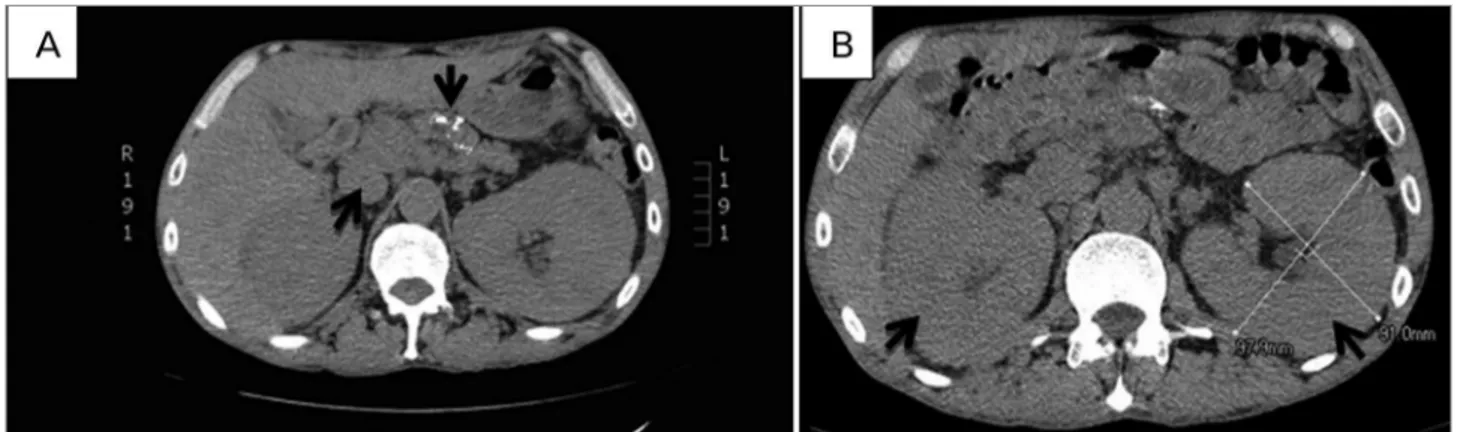
Chest computed tomography scans showed enlarged mediastinal lymph nodes, the greater measuring 2.7 x 1.2 cm; abdomen scans (Figure 1) revealed enlarged kidneys (right kidney measuring 14 cm and left kidney 15.7 cm), signs of chronic pancreatitis with gross calcification in the body and tail of the pancreas, and several enlarged abdominal nodes measuring up to 1.3 cm in diameter; enlarged nodes were detected in the hepatic hilum, close to the celiac trunk and the aorta, and in the iliac chains.
Bone marrow, skin, renal, and inguinal lymph node biopsies were carried out. All the specimens were negative in Congo red staining. Biopsies showed the bone marrow was hypocellular e hypoproliferative, and immunophenotyping yielded normal test results. Skin biopsy revealed moderate, predominantly plasmacytic chronic inflammation in a medium artery.
Renal biopsy showed mesangial matrix expansion within the renal parenchyma, mild mesangial hypercellularity, and presence of a dense polyclonal plasmacytic infiltrate and eosinophils characteristic of acute TIN, with stromal fibrosis in a storiform pattern and extensive tubular atrophy, and focal intraluminal giant cell response (Figure 2). Involvement by interstitial fibrosis and tubular atrophy was estimated at 70%. Immunofluorescence did not reveal immune deposits. Immunohistochemistry of renal tissue for IgG4-positive cells was inconclusive, and the test could not be repeated because the samples were worn out.
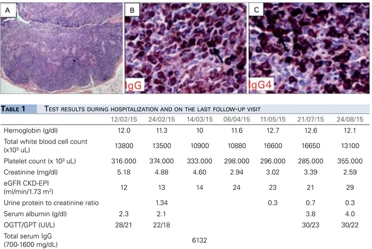
Lymph node biopsy showed paracortical expansion at the expense of abundant plasma cells and sites with fibrosis. Lymph node immunohistochemistry assays revealed a mixed lymphoid population positive for CD20+ B-cells, CD3+ T-cells, Kappa and Lambda light chains, CD138+ plasma cells, and IgG e IgG4. Among plasma cells, more than 100 positive cells were found per field, a significant increase for IgG4+, yielding an IgG4+/ IgG+ ratio > 80% (Figure 3).
The diagnosis agreed upon after the analysis of clinical findings and workup was IgG4-RD with extensive renal, lymph node, and pancreatic involvement. The patient was started on prednisone 60 mg/day. Two months after the start of treatment his creatinine had dropped to 3.02 mg/dL; five months into treatment creatinine levels had decreased to 2.59 mg/dL and the urine protein
Figure 2. acute tubulointerstitial nephritis with storiform fibrosis. A. Inflammatory infiltrate predominantly with plasma cells, interstitial fibrosis, and tubular atrophy, glomerular mesangial matrix expansion (HE, 200x magnification). B. Tubular atrophy and storiform interstitial fibrosis (PAS, 400x magnification). C. Extensive plasmacytic infiltrate associated with stromal fibrosis (HE, 100x magnification). D. Immunohistochemistry test labeled positive for CD138+ plasma cells
in renal tissue (DAB, 200x magnification).
Figure 3. Lymph node biopsy positive for IgG4. A. Parafollicular expansion presenting mixed lymphoid population and various aggregates of polyclonal plasma cells with areas of fibrosis. Specimens subsequently tested with immunohistochemistry were positive for CD20+ B-cells, CD3+
T-cells, Kappa and Lambda light chains, and CD138+ plasma cells. B. Immunohistochemistry test positive for IgG. C. Immunohistochemistry test
positive for IgG4, with > 100 positive cells/high-power field. IgG4+/IgG+ ratio > 80% (Courtesy of Laboratório Bacchi, São Paulo).
to creatinine ratio was at 0.3 (Table 1). The patient responded only partially to therapy with steroids, and was thus started on mycophenolate sodium 720 mg twice a day. The patient is clinically stable with stage-4 chronic kidney disease.
D
ISCUSSIONIgG4-RD is a multi-systemic condition with a varied range of clinical manifestations, depending on the organ or system involved. Reported involved sites include the biliary tract, the salivary glands, periorbital tissues, the kidneys, the lungs, the retroperitoneal space, the thyroid gland, the mediastinum, lymph nodes, the meninges, the aorta, the prostate, the skin, and the pericardium, to name a few.1,3-12 Patients are usually found to have masses characterized by subacute and often pronounced growth (e.g.: orbital pseudotumor), retroperitoneal fibrosis,1,3,8 nephromegaly,10,11 and bone lysis in rare cases.13
12/02/15 24/02/15 14/03/15 06/04/15 11/05/15 21/07/15 24/08/15
Hemoglobin (g/dl) 12.0 11.3 10 11.6 12.7 12.6 12.1 Total white blood cell count
(x10³ uL) 13800 13500 10900 10880 16600 16650 13100 Platelet count (x 10³ uL) 316.000 374.000 333.000 298.000 296.000 285.000 355.000 Creatinine (mg/dl) 5.18 4.88 4.60 2.94 3.02 3.39 2.59 eGFR CKD-EPI
(ml/min/1.73 m2) 12 13 14 24 23 21 29
Urine protein to creatinine ratio 1.34 0.3 0.7 0.3
Serum albumin (g/dl) 2.3 2.1 3.8 4.0
OGTT/GPT (UI/L) 28/21 22/18 30/23 30/22
Total serum IgG
(700-1600 mg/dL) 6132
Within months, many patients with extrarenal involvement present with multi-systemic involvement and subacute progression to organ dysfunction (e.g.: kidney6 or liver7 failure). In our case, the patient underwent extensive testing for an occult cancer, possibly hematologic, before a firm diagnosis was established.
Increased levels of serum and tissue IgG4 offer useful hints in diagnosing the condition, but may not be understood as a specific marker for it. The most important traits are comprised in the disease’s classical histologic finings, which dense lymphoplasmacytic infiltrate organized in a storiform pattern, obliterative phlebitis, and mild to moderate eosinophilic infiltrate.1 Diagnosis requires the presence of at least two of these signs, which combined yield a strong correlation between clinical and pathology findings.9
This pattern, however, may be subject to variation depending on the affected organ; the kidneys, for instance, may present with acute TIN and other glomerular lesion types, particularly in individuals with membranous glomerulonephritis negative for anti-PLA2R,14 as described in recent reviews.1,3,9,10 The histologic pattern of TIN may be related to the stage of the disease, como (a) acute TIN with minimal fibrosis, (b) a more cellular inflammatory pattern with storiform fibrosis, or (c) paucicellular fibrosis.10 The second pattern of disease was seen in the case reported, suggesting disease in an intermediate stage of progression, despite extensive fibrosis.
Although this is not a pathognomonic finding or a finding that would allow the condition to be ruled out if absent, tissue immunohistochemistry was positive for IgG4, with an IgG4/total IgG ratio > 40% and a presence of 10 or more IgG4-producing cells by high-power field (HPF), which aided in the diagnosis of the disease.1,3,9,10 Cheuk et al.12 suggested that morphologic findings consistent with an absolute number of IgG4+ cells > 50/HPF and a IgG4/IgG ratio > 40% are a match for IgG4-RD. These diagnostic cutoff points, however, may vary depending on which organs have been affected.3 Diagnosis may be more challenging in advanced cases, with extensive fibrosis and few plasma cells.
Other findings may include peripheral eosinophilia, high IgE levels, and atopic manifestations. Serum IgG4 levels are increased (> 135 mg/dL) in 70% of
levels may predict disease activity during treatment,2 with yet unknown levels of accuracy.1 There appears to be a correlation between serum IgG4 and the number of involved organs.9 Flow cytometry may reveal increased plasmablast counts; 20-30% of the cases will be positive for antinuclear antibodies and rheumatoid factor.3
The treatment of IgG4-RD depends on the type of tissue involvement. While some patients have indolent disease (adenopathy and parotitis), others present severe multiple organ involvement.1,2,10 An international guideline for the treatment of IgG4-RD was recently published.2 When vital organs are affected, aggressive therapy must be started promptly due to the risk of organ failure even in subclinical cases. Patients with extensive fibrosis may not benefit much from therapy in terms of retrieving organ function.1,2 Additionally, the disease reoccurs frequently, which requires immunosuppressant therapy.1
Many treatment schemes have been proposed. Steroids have had a good record in dealing with acute inflammatory disease. An initial scheme advocates the use of 0.6 mg/kg/day of prednisone for 2-4 weeks, followed by a dose reduction to 5 mg/day in six months and maintenance therapy with 2.5-5 mg/day of the medication for three years. Antiproliferative agents such as mycophenolate mofetil or azathioprine combined with methotrexate may be used to prevent or reduce the incidence of steroid-related adverse events,1,2,10,11 but their efficacy has not been tested in clinical trials. Rituximab has been prescribed to refractory patients,1,2 with isolated reports of success with long-term treatment.15 In an open trial, Carruthers et al.16 described the efficacy of rituximab in 30 patients with IgG4-RD, with 47% achieving complete remission after six months and 40% in remission after 12 months with two doses of 1000 mg of rituximab without a concurrent prescription of steroids.
the medical community and in the implications it may have in causing organ failure when not diagnosed early enough. This multi-systemic disease must be included in the differential diagnosis when symptoms and signs are present in multiple organs. Early diagnosis and treatment improve the prognosis of affected patients.
R
EFERENCES1. Stone JH, Zen Y, Deshpande V. IgG4-related disease. N Engl J Med 2012;366:539-51. DOI: http://dx.doi.org/10.1056/NE-JMra1104650
2. Khosroshahi A, Wallace ZS, Crowe JL, Akamizu T, Azumi A, Carruthers MN, et al.; Second International Symposium on IgG4-Related Disease. International Consensus Guidance Sta-tement on the Management and Treatment of IgG4-Related Disease. Arthritis Rheumatol 2015;67:1688-99. DOI: http:// dx.doi.org/10.1002/art.39132
3. Umehara H, Okazaki K, Masaki Y, Kawano M, Yamamoto M, Saeki T, et al.; Research Program for Intractable Disease by Ministry of Health, Labor and Welfare (MHLW) Japan G4 team. A novel clinical entity, IgG4-related disease (IgG4RD): general concept and details. Mod Rheumatol 2012;22:1-14. DOI: http://dx.doi.org/10.3109/s10165-011-0508-6
4. Shimizu M, Okamura K, Kise Y, Takeshita Y, Furuhashi H, Wee-rawanich W, et al. Effectiveness of imaging modalities for screening IgG4-related dacryoadenitis and sialadenitis (Mikulicz's disease) and for differentiating it from Sjögren's syndrome (SS), with an emphasis on sonography. Arthritis Res Ther 2015;17:223. PMID: 26298875 DOI: http://dx.doi.org/10.1186/s13075-015-0751-x 5. Tran MN, Langguth D, Hart G, Heiner M, Rafter A, Fleming
SJ, et al. IgG4-related systemic disease with coronary arteritis and aortitis, causing recurring critical coronary ischemia. Int J Cardiol 2015;201:33-4. DOI: http://dx.doi.org/10.1016/j.ij-card.2015.08.014
6. Fernández Lorente L, Álvarez DL, López VG, Kollros VA, Ari-za A, Gálvez A, et al. IgG4-related disease: description of a case with pulmonary lesions, mediastinal lymphadenopathies and rapidly progressive renal failure. Nefrologia 2015;35:218-23. DOI:http://dx.doi.org/10.1016/j.nefroe.2015.05.012
7. Björnsson E. Immunoglobulin G4-associated cholangi-tis. Curr Opin Gastroenterol 2008;24:389-94. DOI:http:// dx.doi.org/10.1097/MOG.0b013e3282f6a7c5
8. Rockey DC, Bell PD, Hill JA. Fibrosis-a common pathway to organ injury and failure. N Engl J Med 2015;372:1138-49.
9. Stone JH, Brito-Zerón P, Bosch X, Ramos-Casals M. Diag-nostic Approach to the Complexity of IgG4-Related Disea-se. Mayo Clin Proc 2015;90:927-39. DOI: http://dx.doi. org/10.1016/j.mayocp.2015.03.020
10. Raissian Y, Nasr SH, Larsen CP, Colvin RB, Smyrk TC, Takahashi N, et al. Diagnosis of IgG4-related tubulointers-titial nephritis. J Am Soc Nephrol 2011;22:1343-52. DOI: http://dx.doi.org/10.1681/ASN.2011010062
11. Kuroda N, Nao T, Fukuhara H, Karashima T, Inoue K, Taniguchi Y, et al. IgG4-related renal disease: clinical and pathological characteristics. Int J Clin Exp Pathol 2014;7:6379-85.
12. Cheuk W, Chan JK. IgG4-related sclerosing disease: a critical appraisal of an evolving clinicopathologic enti-ty. Adv Anat Pathol 2010;17:303-32. DOI: http://dx.doi. org/10.1097/PAP.0b013e3181ee63ce
13. Pace C, Ward S. A rare case of IgG4-related sclerosing di-sease of the maxillary sinus associated with bone destruc-tion. J Oral Maxillofac Surg 2010;68:2591-3. DOI: http:// dx.doi.org/10.1016/j.joms.2009.07.073
14. Alexander MP, Larsen CP, Gibson IW, Nasr SH, Sethi S, Fidler ME, et al. Membranous glomerulonephritis is a mani-festation of IgG4-related disease. Kidney Int 2013;83:455-62. DOI: http://dx.doi.org/10.1038/ki.2012.382
15. Yamamoto M, Awakawa T, Takahashi H. Is rituximab effective for IgG4-related disease in the long term? Ex-perience of cases treated with rituximab for 4 years. Ann Rheum Dis 2015;74:e46. DOI: http://dx.doi.org/10.1136/ annrheumdis-2015-207625