R E V B R A S R E U M A T O L . 2 0 1 4 ;5 4 ( 3 ): 2 3 1 – 2 3 3
www.reumatologia.com.br
REVISTA BRASILEIRA DE
REUMATOLOGIA
Case report
IgA nephropathy and polymyositis: a rare association
Thiago Bitar Moraes Barros
a, Fernando Henrique Carlos de Souza
a,
Denise Maria Avancini Costa Malheiros
b, Mauricio Levy-Neto
a, Samuel Katsuyuki Shinjo
a,*
a Service of Rheumatology, Hospital das Clínicas, Medicine Faculty, Universidade de São Paulo, São Paulo, SP, Brazil b Service of Pathology, Hospital das Clínicas, Medicine Faculty, Universidade de São Paulo, São Paulo, SP, Brazil
a r t i c l e i n f o
Article history:
Received on 30 November 2012 Accepted on 30 November 2012
Keywords:
Inlammatory myopathy IgA nephropathy Polymyositis Case report
a b s t r a c t
Polymyositis is a systemic and idiopathic inlammatory myopathy that, besides muscle manifestation, may occur with respiratory involvement, gastrointestinal tract and rarely renal involvement. In this latter, there are only two cases of IgA nephropathy, but both in dermatomyositis. On the other hand, we reported, for the irst time, a case of IgA nephro-pathy in polymyositis.
© 2014 Sociedade Brasileira de Reumatologia. Published by Elsevier Editora Ltda. All rights reserved.
* Corresponding author.
E-mail: [email protected] (S.K. Shinjo).
0482-5004/$ - see front matter. © 2014 Sociedade Brasileira de Reumatologia. Published by Elsevier Editora Ltda. All rights reserved. http://dx.doi.org/10.1016/j.rbre.2012.11.001
Nefropatia por IgA e polimiosite: uma rara associação
Palavras-chave:
Miopatia inlamatória Nefropatia por IgA Polimiosite Relato de caso
r e s u m o
A polimiosite é uma miopatia inlamatória idiopática sistêmica que, além da manifestação muscular, pode eventualmente cursar com acometimento respiratório, do trato gastrintes-tinal e, raramente, renal. Neste último caso, há descrição de apenas dois casos de nefro-patia por IgA em pacientes com mionefro-patia, ambos em dermatomiosite. Em contrapartida, relatamos pela primeira vez esta rara associação em polimiosite.
© 2014 Sociedade Brasileira de Reumatologia. Publicado por Elsevier Editora Ltda. Todos os direitos reservados.
Introduction
Polymyositis is classiied within the spectrum of idiopathic inlammatory myopathies, together with dermatomyositis. It is clinically characterized by progressive proximal muscle weakness of the limbs, leading to high morbidity and func-tional disability.
Among the extramuscular manifestations found, the most common are the respiratory and gastrointestinal involvement respiratory and of gastrointestinal tract involvement.1,2 Renal
in-volvement in polymyositis is uncommon, with rare descriptions of nephropathy associated with acute tubular necrosis second-ary to rhabdomyolysis and chronic glomerulonephritis.3-9
232
R E V B R A S R E U M A T O L . 2 0 1 4 ;5 4 ( 3 ): 2 3 1 – 2 3 3Approximately one-third of patients with this type of renal man-ifestation progresses to chronic renal failure after 20-25 years of disease.10 Among the systemic diseases, IgA nephropathy is
most commonly associated with Henoch-Schönlein purpura.11
However, its association with idiopathic inlammatory myopa-thies is extremely rare, with only two cases described, both in dermatomyositis,12,13 which motivated us to present this case.
Clinical case
Male patient, 35 years-old, white, trader, born in São Paulo. Previously healthy, presented six months ago with progres-sive proximal muscle weakness of all four limbs without ap-parent cause and without constitutional symptoms. When the patient came to our hospital service, the physical exami-nation showed grade III proximal muscle strength in all four limbs without skin lesions and/or involvement of other or-gans, such as lung or kidney. Laboratory tests showed serum creatine kinase (CK) = 3,545 IU/L (normal range: 24-204 IU/L), aldolase = 33 IU/L (reference value: 7.5 < IU/L), antinuclear fac-tor and anti-Jo-1 negative, C-reactive protein = 7.8 µg/mL (ref-erence value: < 5 µg/mL), erythrocyte sedimentation rate = 17 mm/1st hour (reference value: < 10 mm/1st hour),
electroneu-romyography suggestive of proximal inlammatory myopathy of all limbs without evidence of associated neuropathy. The muscle biopsy (biceps brachii) revealed the presence of an endomysial and perimysial inlammatory iniltrate and also necrosis of the muscle ibers and macrophage invasion, thus suggesting inlammatory myopathy. During the clinical inves-tigation, neoplastic and metabolic causes were ruled out.
With the presumptive diagnosis of polymyositis, accord-ing to the criteria of Bohan and Peter,14 corticosteroids
(pred-nisne 0,5mg/kg/day) and methotrexate (maximum dose 20 mg/week) were initiated. After six months of treatment, the patient developed acute lung injury secondary to pneu-mopathy requiring endotracheal intubation and mechanical ventilation for three weeks. Infection and/or disease activ-ity were discarded. In face of the possibilactiv-ity of lung disease secondary to methotrexate, this medication was discontin-ued, and the prednisone dose was optimized. The patient developed progressive pulmonary improvement without sequelae. Subsequently, as a steroid sparing drug, azathio-prine was introduced (maximum dose of 3 mg/kg/day). Due to the stability of the disease and to the pulmonary improve-ment, the dose of prednisone (which at that time was 0.2 mg/kg/day) was gradually reduced. However, with no appar-ent cause the patiappar-ent began to show macroscopic hematu-ria, edema of lower limbs (2+/4+) without hemodynamic and pressure repercussion. The laboratory examination showed: urinalysis with pyuria (> 100 leukocytes/ield), hematuria (> 100 erythrocytes/ield), moderate erythrocyte dysmorphism and lipoid casts; 24h-proteinuria = 1.76 g, serum albumin = 3.6 g/dL, serum creatinine = 0.9 mg/dL and negative urine cul-ture. There was no sign of clinical-laboratorial activity of PM at that time.
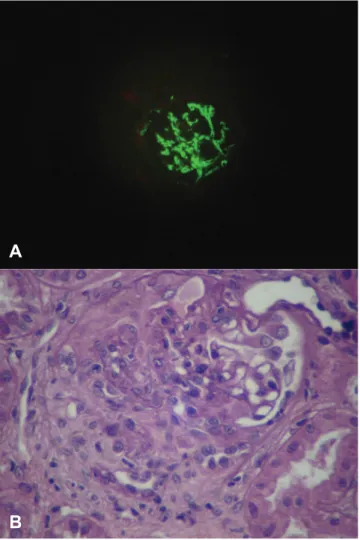
We decided then to perform a renal biopsy which showed 14 glomeruli with diffuse segmental endocapillar cell prolif-eration. One glomerulus showed one cellular crescent and two ibroblastic crescents. Moreover, the glomerulus
exhib-ited mild tubulointerstitial change, focal tubular atrophy and interstitial ibrosis. An interlobular artery showed intimal i-brosis. Immunoluorescence showed diffuse deposits of only IgA in the mesangial region and in the peripheral capillary wall of the glomerulus (ig. 1), suggestive of IgA nephropathy.
The dose of prednisone (0.2 mg/kg/day) was maintained, azathioprine was discontinued, and monthly cyclophospha-mide (0.75 mg/m2, IV) was initiated for 12 months; with the
patient achieving complete remission of the renal disease. disease. Subsequently, as a maintenance drug, azathioprine was reintroduced (maximum dose of 2.5 mg/kg/day). Cur-rently, the patient denotes stability, both from the point of view of polymyositis as of nephropathy, and without corti-costeroid therapy.
Discussion
To our knowledge, this is the irst case in the literature that reports an IgA nephropathy in idiopathic inlammatory my-opathy, particularly polymyositis.
Fig. 1 – Renal biopsy
(A) Optical microscopy. A glomerulus with diffuse proliferative glomerulonephritis. PAS staining,
(B) Immunoluorescence demonstrating diffuse IgA deposit in mesangial region and the peripheral capillary wall of the glomerulus.
A
233
R E V B R A S R E U M A T O L . 2 0 1 4 ;5 4 ( 3 ): 2 3 1 – 2 3 3Renal involvement in idiopathic inlammatory myopathy is uncommon, including acute tubular necrosis and glomeru-lonephritis.3-9 In the latter case, Takizawa et al.4 demonstrated
by renal biopsy in a series of 21 cases of dermatomyositis/ polymyositis, that the presence of nephritis was associated with membranous and proliferative mesangial glomerulone-phritis, respectively, in polymyositis and dermatomyositis.
The relationship between IgA nephropathy and idiopathic inlammatory myopathies is extremely rare. To date, there are only two case reports, both occuring in dermatomyositis.12,13
Civilibal et al.13 reported a case of newly diagnosed juvenile
dermatomyositis, with hematuria and nephrotic proteinuria in course, without loss of kidney function. A percutaneous re-nal biopsy allowed the diagnosis of IgA nephropathy. There was a favourable response both from the point of view of in-lammatory myopathy as of nephropathy, with the mainte-nance of methotrexate associated with oral corticosteroids. On the other hand, Yen et al.12 described a young woman who
developed IgA nephropathy after 1.5 years of an established diagnosis of dermatomyositis while using both azathioprine and corticosteroids.
Unlike these two cases, in the present trial we present a male patient with conirmed diagnosis of polymyositis. How-ever, like the case reported by Yen et al.,12 the patient had been
using corticosteroids and immunosuppressive medication, when evolved with renal dysfunction after 1.5 years, being subsequently diagnosed with IgA nephropathy.
The association between dermatomyositis and IgA ne-phropathy is plausible, since both diseases share the involve-ment of humoral immunity (immunecomplexes).3,15 On the
other hand, the relationship with polymyositis is remote, be-cause in the latter case there is a predominance of cellular immunity. Thus, in the present clinical case, we strongly sug-gest the existence of two distinct morbidities.
In short, to our knowledge, we reported for the irst time a case of IgA nephropathy in a patient with polymyositis.
Conlicts of interest
The authors declare no conlicts of interest.
R E F E R E N C E S
1. Marie I, Hachulla E, Cherin P, Dominique S, Hatron PY, Devulder B et al. Interstitial lung disease in polymyositis and dermatomyositis. Arthritis Rheum. 2002;47:614-22.
2. de Merieux P, Verity MA, Clements PJ, Paulus HE. Esophageal abnormalities and dysphagia in polymyositis and
dermatomyositis. Arthritis Rheum. 1983;26:961-8.
3. Yen TH, Lai PC, Chen CC, Hsueh S, Huang JY. Renal involvement in patient with polymyositis and dermatomyositis. Int J Clin Pract. 2005;59:188-93.
4. Takizawa Y, Kanda H, Sato K, Kawahata K, Yamaguchi A, Uozaki H et al. Polymyositis associated with focal mesangial proliferative glomerulonephritis with depositions of immune complexes. Clin Rheumatol. 2007;26:792-6.
5. Dyck RF, Katz A, Gordon DA, Johnson M, Shainhouse Z, Cardella CJ et al. Glomerulonephritis associated with polymyositis. J Rheumatol. 1979;6:336-44.
6. Pirovino M, Neff MS, Sharon E. Myoglobulinuria and acute renal failure with acute polymyositis. NY State J Med. 1979;79:764-7. 7. Tsunemi M, Ishimura E, Tsumura K, Shoji S, Sugimura T,
Nishizawa Y et al. A case of crescentic glomerulonephritis associated with polymyositis. Nephron. 1993;64:488-9.
8. Valenzuela OF, Reiser IW, Porush JG. Idiopathic polymyositis and glomerulonephritis. J Nephrol. 2001;14:120-4.
9. Makino H, Hirata K, Matsuda M, Amano T, Ota Z. Membranous nephropathy developing during the course of dermatomyositis. J Rheumatol. 1994;21:1377-8.
10. D’Amico G. Natural history of idiopathic IgA nephropathy: role of clinical and histological prognostic factors. Am J Kidney Dis. 2000;36:227-37.
11. Sanders JT, Wyatt RJ. IgA nephropathy and Henoch-Schönlein purpura nephritis. Curr Opin Pediatr 2008;20:163-70.
12. Yen TH, Huang JY, Chen CY. Unexpected IgA nephropathy during the treatment of a young woman with idiopathic dermatomyositis: case report and review of the literature. J Nephrol. 2003;16:148-53.
13. Civilibal M, Selcuk Duru N, Ozagari A, Durali K, Elevli M. Immunoglobulin A nephropathy associated with juvenile dermatomyositis. Pediatr Nephrol. 2009;24:2073-5.
14. Bohan A, Peter JB. Polymyositis and dermatomyositis. N Engl J Med. 1975;292:344-7.