Original Article
Artigo Original
Speech profile of patients undergoing
primary palatoplasty
Perfil da fala de pacientes submetidos à
palatoplastia primária
Katia Ignacio Menegueti1 Laura Davison Mangilli2 Nivaldo Alonso1 Claudia Regina Furquim de
Andrade1
Keywords
Cleft Palate Speech Surgery Velopharyngeal Insuficiency Speech, Language and Hearing
Sciences
Descritores
Fissura palatina Fala Cirurgia Insuiciência velofaríngea Fonoaudiologia
Correspondence address: Claudia Regina Furquim de Andrade Rua Cipotânea, 51, Cidade
Universitária, São Paulo (SP), Brazil, CEP: 05360-160.
E-mail: [email protected]
Received: September 02, 2016
Accepted: April 30, 2017
Study carried out at Departamento de Fisioterapia, Fonoaudiologia e Terapia Ocupacional, Faculdade de Medicina, Universidade de São Paulo – USP - São Paulo (SP), Brazil.
1 Universidade de São Paulo – USP - São Paulo (SP), Brazil.
2 Universidade de Brasília – UnB - Brasília (DF), Brazil.
Financial support: nothing to declare. Conlict of interests: nothing to declare.
ABSTRACT
Purpose: To characterize the proile and speech characteristics of patients undergoing primary palatoplasty in a Brazilian university hospital, considering the time of intervention (early, before two years of age; late, after two years of age). Methods: Participants were 97 patients of both genders with cleft palate and/or cleft and lip palate, assigned to the Speech-language Pathology Department, who had been submitted to primary palatoplasty and presented no prior history of speech-language therapy. Patients were divided into two groups: early intervention group (EIG) – 43 patients undergoing primary palatoplasty before 2 years of age and late intervention group (LIG) – 54 patients undergoing primary palatoplasty after 2 years of age. All patients underwent speech-language pathology assessment. The following parameters were assessed: resonance classiication, presence of nasal turbulence, presence of weak intraoral air pressure, presence of audible nasal air emission, speech understandability, and compensatory articulation disorder (CAD). Results: At statistical signiicance level of 5% (p≤0.05), no signiicant difference was observed between the groups in the following parameters: resonance
classiication (p=0.067); level of hypernasality (p=0.113), presence of nasal turbulence (p=0.179); presence of weak intraoral air pressure (p=0.152); presence of nasal air emission (p=0.369), and speech understandability (p=0.113). The groups differed with respect to presence of compensatory articulation disorders (p=0.020), with
the LIG presenting higher occurrence of altered phonemes. Conclusion: It was possible to assess the general proile and speech characteristics of the study participants. Patients submitted to early primary palatoplasty present better speech proile.
RESUMO
INTRODUCTION
Cleft lip and palate is the most common congenital anomaly
in the human race. It may occur in isolation or associated with
other complex malformations, bringing aesthetic and functional
consequences which require lifelong rehabilitation(1,2). It is the
most frequently treated congenital anomaly in craniofacial
surgery centers, with incidence of 1:600 live births(3). Several
anatomical and functional changes are observed as a consequence of cleft lip and palate, causing alterations in midface growth, hearing, and velopharyngeal sphincter function, with the latter interfering directly with the functions of sucking, swallowing, and speaking(4).
One of the main objectives of cleft palate repair is to establish adequate velopharyngeal function, so that communication development can occur as most appropriately as possible. The chances of adequate speech development decrease signiicantly when late repair occurs(5-7). A previous study(6)
reported statistically signiicant difference between early and late operated individuals with respect to presence of phonological processes, with these processes appearing less frequently in the group that underwent early surgery.
Failure in velopharyngeal closure, either due to the presence of cleft or velopharyngeal dysfunction, with respect to speech-related aspects, results in loss of part of the airway to the nasal cavity,
leading to the appearance of symptoms that may directly or indirectly impair speech understandability(8). Hypernasality,
audible nasal air emission, and weak intraoral air pressure are considered direct or primary consequences of velopharyngeal
closure failure(6,8), whereas compensatory articulation disorders
(CAD) - strategies used by individuals with the objective of compensating in other points of the vocal tract the inability to impose pressure to the oral cavity - are deined as indirect or
secondary consequences(6,8).
During adequate functioning of the velopharyngeal
mechanism, it is necessary that the soft palate and the posterior
and lateral walls of the pharynx perform a wide and synchronized movement, allowing contact between these structures and ensuring total separation between the oral and nasal cavities
during the production of oral sounds(9). Inadequacy of this
function causes infants with cleft lip and palate, even in the irst attempts of vocalization in the second and third months of life, to produce sounds differently compared with infants without cleft. At 6 months of age, infants begin to produce their irst anterior consonants and, in the case of children with cleft, there may be dificulties in performing this task, leading to glottal or
pharyngeal articulations(10).
The irst year of a child is of fundamental importance for
the acquisition of speech and language(10). The speciic scientiic
literature clearly reports that surgical repair of the cleft palate
should be performed early, before two years of age(5,6,11), especially
when considering speech(5,12). The surgical treatment aims to
provide adequate velopharyngeal function, which is essential in
early childhood(6). To this end, different palatoplasty techniques
have been developed over time aiming to achieve adequate
palate length, in order to allow velopharyngeal closure. Another aspect addressed is the reorientation of the muscle ibers of the palate, creating a muscle strap that will allow better mobility
of the soft palate(5).
Nevertheless, despite the progress made in this area, it is still dificult to predict the eficiency of muscle function for speech
after palatoplasty(13,14). Although there is recognized effort to
establish satisfactory velopharyngeal function, a study using magnetic resonance imaging and computerized reconstruction showed that up to 35% of individuals with cleft palate may remain with velopharyngeal insuficiency after surgery(13).
This insuficiency may be due to inadequate dissection of the palatine musculature, insuficient length of the palate, or anatomical changes of the pharynx posterior wall(13). Another study showed
that approximately 30% of patients might remain with changes
in articulation and speech resonance(14) after palatoplasty.
In Brazil, as in other developing countries, it is not always possible to follow the stages of cleft lip and palate treatment advocated and conducted by most international and reference national centers, because as previously mentioned, due to socioeconomic and cultural factors, parents seek treatment when
the child is already at an older age, past the period of language acquisition, often in adult age(2,15,16).
Speech alterations associated with velopharyngeal dysfunction would be identiied through clinical speech assessment performed by speech-language pathologists and by instrumental evaluation of the velopharyngeal function. The results of these evaluations allow determination of better treatment and prognosis to the
case(17).
Perceptual clinical evaluation of speech is considered the main indicator of velopharyngeal dysfunction, and it is an
essential part of clinical diagnostics(18). Although subjectively,
this assessment allows identiication of changes, measurement of severity, and evaluation of effectiveness of the treatments
performed(18,19). Over time, studies(19) have been concerned with
improving auditory-perceptual assessment in order to make it less susceptible to errors arising from its subjectivity. Thus,
adoption of criteria of scores to represent the speech-language
pathologist’s judgment is suggested. Among the different scales developed to measure resonance and other characteristics of speech, the most popular is the scale with equal intervals, in which the rater assigns a score to the aspect analyzed in a linear scale, where the smallest number indicates absence of
alterations and the highest one denotes the maximum degree of change. Therefore, on a six-point scale in the assessment of hypernasality, a score of one indicates absence of hypernasality
and a score of six shows its most severe degree(20).
Two domestic studies(6,21) veriied a relation between speech
alterations and time of palatoplasty, both considering the best time
for surgery until 18 months of age. Palandi and Guedes (2011)(6)
concluded that individuals who underwent late intervention showed statistically signiicant difference regarding the presence
of some phonological processes, and considered that the age
by Mituuti et al. (2010)(21) indicate that few individuals present
speech changes after cleft repair (ranging from a minimum of
8%, with poor intraoral air pressure, to a maximum of 26%, with CADs).
In view of the presented information, the objective of this study is to characterize the proile and speech characteristics of patients undergoing primary palatoplasty in a university hospital (HCFMUSP) in the city of Sao Paulo, Brazil, considering the time of intervention (early, before two years of age; late, after two years of age). It is a national referral center for cases of cleft lip and palate which receives a large number of new cases
annually. Some of these patients start their treatment at the
institution, whereas others choose the hospital to continue care. Due to the great demand, the institution identiied the necessity to characterize the patients’ speech changes, regardless of time of intervention, in order to initiate discussion and propose improvements in the care of these individuals.
METHODS
This is an observational, cross-sectional study whose procedures
of selection and assessment of participants only started after the
pertinent ethical processes had been completed: approval by the
Ethics Committee on Human Research of the aforementioned Institution (CAPPesq HCFMUSP 513.535) and signing of an Informed Consent Form (ICF) by the parents and/or guardians.
Study participants were 97 individuals with diagnosis of non-syndromic cleft palate and/or cleft lip and palate who were referred to speech-language pathology evaluation between May 2011 and July 2015 according to the service’s demand. The participants were divided into two groups: Early Intervention Group (EIG) and Late Interventiojn Group (LIG). The EIG was composed of individuals who had undergone primary palatoplasty up to the second year of life and the LIG comprised individuals who had been submitted intervention after the age of two(6).
Medical diagnosis and monitoring of the participants were
conducted by the Craniomaxillofacial Surgery Team of the
HCFMUSP’s Department of Plastic Surgery and Burns as a collaboration which evolves the assistance of plastic surgeons
and otolaryngologists.
Study inclusion criteria were as follows: individuals 1) of both genders; 2) with minimum age of six years, and no maximum age restriction; 3) diagnosed with non-syndromic cleft palate
and/or cleft lip and palate; 4) submitted to palatoplasty at least three months prior to speech-language pathology assessment;
5) who had not undergone previous speech-language pathology therapy and/or treatment with prostheses.
Minimum age of the participants was established based on
the studies by Andrade et al. (2004)(22), who advocate that the
phonological processes and development of oral language in
children are already completed at this age. The use of this criterion
aims to exclude the language acquisition variable in this study.
Exclusion criteria comprised: 1) minor surgeries on the palate; 2) presence of neurological disorders; 3) history of face trauma; 4) presence of respiratory diseases; 5) presence
of palate istulas of any length; 6) presence of speech-language
pathology comorbidities (language and auditory complaints
and deicits); 7) presence of cognitive impairment or level of consciousness that would prevent evaluators from understanding the verbal information required for speech-language assessment
and treatment.
As previously mentioned, some of the study participants were operated at the HCFMUSP and some underwent surgery elsewhere; therefore, it was not possible to control the variables type of surgical procedure and/or number of surgeons involved
in these procedures.
All participants were submitted to routine speech-language
assessment at the Speech-language Pathology (SLP) Clinic of
the HCFMUSP. The evaluations were conducted by a trained speech-language therapist experienced in the area, who belonged
to the SLP Team responsible for assistance at the Cleft Lip and Palate Outpatient Clinic.
The data used in this study comprise the range of aspects
evaluated during the routine assessment, more speciically the Auditory-perceptual Assessment of Speech. A protocol speciically developed by the Clinic is used in these evaluations. It was prepared by the SLP Clinic after extensive literature review and participation in thematic meetings aimed at standardizing
assessment protocols for cleft palate patients. This protocol
includes an evaluation of the morphological aspects of the oral
motor system, occlusion, and speech. The latter is assessed
through analysis of the speech samples, which comprise segments of directed speech (repetition of words and sentences) with
recurrence of the target sound, containing all the phonemes of
Brazilian Portuguese; automatic speech during counting from
one to 10; and spontaneously speech, requesting the patient
to report a daily event. To verify the occurrence of nasal air
escape during speech, a nasal air emission test is conducted
using a Glatzel mirror.
Samples were collected in a SLP room with environment speciically treated for this purpose. The participants remain seated and the voice samples are recorded using a unidirectional microphone positioned at the standard distance of 10 cm away
from their mouths at a 45° angle. The stimuli captured by the
microphone are digitized using the Praat.wav software with 22.050 Hz sampling frequency and 16-bit resolution.
The following parameters are assessed by the protocol: presence of alterations in resonance, severity of resonance
alteration, presence of compensatory articulation disorders
(CAD), presence of nasal turbulence, presence of weak intraoral
air pressure, presence of audible air emission, and rating of speech
understandability - variables considered in the present study. The samples were analyzed according to the following criteria: 1) presence or absence of hypernasality, with severity rating when present: mild, moderate, or severe; 2) presence or absence of hyponasality, with severity rating when present: mild, moderate, or severe; 3) presence or absence of CADs; 4) determination of
the types of CADs present and the number of occurring altered
phonemes; 5) presence of nasal turbulence; 6) presence of weak
air emission; 7) speech understandability rating: adequate or
inadequate, with severity ratings: mild, moderate, or severe. Hypernasality was considered present when excessive nasal resonance was veriied during production of oral sounds(8) in at
least one phoneme of each of the analyzed samples. Hyponasality was identiied when decreased or insuficient nasal resonance was veriied during production of nasal sounds(8) in at least one
phoneme of each of the analyzed samples.
CADs are called compensatory because they are an attempt
to compensate for the dificulty in producing a phoneme at
the appropriate place of articulation. They are considered
learning disorders related to structural changes, especially with the presence of weak intraoral air pressure for production of plosive, fricative and affricate consonants. In this study, CADs were considered present when there was replacement of orally articulated sounds with sounds articulated at points short of
the place of failure, in an attempt to approximate the acoustic
result as close as possible to what is considered as the standard
sound(23) in at least one speech phoneme of the participant.
The main CADs are glottal stop, pharyngeal plosive, mid-dorsum palatal plosive, pharyngeal fricative, velar fricative, posterior nasal fricative, and nasal fricative(24).
Presence of nasal turbulence was deined as the result of air friction between the soft palate and the pharyngeal walls owing to non-closure of the velopharynx(24) in at least one phoneme
of each of the analyzed samples.
Weak intraoral air pressure was considered present when reduced air pressure was maintained in the oral cavity during
production of speech oral sounds(19) in at least one phoneme of
each of the analyzed samples.
Nasal air emission was considered present when audible escape of air to the nasal cavity was observed during speech
production(19) in at least one phoneme of each of the analyzed
samples.
For speech understandability analysis, all speech samples
of the participants were considered: directed speech, automatic
speech, and mainly spontaneous speech.
Inter-reliability of the speech-language pathologists responsible for the Cleft Lip and Palate Outpatient Clinic
All speech-language pathologists responsible for assistance
at the Cleft Lip and Palate Outpatient Clinic attended speciic
training at the Speech-language Pathology and Audiology
Service of the HCFMUSP, and only started their practice after achieving a high degree of agreement with their peers.
However, to ensure reliability to the study, a randomly selected percentage of participants (30%, n=31) were reevaluated by the other speech-language therapists involved in the Cleft Lip and Palate Outpatient Clinic, following the same criteria previously described. Each rater did not have access to the previously conducted evaluations, and the results were used to determine the reliability between them.
Table 1 shows the analysis of the results presented independently by the three speech-language pathologists responsible for the
consultations, referring to resonance and speech understandability. The inter-rater comparison indicated high agreement for all
variables considered, conirming the reliability of the analysis. The inter-rater concordance analysis was calculated by the Kappa coeficient.
Data analysis
Data were submitted to statistical analysis using the SPSS.22 software. Descriptive analysis was performed by the
frequency and percentage of distribution for the categorical
variables using mean, standard deviation, minimum and maximum values, median, and quartiles for the numeric variables. Inferential analysis was conducted by employing the
non-parametric Mann-Whitney test for interrupted comparisons
of the numerical variables and the Chi-square or Fisher’s exact tests for the categorical variables. A signiicance level of 5% was adopted for all statistical analyses.
RESULTS
General proile of the participants
Table 2 shows the age of participants in the early (EIG)
and late (LIG) intervention groups at the time of palatoplasty and speech-language assessment, where it is possible to observe that a larger number of participants underwent late cleft palate repair. A wide variation in age at the time of palatoplasty can also be observed, including surgeries
performed in adulthood.
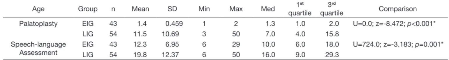
To conirm the age difference at the time of primary
palatoplasty, the median age of the participants in both groups
was compared at the times of surgery and of speech-language assessment. In both comparisons, participants in the LIG presented higher median age, with statistically signiicant
difference.
No statistically signiicant difference was observed between the groups with respect to participants’ gender (Table 3).
No statistically signiicant difference was found between the groups regarding the type of cleft, with a larger number of
cases of cleft lip and palate in both groups (Table 4).
Table 1. Inter-rater reliability analysis – Kappa coefficient
Variable K p
Kappa confidence
interval Hypernasality 1.00 <0.001 0.76-1.0 Severity Rating 0.730 <0.001 0.56-0.90
Hyponasality 1.00 <0.001 0.76-1.0 Severity Rating 0.573 <0.001 0.41-0.74 Speech understandability 1.00 <0.001 0.76-1.0
Rating 0.909 <0.001 0.75-1.0
Speech characterization of participants - general and group analysis
As for the parameter resonance classiication in both groups (EIG and LIG), Table 5 shows that most participants presented inadequate results. Language assessment related to speech
production did not present statistical difference between the groups for resonance classiication, but the p-value was marginal (close to 0.05), which can be interpreted as a trend in Biostatistics. Considering this trend, it is possible to state that the LIG would have more participants classiied with inadequate resonance.
Regarding the analysis for resonance alteration in both
groups, a larger number of participants with hypernasality are
observed (Table 6). As for hyponasality, it was present at mild
severity in only four participants of the LIG.
No difference between the groups was found with respect to the presence or absence of hypernasality, but when the frequency distribution for each severity level was veriied, the EIG presented a higher percentage of participants with mild level compared with that of the LIG, whereas the LIG showed a higher percentage of participants with severe level than that of the EIG (Table 7).
No differences between the groups were observed for the parameters nasal turbulence, weak intraoral air pressure, and
audible nasal air emission (Table 8). With respect to speech
understandability, there was no statistically signiicant difference
Table 2. Age of participants in the early (EIG) and late (LIG) intervention groups at the time of palatoplasty and speech-language assessment
Age Group n Mean SD Min Max Med 1
st
quartile 3rd
quartile Comparison Palatoplasty EIG 43 1.4 0.459 1 2 1.3 1.0 2.0 U=0.0; z=-8.472; p<0.001*
LIG 54 11.5 10.69 3 50 7.0 4.0 15.8
Speech-language Assessment
EIG 43 12.3 6.95 6 29 10.0 6.0 18.0 U=724.0; z=-3.183; p=0.001* LIG 54 19.8 12.37 6 50 16.0 9.0 29.3
*statistical significance (p<0.05)
Caption: n = number of participants; min = minimum age; max = maximum age; med = median; SD = standard deviation; EIG = early intervention group; LIG = late intervention group
Table 3. Gender comparison between participants in each group
Group Gender Total Statistics
Female Male
EIG 19 24 43 X2=0.867; df=1; p=0.352*
LIG 29 25 54
*statistical significance (p<0.05) - Chi-square test
Caption: EIG = early intervention group; LIG = late intervention group
Table 4. Comparison of type of cleft between the groups
Type of cleft Group Total Statistics
EIG LIG
Cleft lip and palate 30 31 61 X2=1.567; df=1 p=0.211*
Cleft palate 13 23 36
*statistical significance (p<0.05) - Chi-square test
Caption: EIG = early intervention group; LIG = late intervention group
Table 5. Resonance classification Resonance
classification
EIG LIG
Total Statistics
Frequency % Frequency %
Adequate 13 30.2 8 14.8 21 X2=3.355; df=1 p=0.067*
Inadequate 30 69.8 46 85.2 76
*statistical significance (p<0.05) - Chi-square test
Caption: EIG = early intervention group; LIG = late intervention group
Table 6. Characterization of hypernasality Resonance
alteration Classification
EIG LIG
Total Statistics
Frequency % Frequency %
Hypernasality Absent 12 27.9 8 14.8 20 X2=2.507; df=1 p=0.113
Present 31 72.1 46 85.2 77
Hyponasality Absent 43 100.00 50 92.6 93 p=0.127
Present (mild) 0 0.0 4 7.4 4
between the groups, with most participants presenting inadequate
speech (Table 9) regardless of age at the time of palatoplasty.
However, high severity ratings are observed in the LIG when analyzing the data according to speech understandability level
(Table 10), with the EIG presenting a higher percentage of
participants with mild level than that of the LIG and the LIG showing a higher percentage of participants with severe level compared with that of the EIG.
Table 11 presents the general characterization of the participants regarding compensatory articulation disorders.
Higher occurrence of participants with inadequate resonance was
observed in the LIG, with glottal stop and pharyngeal fricative
as the most common CADs (Table 12). In the LIG, glottal stop
also occurred with greater frequency among the other CADs.
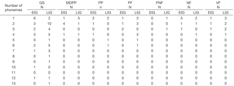
Table 13 shows the number of phonemes with identiication of CADs according to the groups. As for glottal stop in the
LIG, the number of altered phonemes in the participants’ speech ranged from one to 12, which indicates variability of the participants’ proile. In the LIG, also for glottal stop, the number of altered phonemes varied from one to 13. Statistically signiicant difference was observed between the groups regarding
the number of altered phonemes (Table 14).
Table 7. Hypernasality rating
Severity rating EIG LIG
Frequency % Frequency %
Mild 15 48.4 15 32.6
Moderate 13 41.9 20 43.5
Severe 3 9.7 11 23.9
Caption: EIG = early intervention group; LIG = late intervention group
Table 8. Presence of nasal turbulence
Variable Classification EIG LIG Total Statistics
Frequency % Frequency %
Nasal turbulence Absent 37 86.0 51 94.4 88 p=0.179
Present 6 14.0 3 5.6 9
Weak intraoral air pressure Absent 30 69.8 30 55.6 60 X2=2.049; df=1; p=0.152
Present 13 30.2 24 44.4 37
Audible nasal air emission Absent 33 76.7 37 68.5 70 X2=0.806; df=1; p=0.369
Present 10 23.3 17 31.5 27
Caption: EIG = early intervention group; LIG = late intervention group
Table 9. Classification of speech understandability Speech
understandability classification
EIG LIG
Total Statistics
Frequency % Frequency %
Adequate 13 30.2 9 16.7 22 X2=2.512; df=1; p=0.113
Inadequate 30 69.8 45 83.3 75
Total 43 100.0 54 100.0 97
Caption: EIG = early intervention group; LIG = late intervention group
Table 10. Speech understandability rating
Severity rating EIG LIG
Frequency % Frequency %
Mild 17 56.7 11 24.4
Moderate 9 30.0 21 46.7
Severe 4 13.3 13 28.9
Total 30 100.0 45 100.0
Caption: EIG = early intervention group; LIG = late intervention group
Table 11. Classification of the compensatory articulation disorders (CAD)
CADs EIG LIG Total
Frequency % Frequency %
Present 24 55.8 41 75.9 65
Absent 19 44.2 13 24.1 32
Total 43 100.0 54 100.0 97
DISCUSSION
In the Early Intervention Group (EIG), the mean age of participants at the time of palatoplasty was 1.4 years. As reported in previous studies(5,7,10,11), this age would be compatible with
that recommended by the national and international protocols,
which afirm that when intervention is performed at this age the chances of individuals presenting speech alterations as a result
of palate cleft are reduced.
The speciic scientiic literature clearly establishes that palate
cleft surgical repair should be conducted early, before the age
of two(5,11), especially with regards to speech(5).
In the Late Intervention Group (LIG), the mean age of participants at the time of primary palatoplasty was 11.5 years.
Studies(16,25) have shown that, in developing countries, lack of
information and dificulty in accessing the health system are the main indicators of late primary intervention, and this a characteristic of the population cared at the HCFMUSP, which
often does not have access to this service according to the age
recommended by the protocols of the area.
The indings of this study are in agreement with those described
in the literature(12) regarding gender, with predominance of males
over females(12,15), although without statistically signiicant results.
With respect to type of cleft, only patients with cleft lip and palate and/or cleft palate were included in this study; those with cleft lip only were excluded. Consequently, comparison with the epidemiological data veriied in the course of this study is impossible for the population investigated.
Regarding the characterization of speech in general, the study identiied a larger number of participants in both groups with
alterations in the parameters hypernasality, speech understandability, and presence of CADs. A smaller but considerable number of
changes were observed for the parameters nasal turbulence, weak intraoral air pressure, and audible nasal air emission.
According to the results obtained in the present study, it is
possible to observe that, even without statistical difference, the data suggest worse performance of the participants who Table 14. Altered phonemes with occurrence of compensatory articulation disorders (CAD)
Group N Mean SD Min Max Med 1st quartile 3rd quartil Comparison
EIG 43 2.6 3.07 0 12 2.0 0.0 4.0 U=847.5;
z=-2.323; p=0.020*
LIG 54 4.4 3.92 0 14 4.0 0.8 7.3
*statistical significance (p<0.05)
Caption: n = number of participants; min = minimum age; max = maximum age; med = median; SD = standard deviation; EIG = early intervention group; LIG = late intervention group
Table 12. Characterization of the compensatory articulation disorders (CAD)
Group
Types of CADs GS
n
MDPP n
PP n
PF N
PNF N
NF N
VF n
EIG 19 6 4 4 0 5 3
LIG 35 5 4 11 2 4 8
Caption: n= number of individuals; CADs = compensatory articulation disorders; GS = glottal stop; MDPP = mid-dorsum palatal plosive; PP = pharyngeal plosive; PF = pharyngeal fricative; PNF = posterior nasal fricative; NF = nasal fricative; VF = velar fricative
Table 13. Number of phonemes with identification of compensatory articulation disorders (CAD) according to the groups (n= 26)
Number of phonemes
GS N
MDPP N
PP n
PF N
PNF N
NF N
VF N EIG LIG EIG LIG EIG LIG EIG LIG EIG LIG EIG LIG EIG LIG
1 6 2 1 3 2 2 1 3 0 1 3 2 1 3
2 3 10 4 1 1 0 1 3 0 0 1 1 1 2
3 2 4 0 0 0 0 2 0 0 1 1 0 1 2
4 3 5 1 1 1 0 0 3 0 0 0 1 0 1
5 0 1 0 0 0 1 0 1 0 0 0 0 0 0
6 2 5 0 0 0 1 0 1 0 0 0 0 0 0
7 1 3 0 0 0 0 0 0 0 0 0 0 0 0
8 0 1 0 0 0 0 0 0 0 0 0 0 0 0
9 0 1 0 0 0 0 0 0 0 0 0 0 0 0
10 1 0 0 0 0 0 0 0 0 0 0 0 0 0
11 0 0 0 0 0 0 0 0 0 0 0 0 0 0
12 1 1 0 0 0 0 0 0 0 0 0 0 0 0
13 0 1 0 0 0 0 0 0 0 0 0 0 0 0
underwent late cleft repair. Clear difference between the groups was observed regarding the presence of CADs, with a trend for difference in hypernasality as well, as already mentioned
in the literature(6,7,9,21). These differences may be related to the
dificulty in velopharyngeal closure, which often occurs in late
palatoplasty(5,6,11).
It is worth noting that this study could not control the type of surgery and the number of surgeons involved in them because of the nature of the patients’ admission to the service.
As for resonance classiication, a more severe rating of hypernasality was observed in the cases of late cleft repair. This aspect would be justiied by the fact that palate closure occurs at a more advanced age, at which the altered speech model would already be established, as well as greater chances of unfavorable anatomical characteristics(6,13,26). It is also possible
to correlate this relationship with an idea widely advocated in
the literature(7,9), that on the importance of conducting early
palatoplasty. Concerning hyponasality, it was observed in only four participants in the present study, which conirms
hypernasality as the most commonly present resonance alteration in the speech of cleft patients(14).
Other characteristics of speech, which are mentioned in the
literature as less common(14,17), were also identiied similarly
in this study. Both groups presented low occurrence of nasal
turbulence and, clinically, the literature describes it as a
seldom-observed parameter(6). Weak intraoral pressure is also not
the most commonly observed change in clinical assessment, and when it occurs, it is generally related to plosive phonemes(2,6).
In this study, its occurrence was relative, but did not reach 50%
of the sample in either group. This result can be correlated to
the high presence of CADs in the study population. Signiicant audible nasal air emission was observed in less than 40% of the participants, with no statistically signiicant difference between
the groups. It is also not considered a common disorder in the speech of cleft patients(6).
With respect to the CADs, higher occurrence was found in the LIG. This result corroborates the indings in the literature(7,9),
and for such changes in speech, speech-language therapy is the most indicated conduct(7,9,25). According to the literature(6-9),
when individuals cannot produce a phoneme at the appropriate
articulation place, they compensate to emit it as closely as possible to the correct production. According to Perry and Kuehn(9), the occurrence of CADs justiies speech-language
therapy, and the cases without velopharyngeal insuficiency (VFI) justify periodic speech-language pathology monitoring in order to avoid speech and articulation alterations. The CADs, along with hypernasality, are the most common changes in the speech of patients with palate cleft(14,18).
Speech understandability inadequacy is another commonly present alteration in cleft patients(5,6,11). Thus, as in the literature,
high occurrence of inadequacy was observed in the participants of this study, with greater severity in the LIG. According to previous studies(14,18), speech understandability inadequacy may
also be associated with the presence of CADs, corroborating the indings of this study.
One of the factors associated with the larger number of
late palatoplasty in the population cared at the HCFMUSP is
the lack of access to information and reference centers for the
treatment of cleft patients(17). The literature(2,15-17) describes this
dificulty and mentions that, in developing countries, there is also the dificulty of registering this population. These aspects are associated with a delay in seeking treatment. Many Brazilian centers have the same proile as the HCFMUSP, admitting patients with lack of information on cleft palate and its genetic
relationships and on the necessary treatments. International reference centers(15,16,26) suggest some proposals to minimize
this problem, such as population awareness of the risk factors and occurrence of cleft palate and the use of standardized
treatment protocols(15,27). A domestic study(28) compared the
results of a health service at two different moments, and reached the conclusion that standardization of care for cleft patients by applying protocols showed improvement in the results found
in short time.
It is possible to observe a signiicant difference in access to information, treatment, and adherence in developed countries.
According to the literature, in countries such as England(12) and
the USA(29), primary palatoplasty occurs up to 1 year of age,
minimizing the effects of cleft palate on speech. In the Brazilian service, many patients only have access to it as adults.
Standardized, controlled and randomized studies on the speech aspects of individuals with cleft lip and palate are scarce. Predictive values of prevalence and incidence of speech disorders are not identiied in the literature, and the results found are variable. With this study, we expect to obtain material for the formulation of further better-structured studies with greater scientiic evidence. It will also allow discussion of clinical protocols, which will directly assist the study population. The determination of evidence-based clinical guidelines and of service indicators would be the high point of speech-language
pathology practice after the compilation of these initial data.
It is believed that determination of the proile of the patients cared at the referred hospital will help relect on speech-language pathology assistance, as well as on the discussion about the best
therapeutics, uni- or multi-disciplinary, that could be offered
to cleft patients. Determination of the proile of the population cared at the HCFMUSP may assist with the organization of the speech-language pathology care provided, contributing to the organization and planning of a service based on evidence and on
the assumptions of the concept of health technology assessment.
CONCLUSION
Based on what has been presented, it was possible to characterize the general proile and speech characteristics of
patients undergoing primary palatoplasty in the aforementioned
university hospital (HCFMUSP) - a national referral center for
treatment of cleft lip and palate located in the city of Sao Paulo.
the early (EIG) and late (LIG) intervention groups. The variable
resonance presented marginal statistical results, indicating a trend in differentiating the groups also in relation to this alteration.
In conclusion, patients submitted to early primary palatoplasty
present better speech proile.
REFERENCES
1. Freitas JAS, Neves LT, Almeida ALPF, Garib DG, Trindade-Suedam IK, Yaedú RYF, et al. Rehabilitative treatment of cleft lip and palate: experience of the Hospital for Rehabilitation of Craniofacial Anomalies/ USP (HRAC/USP) Part 1: overall aspects. J Appl Oral Sci. 2012;20(1):9-15. PMid:22437671. http://dx.doi.org/10.1590/S1678-77572012000100003. 2. Souza J, Raskin S. Estudo clínico e epidemiológico de fissuras orofaciais. J Pediatr. 2013;89(2):137-44. PMid:23642423. http://dx.doi.org/10.1016/j. jped.2013.03.010.
3. Cox TC. Taking it to the max: the genetic and developmental mechanisms coordinating midfacial morphogenesis and dysmorphology. Clin Genet. 2004;65(3):163-76. PMid:14756664. http://dx.doi.org/10.1111/j.0009-9163.2004.00225.x.
4. Nahai FR, Williams JK, Burstein FD, Martin J, Thomas J. The management of cleft lip and palate: pathways for treatment and longitudinal assessment. Semin Plast Surg. 2005;19(4):275-85. http://dx.doi.org/10.1055/s-2005-925900.
5. Rohrich RJ, Love EJ, Byrd HS, Johns DF. Optimal timing of cleft palate closure. Plast Reconstr Surg. 2000;106(2):413-21, quiz 422, discussion 423-5. PMid:10946942. http://dx.doi.org/10.1097/00006534-200008000-00026.
6. Palandi BBB, Guedes ZCF. Aspecto de fala de indivíduos com fissura palatina e labial, corrigida em diferentes idades. Rev CEFAC. 2011;13(1):8-16. http://dx.doi.org/10.1590/S1516-18462011005000012.
7. Abdel-Aziz M. Speech outcome after early repair of cleft soft palate using Furlow technique. Int J Pediatr Otorhinolaryngol. 2013;77(1):85-8. PMid:23116906. http://dx.doi.org/10.1016/j.ijporl.2012.09.038. 8. Peterson-Falzone SJ, Trost-Cardamone JE, Karnell MP, Hardin-Jones M.
The clinican’s guide to treating cleft palate speech. St. Louis: Mosby; 2006. Effects of cleft and non-cleft VPI on speech in older children. p. 17-39.
9. Perry JL, Kuehn DP. Magnetic resonance imaging and computer reconstruction of the velopharyngeal mechanism. J Craniofac Surg. 2009;20(2, Suppl 2):1739-46. PMid:19816343. http://dx.doi.org/10.1097/ SCS.0b013e3181b5cf46.
10. Hortis-Dzierzbicka M, Radkowska E, Fudalej P. Speech outcomes in 10-year-old children with complete unilateral cleft lip and palate after one-stage lip and palate repair in the first year of life. J Plast Reconstr Aesthet Surg. 2012;65(2):175-81. PMid:21978731. http://dx.doi.org/10.1016/j. bjps.2011.09.015.
11. Chapman KL, Hardin-Jones MA, Goldstein JA, Halter KA, Havlik RJ, Schulte J. Timing of palatal surgery and speech outcome. Cleft Palate Craniofac J. 2008;45(3):297-308. PMid:18452355. http://dx.doi.org/10.1597/06-244. 12. Fitzsimons KJ, Copley LP, Deacon SA, van der Meulen JA. Hospital care of children with a cleft in England. Arch Dis Child. 2013;98(12):970-4. PMid:23968774. http://dx.doi.org/10.1136/archdischild-2013-304271.
13. Perry JL, Kuehn DP. Magnetic resonance imaging and computer reconstruction of the velopharyngeal mechanism. J Craniofac Surg. 2009;20(2, Suppl 2):1739-46. PMid:19816343. http://dx.doi.org/10.1097/ SCS.0b013e3181b5cf46.
14. Lima MRF, Leal FB, Araújo SVS, Matos EF, Di Ninno CQMS, Britto ATBO. Atendimento fonoaudiológico intensivo em pacientes operados de fissura labiopalatina: relato de casos. Rev Soc Bras Fonoaudiol. 2007;12(3):240-6. http://dx.doi.org/10.1590/S1516-80342007000300012.
15. Shapira Y, Haklai Z, Blum I, Shpack N, Amitai Y. Prevalence of non-syndromic orofacial clefts among jews and arabs, by type, site, gender and geography: a multi-center study in Israel. Isr Med Assoc J. 2014;16(2):759-63. PMid:25630204.
16. Ajike SO, Adebola RA, Efunkoya A, Adeoye J, Akitoye O, Veror N. Epidemiology of adult cleft patients in North-western Nigeria: our experience. Ann Afr Med. 2013;12(1):11-5. PMid:23480989. http://dx.doi. org/10.4103/1596-3519.108243.
17. Penido FA, Noronha RMS, Caetano KI, Jesus MSV, Di Ninno CQMS, Britto ATBO. Correlação entre os achados do teste de emissão de ar nasal e da nasofaringoscopia em pacientes com fissura labiopalatina operada. Rev Soc Bras Fonoaudiol. 2007;12(2):126-34. http://dx.doi.org/10.1590/ S1516-80342007000200010.
18. Prandini EL, Pegoraro-Krook MI, Dutka JCR, Marino VCC. Occurrence of consonant production errors in liquid phonemes in children with operated cleft lip and palate. J Appl Oral Sci. 2011;19(6):579-85. PMid:22230991. http://dx.doi.org/10.1590/S1678-77572011000600007.
19. Schuster M, Maier A, Bocklet T, Nkenke E, Holst A, Eysholdt U, et al. Automatically evaluated degree of intelligibility of children with different cleft type from preschool and elementary school measured by automatic speech recognition. Int J Pediatr Otorhinolaryngol. 2012;76(3):362-9. PMid:22236457. http://dx.doi.org/10.1016/j.ijporl.2011.12.010.
20. Kuehn D, Moller KT. Speech and language issues in the cleft palate population: the state of the art. Cleft Palate Craniofac J. 2000;37(4):348. http://dx.doi.org/10.1597/1545-1569(2000)037<0348:SALIIT>2.3.CO;2. 21. Mituuti CT, Piazentin-Penna SHA, Brandão GR, Bento-Gonçalves CGA. Caracterização da fala de indivíduos submetidos à palatoplastia primária. Rev Soc Bras Fonoaudiol. 2010;15(3):355-61. http://dx.doi.org/10.1590/ S1516-80342010000300008.
22. Andrade CRF, Befi-Lopes DM, Fernandes FDM, Wertzner HF. ABFW Teste de Linguagem Infantil nas áreas de fonologia, vocabulário, fluência e pragmática. São Paulo: Ed. Pró Fono; 2004.
23. Henningsson G, Kuehn DP, Sell D, Sweeney T, Trost-Cardamone JE, Whitehill TL. Universal parameters for reporting speech outcomes in individuals with cleft palate. Cleft Palate Craniofac J. 2008;45(1):1-17. PMid:18215095. http://dx.doi.org/10.1597/06-086.1.
24. Trindade IEK, Genaro KF, Yamashita RP, Miguel HC, Fukushiro AP. Proposal for velopharyngeal function rating in a speech perceptual assessment. Pro Fono. 2005;17(2):259-62. PMid:16909536. http://dx.doi. org/10.1590/S0104-56872005000200015.
26. Vogel AP, Ibrahim HM, Reilly S, Kilpatrick N. A comparative study of two acoustic measures of hipernasality. J Speech Lang Hear Res. 2009;52(6):1640-51. PMid:19951929. http://dx.doi.org/10.1044/1092-4388(2009/08-0161).
27. Owotade FJ, Ogundipe OK, Ugboko VI, Okoje VN, Olasoji HO, Makinde ON, et al. Awareness, knowledge and attitude on cleft lip and palate among antenatal clinic attendees of tertiary hospitals in Nigeria. Niger J Clin Pract. 2014;17(1):6-9. PMid:24326798. http://dx.doi.org/10.4103/1119-3077.122822.
28. Alonso N, Tanikawa DYS, Lima JE Jr, Ferreira MC. Avaliação comparativa e evolutiva dos protocolos de atendimento dos pacientes fissurados.
Rev Bras Cir Plast. 2010;25(3):434-8. http://dx.doi.org/10.1590/S1983-51752010000300006.
29. Basseri B, Kianmahd BD, Roostaeian J, Kohan E, Wasson KL, Basseri RB, et al. Current national incidence, trends, and health care resource utilization of cleft lip-cleft palate. Plast Reconstr Surg. 2011;127(3):1255-62. PMid:21364426. http://dx.doi.org/10.1097/PRS.0b013e3182043af6.
Author contributions