*e-mail: [email protected]
Microhardness and EBSD Microstructure Mapping
in Partially-pressed Al and Cu through 90º ECAP Die
Alexander P. Zhilyaeva,b*, Terence G. Langdona,c
aMaterials Research Group, Faculty of Engineering and the Environment,
University of Southampton, Southampton SO17 1BJ, UK
bInstitute for Metals Superplasticity Problems, Russian Academy of Science,
39 Khalturina, Ufa, 450001, Russia
cDepartments of Aerospace & Mechanical Engineering and Materials Science,
University of Southern California, Los Angeles, CA 90089-1453, USA
Received: September 27, 2012; Revised: November 14, 2012
Equal-channel angular pressing (ECAP) is widely recognized as an effective method for processing ultrafine-grained and even nanostructured materials. Important details on processes occurring in the die intersections can be obtained by mapping the microhardness and EBSD microstructures in partially-pressed aluminum and copper through the 90° die of ECAP. Precise measurements were made using grids of partially-pressed Al and Cu and detailed color maps were plotted and compared with EBSD maps. A narrow region along the bisector of two channels reflects altering in microhardness level of the material subjected to simple shear. The microstructural evolution suggests significant refining of the grain structure but there is noticeable inhomogeneity in the microhardness and microstructure for both materials. Factors contributing to the inhomogeneous hardness distribution include the coarse initial grain size, and inhomogeneous deformation across the plane of the die channel intersection due to die wall friction and the formation of a dead zone at the outer corner.
Keywords: equal channel angular pressing, microstructure, EBSD, aluminium, copper
1. Introduction
Refinement and homogenization of microstructure are highly beneficial to the mechanical properties of engineering materials. Conventional thermomechanical treatments for this purpose typically include deformation processing to von Mises equivalent strains ≤5 but recent severe plastic deformation (SPD) processing methods1 have enabled
systematic investigations of equivalent strains >10. Among many SPD techniques, equal-channel angular pressing (ECAP)2 and high-pressure torsion (HPT)3 have been
widely used for producing bulk metallic materials that have ultra-fine grain (sometimes nanograin) size and enhanced strength4. Pure metals such as copper and aluminium can
be processed by ECAP at room temperature5-9 and they can
serve as ideal model metals for studying microstructure evolution at very early stage of deformation.
In the context of SPD processing, “refinement” usually refers to the production of equi-axed ultrafine (≤1 µm) or nanoscale (≤100 nm) microstructure features throughout the bulk material. Elongated, ribbon-like dislocation cells or subgrain structures form at smaller equivalent strains during SPD at low homologous temperatures and then evolve into highly refined equi-axed structures at larger strains. These structures apparently develop by recovery-dominated processes in the absence of high-angle boundary migration, and their evolution is also strain-path dependent.
Interfaces in such structures are often high-angle in nature (disorientations ≥15°) but are not disordered high-angle boundaries. Predictive models of the mechanisms by which SPD-induced microstructures evolve and transform into ultrafine grain structures with disordered high-angle boundaries have not been established. Indeed, the current understanding of microstructure control during conventional deformation processing and recrystallization treatments is largely empirical in nature and therefore our ability to design processes to achieve particular microstructure outcomes is limited.
Advances in the characterization and understanding of such microstructures have involved grain-specific orientation determinations utilizing convergent beam electron diffraction methods in transmission electron microscopy. These methods have high spatial and orientation resolution, but provide limited amounts of data. In contrast, electron backscatter diffraction analysis (EBSD) and orientation imaging microscopy (OIM)10,11 methods have
2. Experimental Material and Procedures
Aluminium (99.7%) and copper (99.9%) plates were machined to provide 8 × 8 × 50 mm3 billets for pressing in
a 90º die. Prior to ECAP, the billets were annealed. From conventional optical microscopy, the initial recrystallized grain size was ~300 µm for aluminium12 and ~100 µm
for copper. Processing by ECAP of the square billets was conducted at room temperature using a die with an angle,
Φ, between channels of 90º and an outer angle of 20°: further details can be found elsewhere13. After pressing
approximately one-half of the billet length, the process was stopped and specimen was extracted from the die. All specimens had L-shape and they were used for scanning electron microscopy (SEM), microhardness measurements and EBSD analysis. Figure 1a shows a montage of SEM images of partially-pressed Cu billet.
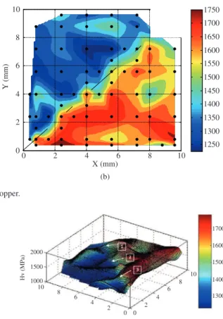
The Vickers microhardness, Hv, was measured on polished mirror-like billet surfaces using a standard microhardness tester equipped with a Vickers indenter. The hardness measurements used a load of 100 gf and dwell times of 10 s. These measurements were taken in a grid pattern on the L-shaped specimens with special attention to the intersection of the channels where the shear is localized. As can be seen from Figure 1b, microhardness values were recorded using a finer mesh near the intersection line. In Figure 1b, each dot of the mesh corresponds to 5 Hv measurements.
Samples for EBSD (OIM) were mechanically polished and then electropolished in a Buehler Electromet 4 apparatus using a 20% perchloric acid –80% ethanol electrolyte cooled to −25 °C. The OIM utilized a Topcon S510 scanning electron microscope operating with a tungsten filament. The minimum step size of 0.05 µm was no more than about 0.1 of the mean (sub)grain size in these materials. Samples were examined along the intersection line of the ECAP channels. The OIM scans were performed approximately on the left and right side of the intersection for Al billet and near the intersection for the copper billet: some additional details are provided elsewhere14. The symmetry of an ECAP billet is monoclinic
and therefore the senses of the coordinate axes in the flow plane were carefully ascertained. The OIM study involved standard clean-up procedures15, as follows: (i) grain dilatation
with a grain tolerance angle (GTA) of 5°; a minimum grain size (MGS) of two pixels; (ii) grain confidence index (CI)
standardization with GTA = 5° and MGS = 2; (iii) neighbour CI correlation with minimum CI of 0.1.
3. Experimental Results and Discussion
3.1.
Microstructure and microhardness evolution
in partially-pressured copper
Figure 1 shows a montage of SEM images (Figure 1a) and color-coded map of microhardness, Hv of partially-pressed copper (Figure 1b). The low magnification of the SEM
Figure 1. SEM montage (a) and Hv map (b) of partially-pressed copper.
Figure 3. Grain size distribution of ECAP copper along the intersection shown in Figure 2.
micrograph in Figure 1a reveals two clearly distinguished areas: a non-deformed area prior to the channel intersection and a sheared area where the copper has moved beyond the intersection. The microstructure prior to compression is not modified significantly and undistorted twins indicated by arrows are clearly observed (Figure 1a). After shearing, the microstructure has a typical distorted structure2. Figure 1b
shows a microhardness color-coded map of Figure 1a at the same magnification and it reveals some important details of the shearing process occurring during the first pass of the copper: (i) shearing is heterogeneous along the intersection; (ii) it appears that lower part of the billet was sheared to a
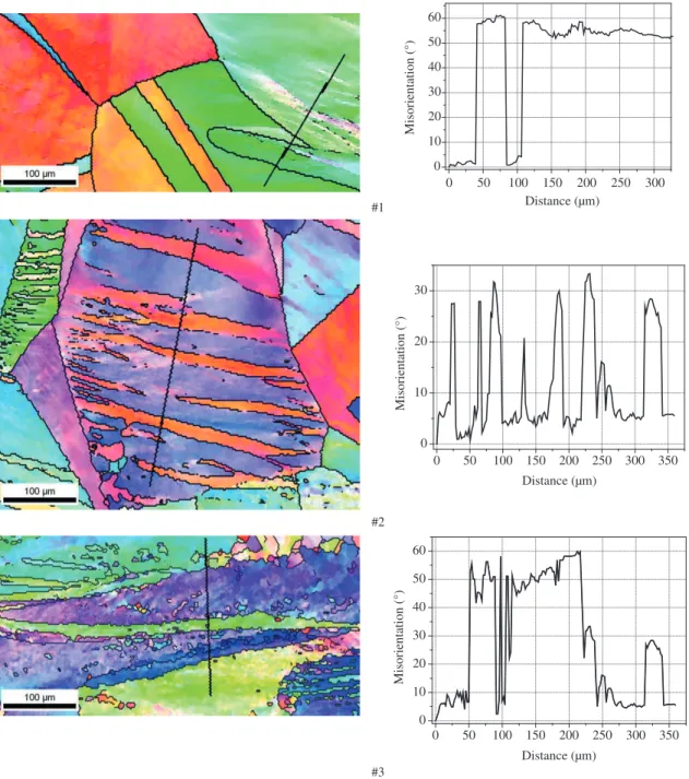
Figure 4. IPF maps taken along the intersection at positions indicated in Figure 2 and corresponding point-to-origin misorientation profiles.
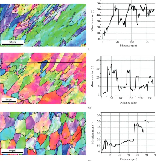
Figure 7. IPF maps of partially-pressed aluminum taken along the intersection at positions indicated in Figure 5 and corresponding point-to-origin misorientation profiles.
higher strain than the upper part; (iii) there is some symmetry in high and low microhardness relative to the intersection as a mirror plane. Figure 2 is a three dimensional representation of Figure 1b and it clearly demonstrates all features of the deformation map including heterogeneity of the shear deformation and symmetry with respect to the intersection. Along the shearing line detailed EBSD mapping was performed and the area averaged grain sizes were plotted in Figure 3 as a function of the distance from the inner corner of the ECAP die. The mean grain size follows a tendency for Hv oscillation along the channel intersection. The most refined region lies in the central part of the ECAP billet.
Representative inverse pole figure (IPF) maps, numbered and indicated by arrows in Figure 2, are shown in Figure 4. The IPF map #1 corresponds to the region close to the inner corner of the ECAP die. It shows almost a non-deformed structure with large grains separated by twins (indicated
intersection and shows significant refinement with some elongated grains of 25 µm in width. The misorientation profile suggests that inside the prior grains there develop subgrains separated by low-angle boundaries (~10°). The IPF map #3 representing the lower part of the shearing region close to the outer angle shows a significantly equiaxed microstructure with grains separated by mostly high-angle boundaries.
4. Summary and Conclusions
• TheapplicationofEBSDandOIMmethodstothe characterization of SPD-induced microstructures has provided valuable insight into the evolution of microstructure during processing by various SPD methods;
• The inhomogeneous distribution of deformation during initial ECAP passes for Cu and Al has been characterized by OIM and microhardness data. The development of elongated structures and the fragmentation of such structures to form highly refined equiaxed structures at very large strains was also investigated by these methods;
• It is concluded that aluminium possesses a more homogeneous sheared microstructure during first pass of ECAP by comparison to copper.
Acknowledgements
Participation at NANOMAT-2012 was made possible through award FAPESP#2011/51245-8 under a cooperation agreement with the University of Southampton. This work was supported by the European Research Council under ERC Grant Agreement No. 267464-SPDMETALS. The authors acknowledge Prof. Terry McNelley (Naval Postgarduate School, CA, USA) for fruitful discussion of results and Dr. G.I. Raab for assistance in sample processing.
by thick black lines). However, the right bottom corner possesses a quite typical sheared area with high density of the low-angle boundaries indicated by thin black lines. The corresponding point-to-origin misorientation profile (shown in the right column) supports these findings. The IPF map #2 corresponds to the middle region of the channel intersection and reflects intensive shearing process that has occurred. It is evident that the large prior grain is split by new shear bands which are separated by high-angle (>15°) boundaries. The misorientation profile clearly demonstrates an oscillation in misorientation angle up to 33 degrees. The IPF map #3 refers to the region close to the outer corner and reflects a shearing along this relief arc. The deformation in this region consists of a combination of shearing and rotation modes and it leads to a quite complex microstructure with a combination of subgrains and curvilinear shear bands.
3.2.
Microstructure and microhardness evolution
in partially-pressured aluminium
Figure 5 is a three dimensional representation of the Hv map plotted using microhardness measurements in the partially-pressed Al billet. It shows clearly a different behaviour compared to the ECAP Cu features. Specifically, the microhardness distribution is less heterogeneous but there remains symmetry with respect to the intersection and the transition line between the non-sheared and the sheared regions is rather straight. The EBSD maps were acquired along the intersection on the left and right side and grain sizes plotted in Figure 6 reflect the oscillating nature of microstructural evolution along the line of the channel intersection.
The IPF maps representing typical microstructures in the area indicated by labels 1-3 in Figure 5 are shown in Figure 7. The IPF map #1 shows microstructure evolution close to the inner corner and it is evident that shearing takes place in this region. The misorientation profile demonstrates a developed refined structure with grains and subgrains. The IPF map #2 corresponds to the middle part of the channel
References
1. Valiev RZ, Islamgaliev RK and Alexandrov IV. Bulk nanostructured materials from severe plastic deformation. Progress in Materials Science. 2000; 45(2):103-189. http:// dx.doi.org/10.1016/S0079-6425(99)00007-9
2. Valiev RZ and Langdon TG. Principles of equal-channel angular pressing as a processing tool for grain refinement. Progress in Materials Science. 2006; 51(7):881-981. http:// dx.doi.org/10.1016/j.pmatsci.2006.02.003
3. Zhilyaev AP and Langdon TG. Using high-pressure torsion for metal processing: Fundamentals and applications. Progress in Materials Science. 2008; 53(6):893-979. http://dx.doi. org/10.1016/j.pmatsci.2008.03.002
4. Valiev RZ, Sabirov I, Zhilyaev AP and Langdon TG. Bulk Nanostructured Metals for Innovative Applications. JOM. 2012; 64(10):1134-1142. http://dx.doi.org/10.1007/ s11837-012-0427-9
5. Komura S, Horita Z, Nemoto M and Langdon TG. Influence of stacking fault energy on microstructural development in equal-channel angular pressing. Journal of Materials Research. 1999; 14:4044-4050. http://dx.doi.org/10.1557/ JMR.1999.0546
6. Mishra A, Richard V, Gregori F, Asaro RJ and Meyers MA. Microstructural evolution in copper processed by severe plastic deformation. Materials Science and Engineering A. 2005; 410-411:290-298. http://dx.doi.org/10.1016/j.msea.2005.08.201
7. Iwahashi Y, Horita Z, Nemoto M and Langdon TG. The process of grain refinement in equal-channel angular pressing. Acta Materialia. 1998; 46(9):3317-3331. http://dx.doi.org/10.1016/ S1359-6454(97)00494-1
Materials Science and Engineering A. 2006; 429(1-2):137-148. http://dx.doi.org/10.1016/j.msea.2006.05.009
13. Zhilyaev AP, Oh-ishi K, Langdon TG and McNelley TR. Influence of ECAP processing parameters on texture and microstructure of commercially pure aluminum. Materials Science and Engineering A. 2006; 441(1-2):245-252. http:// dx.doi.org/10.1016/j.msea.2006.08.029
14. McNelley TR, Zhilyaev AP, Swaminathan S, Su J and Menon ES. Application of EBSD Methods to Severe Plastic Deformation (SPD) and Related Processing Methods. In: Schwartz AJ, Kumar M, Adams BL, Field DP, editors. Electron Backscatter Diffraction in Materials Science. Springer; 2009. chap. 20, p. 277-288. Pergamon Materials Series.
15. OIM. User’s Manual. EDAX-TSL. E. Mahway; 2005. 9. Lugo N, Llorca N, Cabrera JM and Horita Z.Microstructures
and mechanical properties of pure copper deformed severely by equal-channel angular pressing and high pressure torsion. Materials Science and Engineering A. 2008; 477(1-2):366-371. http://dx.doi.org/10.1016/j.msea.2007.05.083
10. Adams BL, Wright SI and Kunze K. Orientation imaging: the emergence of a new microscopy. Metallurgical and Materials Transactions A. 1993; 24(4):819-831. http://dx.doi. org/10.1007/BF02656503
11. Randle V and Engler O. Texture analysis: macrotexture, microtexture & orientation mapping. Amsterdam: Gordon and Breach; 2000.