nucleossomo
BRASÍLIA-DF 2020
Estudos biofísicos e in vivo de moléculas ligantes de nucleossomo
KAIAN AMORIM TELES
BRASÍLIA-DF 2020
Estudos biofísicos e in vivo de moléculas ligantes de nucleossomo
Tese de doutorado apresentada à Universidade de Brasília como parte das exigências do Programa de Pós-Graduação em Patologia Molecular, para a obtenção do título de Doutor.
Prof. Dr. Guilherme Martins Santos
BRASÍLIA-DF 2020
Estudos biofísicos e in vivo de moléculas ligantes de nucleossomo
Tese de doutorado apresentada à Universidade de Brasília como parte das exigências do Programa de Pós-Graduação em Patologia Molecular, para a obtenção do título de Doutor.
Aprovada em de
Prof. Dr. Francisco de Assis Rocha Neves – UnB
Prof. Dra. Angélica Amorim Amato – UnB
Prof. Dra. Andreza Fabro de Bem – UnB
Prof. Dra. Ana Carolina Migliorini Figueira – LNBio
Prof. Dr. Guilherme Martins Santos – UnB
BRASÍLIA-DF 2020
Trabalho desenvolvido no Laboratório de Farmacologia Molecular, Universidade de Brasília, sob a orientação do Prof. Guilherme Martins Santos
Aos meus pais, Ricardo e Ana Maria, pelo amor, carinho e fé que sempre tiveram em mim, sem vocês essa conquista não aconteceria.
À minha esposa Bruna, que sempre me ajudou a sonhar mais alto e me ajudou a crescer muito mais do que pensei que conseguiria.
Às criaturas pequenas da minha vida Caio e Alice, que nessas páginas tenha algo que ajude a fazer o mundo de vocês pouco melhor.
Ao meu orientador, Guilherme Santos que me mostrou os caminhos dessa profissão que tanto amo e foi muito mais que um orientador para mim. Agradeço também sua familía, Daniela, Clara e Caio, por terem me recebido tão bem em sua casa e ter me feito sentir parte da família.
Aos meus colegas do meu grupo de pesquisa, Vinícius Fernandes e Natália Montenegro, que são cientístas incríveis, além de grandes amigos, espero trabalhar com vocês em mais mil projetos.
Aos ex participantes do meu grupo de pesquisa, Bel, Manu, Camyla, e Wanessa, aprendi muito com todas vocês.
Aos professores, Hugo van Ingen e Alexandre Gingras, por terem aberto as portas de seus laboratórios para mim, me porporcionando grandes aprendizados além grandes experiências de vida.
Ao professor César Grisólia e seu aluno Diego Souza, por todo os ensinamentos e ajuda com os experimentos em animais.
Ao professor Werner Treptow, por toda a ajuda com os trabalhos in silico.
À Amandda, Marielly e Bruna pela ajuda com os experimentos de citometria de fluxo. À Rilva, que e sempre me ajdou com tudo que precisei.
À Simone e Isadora, por todas as breves conversas que tivemos.
Aos meus colegas de FarMol, Alessandra, Daniela, Hanna, Mariella, as Caróis, Délia, Louise, Paloma, Natália, Karla, Paulo, Luma e todos do FarMol por dividirem tantos momentos comigo.
Ao professor Francisco Neves, por conversas que me ajudaram a trilhar meu caminho. Às professoras, Angélia Amato, Fátima Borin e Djane Braz por deixarem sempre suas portas abertas para mim, me dando espaço para tirar dúvidas e me ajudaram a crescer, além de serem grande companhias para cafés.
À todos os professores do FarMol, Marie Tagashi, Luiz Simeone, Ingrid Metzger, Michella Soares, Flora Milton e Carine Roiter pelos ensinamentos e por ajudar a criar um ambiente de trabalho amistoso.
Ao professor Carlos Pantoja por ter sido um grande orientador adotivo.
Aos meus colegas de Shisha e viagens, Natália, Isadora, Djane, Henrique, Sid, Simoni, Manuel, Marcela, Cínthia, e Luís por todas boas conversas e risadas.
sonhos. Eu te amo!
Aos meus irmãos, Alice e Marco, que me fizeram buscar sempre mais.
“It's the questions we can't answer that teach us the most. They teach us how to think. If you give a man an answer, all he gains is a little fact. But give him a question and he'll look for his own answers” (Patrick Rothfuss)
PREFACE
This work explores the results that I obtained during my PhD. Herein, I will present the results of the three projects I worked, (i) Biophyisical and in vivo characterization of Nucleosome Binding Peptides (NBPeps), (ii) Understanding the role of lipids on chromatin, and (iii) Investigation of peptide derived from H4 tail in phase separation. Firstly, I make a general review the many aspects of chromatin and its dynamics, followed by introduction, materials and methods, results, and discussions of the projects. (i) Part I comprises the in vitro, cell based and in vivo approach used to understand the role of NBPeps. This work suggests that chromatin can be modified by NBPeps, highlighting the importance of the nucleosome surface as a new pharmacological target and pharmacological tool. (ii) In part II, I review the current literature about lipids prevalence in the cell nucleus and the potential role of these molecules on chromatin. In this opinion type review, it is proposed new roles for lipids on chromatin. (iii) In part III, I explore findings that I obtained while investigating the role of NBPeps on the nucleosome, that suggested that one of the NBPeps, H4pep, could phase separate.
It is also presented at the end of this thesis, a paper that I was able to publish during my PhD period unrelated to my main projects, that originated during my Postgraduate education, titled Cyclophosphamide administration routine in autoimmune rheumatic diseases: a review.
A modulação da cromatina é feita em grande parte por Moléculas Ligantes de Nucleossomo (NBMs) que atuam interagindo com a superfície do nucleossomo, alterando a arquitetura da cromatina. Portanto, foi avaliado se Peptídeos Ligantes de Nucleossomo (NBPeps) seriam capazes de interagir com a superfície do nucleossomo diretamente e modular a cromatina, induzindo desfechos fenotípicos na célula. Para entender como NBPeps afetam a estrutura do nucleossomo, foram realizados diversos ensaios bioquímicos, que indicaram que NBPeps diferentes afetam de forma específica a estrutura do complexo, apesar de sítios de ligação semelhantes. Ensaios realizados em cultura de células mostraram que os NBPeps são captados por células Hela através de mecanismo ativo e apresentam toxicidade seletiva para células Hela, quando comparadas com células CCD10595K. Usando zebrafish (Danio rerio) como modelo animal demonstramos que NBPeps podem atingir o ambiente nuclear em eritrócitos de zebrafish adultos, além disso,penetram diferentes tecidos diferente da larva do peixe e induzem alterações fenotípicas em embriões, causando alterações no desenvolvimento, inibindo a produção de melanócitos, alterando a taxa de eclosão, e apresentam uma baixa taxa de letalidade. Nós concluímos que NBPeps causam desfechos fenotípicos em embriões de zebrafish, apesar de ter o sítio de ligação semelhante. Nossos resultados sugerem que NBPeps podem ter funções terapêuticas importantes.
Também foi feita uma revisão sobre os impactos de lipídios no ambiente nuclear com ênfase na modulação da cromatina, foi discutindo o possível papel do nucleossomo como reservatório de lipídeos no núcleo e seu papel na modulação da cromatina.
Além disso, foi investigado a capacidade de um NBPeps (H4pep) em induzir separação de fase
in vitro. Foi feito ensaio de gota, mostrando que H4pep precisa de DNA para criar a separação
de fase, também foi demonstrado que o complexo DNA:H4pep em altas concentrações pode alterar o estado de separação de fase, criando uma separação do tipo gel-liquida. Realizando o ensaio de resistência ao sal, foi demonstrado que H4pep separa de fase em condições fisiológicas de sal. A relevância biológica desses dados ainda não foi determinada.
ABSTRACT
The modulation of chromatin is known to be orchestrated by Nucleosome Binding Molecules (NBMs), which would act on the nucleosome surface to modulate chromatin architecture. Herein, we evaluated if Nucleosome Binding Peptides (NBPeps) would be able to occupy the nucleosome surface directly, thereby modulating chromatin status and influencing phenotypic outcomes. To understand how the nucleosome structure is affected by NBPeps, we performed biochemical assays indicating that NBPeps present differential actions on the nucleosome structure despite binding to similar target regions on the nucleosome. Cell-based assays showed that NBPeps are uptake by HeLa cells by an active mechanism and have selective toxicity for HeLa cells when compared to CCD10595K cells. Using Zebrafish models we demonstrated that NBPeps penetrated different tissues, showing specific effects on cell physiology and phenotypic outcome, altering the development of zebrafish embryos, inhibiting the development of melanocytes, changing hatching patterns and inducing death at a small rate. Moreover, analysis in adult zebrafish showed that NBPeps can reach the nuclear environment of erythrocytes in vivo. We concluded that NBPeps present specific phenotypic outcomes in zebrafish embryos despite having a similar binding sites. Taken together, our data suggests that NBPeps might have important therapeutic implications.
Herein, we also reviewed the impacts of lipids on the nuclear environment, discussing the potential role of nucleosome as a reservoir of lipids in the nucleus and, also emphasizing the lipids as a modulator of chromatin architecture.
Finally, we investigated the capacity of a NBPep (H4pep) in creating phase separation in vitro. Droplet assay shows that H4pep requires DNA to crate phase separation, also that the complex DNA:H4pep at high concentrations can change phase creating a gel-liquid phase separation. Salt resistance assay shows that H4pep creates phase separation at a physiological concentration of NaCl. The relevance biological relevance of this data is yet to be determined.
TABLE OF CONTENTS Contents AGRADECIMENTOS ... 7 PREFACE ... 10 RESUMO... 11 ABSTRACT ... I CHAPTER I ... 18 1. GENERAL INTRODUCTION ... 18
1.1.THE NUCLEOSOME AND THE CHROMATIN ... 18
1.2.CHROMATIN DYNAMICS ... 20
2. PART I - NBPEPS ... 22
2.1. NUCLEOSOME BINDING PROTEINS ... 22
2.2.THE ACIDIC PATCH AND NBPEPS ... 28
3. AIMS PART I (NBPEPS) ... 33
3.1.PRIMARYAIM ... 33
3.2.SECONDARYAIMS ... 33
4. METHODS ... 34
5. RESULTS AND DISCUSSION (NBPEPS) ... 38
5.1.NBPEPS SECONDARY STRUCTURE CHARACTERIZATION ... 38
5.2.GMIP1 BINDING TO THE NUCLEOSOME IN VITRO ... 39
5.3NBPEPS BINDING TO THE NUCLEOSOME ... 40
5.4.NBPEPS INDUCE PRECIPITATION OF THE NUCLEOSOME IN VITRO ... 42
5.5.NBPEPS UPTAKE BY CELLS ... 43
5.6.NBPEPS INDUCE CYTOTOXICITY IN A SPECIFIC MANNER ... 44
5.7.NBPEPS CAN PENETRATE CELL NUCLEUS IN VIVO ... 45
5.8.NBPEPS PENETRATED DIFFERENT TISSUES OF THE ZEBRAFISH LARVAE ... 46
5.9.NBPEPS INDUCES ABNORMALITIES IN ZEBRAFISH EMBRYOS DEVELOPMENT ... 47
6. CONCLUSION ... 52
7. REFERENCES ... 53
CHAPTER II ... 60
8. INTRODUCTION PART II - LIPIDS ... 60
8.1.LIPIDSINTHENUCLEUS ... 60
8.2.LIPIDS,DNA,ANDCHROMATIN ... 62
9. AIMS PART II - LIPIDS ... 64
9.1.PRIMARYAIMS ... 64
9.2.SECONDARYAIMS ... 64
10. METHODS ... 65
11. RESULTS AND DISCUSSION (LIPIDS) ... 66
11.1.LIPIDS IN THE NUCLEAR ENVIRONMENT: LIPIDS AND CHROMATIN ... 66
13. REFERENCES ... 70
CHAPTER III ... 74
14. INTRODUCTION PART III - PHASE SEPARATION ... 74
14.1PHASE SEPARATION ... 74
14.2HISTONE LIKE MOTIFS ... 76
15.AIMS PART III – PHASE SEPARATION ... 77
15.1.PRIMARYAIMS ... 77
15.2.SECONDARYAIMS ... 77
16.METHODS ... 78
17. RESULTS AND DISCUSSION ... 79
17.1H4PEP IS DEPENDENT ON DNA TO THE FORMATION OF PHASE SEPARATION ... 79
17.2KINETICS OF PHASE SEPARATION OF H4PEP ... 80
17.3.SALT-RESISTANCE ASSAY ... 82
18. CONCLUSION ... 84
19. REFERENCES ... 85
20. APPENDIX A ... 89
21. APPENDIX B ... 98
LIST OF FIGURES
Figure 1: Side and front view of the first high-resolution crystal structure of the nucleosome. .. 18
Figure 2: Chromatin compaction schematics ... 20
Figure 3: Chromatin phase separation and compaction ... 21
Figure 4: Chromatin regulation by phase separation. ... 22
Figure 5: Acidic patch is a docking hub for NBPs. ... 28
Figure 6: Kinetics of drug receptor for the nucleosome. ... 29
Figure 7: Mode of binding of RCC1 and GMIP1 design... 31
Figure 8: Circular dichroism of NBPeps for determination of secondary structure. ... 38
Figure 9: NMR: HQSC spectra of [C13,N15]H2A-H2B dimers (black) and with GMIP1 (red). 39 Figure 10: NBPeps interaction assay. ... 41
Figure 11: GMIP1 binding assay with nucleosome. ... 42
Figure 12: Nucleosome precipitation assay with NBPeps. ... 43
Figure 13: NBPeps cell penetration. ... 44
Figure 14: Cytotoxicity evaluation of NBPeps. ... 45
Figure 15: NBPeps distribution in vivo. ... 46
Figure 16: NBPeps distribution in zebrafish larvae. ... 47
Figure 17: Fish Embryo Toxicity (FET) with NBPeps. ... 50
Figure 18: Distribution of lipids in the nucleus. ... 62
Figure 19: Lipidic profile of nuclear compartments in liver, thymus, and embryonic cells. ... 67
Figure 20: Hypothetical role of lipids on chromatin. ... 69
Figure 21: Phase separation and its physical states. ... 75
Figure 22: Visualization chamber for droplet assay. ... 79
Figure 23: Droplet assays... 80
Figure 24: Effects overtime of phase separation induced by H4pep. ... 81
LIST OF TABLES
Table 1: List of all NBPeps used in this work. ... 30 Table 2: Fish Embryo Toxicity assay. ... 48
LIST OF ABBREVIATIONS
ALS Amyotrophic Lateral Sclerosis
ASH2L Histone lysine methyltransferase complex subunit
ATP Adenosine Triphosphate
AUC Analytical Ultra Centrifugation
CCAN Constitutive Centromere Associated Network
CD Circular Dichroism
CENP-A Centromere protein A
CENP-C Centromere protein C
CENP-N Centromere Protein N
Chd1 Chromodomain-helicase-DNA-binding protein 1
cLD cytoplasmatic Lipids Droplets
COMPASS Complex of Proteins Associated with Set1
Cryo-EM Cryogenic Electron Microscopy
DIC Differential interference contrast
DNA Deoxyribonucleic acid
DMEM Dulbecco's Modified Eagle Medium
DNMT DNA methyltransferase
Dot1L Disruptor of Telomeric Silencing 1-Like
DUB Deubiquitinating enzymes
EDTA Ethylenediaminetetraacetic acid
EPA Eicosapentaenoic Acid
FACT Facilitates Chromatin Transcription
FET Fish Embryo Toxicity
FTD Frontotemporal Dementia
GCN4 General control protein GCN4
GMIP1 Genetic Modified Inducible Peptide 1
HAT Histone Acetyl Transferase
HDAC Histone deacetylases
HDACi Histone deacetylases inhibitor
HGPS Hutchison-Gilford Progeria Syndrome
HMGN2 High Mobility Group Nucleosomal 2
HO Histone Octamers
HP1α Heterochromatin protein 1 alpha
HSQC Heteronuclear Single Quantum Coherence
IDRs Intrinsically disordered regions
iMEF immortalized murine embryonic fibroblasts
INO80 Inositol-requiring 80
ISWI Imitation SWI
KSHV Kaposi's Sarcoma Herpesvirus
LANA Latency Associated Nuclear Antigen
LC-MS Liquid Chromatography Mass Spectotmetry
LEDGF Lens epithelium–derived growth factor p75 splice variant
LLPS Liquid Liquid Phase Separation
MBM Minimum Binding Motif
MLL1 Mixed linage leukemia
MTT 3-(4,5-dimethylthiazol-2-yl)-2,5-diphenyltetrazolium bromide
NBP Nucleosome Binding Proteins
NBPeps Nucleosome Binding Peptides
nLD nuclear Lipids Droplets
NMR Nuclear Magnetic Resonance
NOE Nuclear Overhauser Effect
OTC4 Octamer-binding transcription factor 4
PEG Polyethylene Glycol
PPAR-γ Peroxisome proliferator-activated receptor gamma
PTM Post Translational Modification
RbBP5 Retinoblastoma-binding protein 5
RCC1 Regulator of Chromosome Condensation 1
RNA Ribonucleic acid
RNAPII RNA polymerase II
RSC Remodeling the Structure of Chromatin)
SAGA Spt-Ada-Gcn5 acetyltransferase
SAXS Small Angle X-ray Scattering
Set2 SET domain-containing protein 2
SG Stress Granules
SIR 3 Silent Information Regulator 3
Snf2 Transcription regulatory protein SNF2
Snf5 SWI/SNF chromatin-remodeling complex subunit SNF5
SOX11 SRY-box transcription factor 11
SOX2 Sex determining region Y
SWI/SNF Switch/Sucrose Non-Fermentable
TAMRA 5-(and-6)-Carboxytetramethylrhodamine
CHAPTER I
1. GENERAL INTRODUCTION
Even before the discovery of the DNA in 1868 (reviewed in (Dahm 2005) and further with its structure determination by Watson and Crick in 1953 (Watson and Crick 1953), the life code was of great interest for scientists, to the point today several fields of science are dedicated to understanding it, making a crucial knowledge field on modern life. Consequential progress has been made and our understanding of how the DNA works inside a cell has improved significantly. For instance, it is known that in eukaryotic cells, the DNA is tightly packed inside the nucleus, and this packing forms a hierarchical structure that is finely regulated.
1.1. The nucleosome and the chromatin
The nucleosome is the first structure in the hierarchy that organizes the DNA inside the cell nucleus. It is composed of DNA and a disk-shaped octamer of proteins known as histones (H2A, H2A, H3, and H4), that works as a scaffold for the DNA to wrap around making 1.7 turns, as seen in Figure 1 (Luger, Mader et al. 1997).
Figure 1: Side and front view of the first high-resolution crystal structure of the nucleosome. In brown and turquoise, 146 bp of DNA wrapping around the octamer of canonical histones; H2A in yellow; H2B in red; H3 in blue and H4 in green. Adapted from (Luger, Mader et al. 1997)
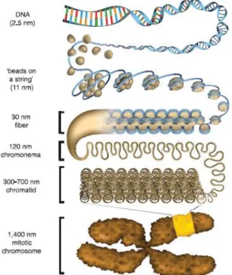
This unit is repeated several times, connected by linker DNA, and forming the second hierarchical structure of DNA packing in the nucleus, the chromatin (Figure 2). This structure can assume different conformations, the first is said to be the beads-on-a-string, or euchromatin. Internucleosomal interactions can occur creating the compacted chromatin, or heterochromatin, also known as the 30 nm fiber. These interactions induce more compacted states going through chromonema, chromatid and finally, during mitosis the chromatin can compact even further forming chromosomes (reviewed in (Bickmore and van Steensel 2013) (Woodcock 2006).
The compaction and relaxation of chromatin are vital for the regulation of a myriad of cellular processes, given that when the chromatin is compacted, the access of the basal transcriptional machinery is blocked, silencing gene expression; when is relaxed, DNA is exposed, allowing the basal transcriptional machinery to interact with the genetic material. For these reasons the chromatin is considered to be the first major transcriptional barrier (Bintu, Ishibashi et al. 2012).
Figure 2: Chromatin compaction schematics
Figure 2: Chromatin compaction schematics. Going from free DNA through the mitotic chromosome and their respective sizes in thickness. Adapted from (Ou, Phan et al. 2017)
1.2. Chromatin Dynamics
The compaction of chromatin, is a major component for gene expression regulation, thus there are a plethora of manners that it can be regulated. The most well-characterized aspect of the compaction and relaxation phenomenon is the histone H4 tail interaction, in this case, internucleosomal interactions can occur, mediated by the N-terminus region of the H4 histone that binds to an acidic region of the neighboring nucleosome, known as acidic patch (Luger, Mader et al. 1997). The histone H4 tail was confirmed by Dorigo et al. to be the only histone tail to be necessary to induce compaction in the chromatin (Dorigo, Schalch et al. 2003). But this interaction can be modified by Post Translational Modifications (PTMs), that are mediated by enzymes such as Histone Acetyl Transferase (HAT), which deposits an acetyl group in the lysine 16 of the H4 histone, neutralizing the net charge of this amino acid, thus preventing the interaction with the acidic patch of the neighbor nucleosome, hence inducing the relaxed state in the chromatin. The
enzyme Histone Deacetylase (HDAC) can undo this process, promoting the compaction of chromatin (Marmorstein and Roth 2001). Furthermore, HDAC inhibitors (HDACi) are a class of drugs, that are used to treat a myriad of diseases, from neurological disorders, such as T-cell lymphoma, multiple myeloma, epilepsy, bipolar disorders and migraine (Eckschlager, Plch et al. 2017), and recently has been proposed as a potential treatment for the COVID-19 infection (Gordon, Jang et al. 2020).
Another factors that induce chromatin compaction is the presence of mono or divalent cations. Using AUC (Analytical Ultra Centrifugation) technique, Korolev and collaborators showed that the higher the cation charge, fewer ions were necessary to induce condensation of the
chromatin, with the best being Mg 2+. This happens due to neutralization of residual charges in the
DNA, facilitating the internucleosomal interaction (Lundberg, Berezhnoy et al. 2010).
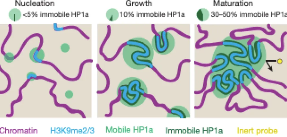
The histone H1 is a non-canonical histone that is not part of the histone octamer and binds to the chromatin. It was thought to bind to the dyad region of the nucleosome (entry and exit points of the DNA in the nucleosome), acting as a clamp, inducing chromatin compaction (Robinson, An et al. 2008, Song, Chen et al. 2014). However, it was revealed a new mechanism of compaction mediated by this histone. Using SAXS (Small Angle X-ray Scattering) and fluorescent microscopy techniques, it was showed that this protein induces a phase separation when bounded to the chromatin, potentially shielding the separated phase from other molecules and inducing compaction, as seen in Figure 3 (Larson, Elnatan et al. 2017, Strom, Emelyanov et al. 2017).
Figure 3: Chromatin phase separation and compaction
Figure 3: Chromatin phase separation and compaction. Histone HP1a recognize PTMs in histones and binds inducing a compacted and phase separation in the chromatin, preventing other molecules
to interact with it. Adapted from (Strom, Emelyanov et al. 2017).
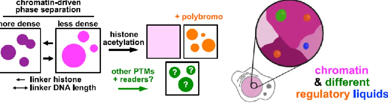
Further studies in the role of phase separation and chromatin dynamics revealed that in fact, reconstituted chromatin undergo histone tail-driven liquid-liquid phase separation, also that linker DNA lengths and histone H1 play an important role in the formation of droplets, impacting over some relevant characteristics. Furthermore, it was revealed that acetylation by p300, antagonizes the formation of phase separation, indeed, in the presence of highly acetylated chromatin, the formation of a different phase-separated environment that is immiscible with the non-acetylated chromatin happens, Figure 4 shows a model of how chromatin dynamics are dictated by phase separation. These recent studies are revealing a whole new mechanics for chromatin regulation that are highly dependent on the phase separation phenomenon.
Figure 4: Chromatin regulation by phase separation.
Figure 4: Chromatin regulation by phase separation. A model for chromatin dynamics regulated by distinct membranelles subdomains. Adapted from (Gibson, Doolittle et al. 2019)
2. PART I - NBPeps
2.1. Nucleosome Binding Proteins
One of the main forms of chromatin remodeling and regulation is mediated by NBPs (Nucleosome Binding Proteins). Several of these proteins have been identified, some with the detailed mechanism of action and atomic levels resolution of interaction with the nucleosome surface, here I summarized some NBPeps that have been identified interacting with the nucleosome, I briefly describe how it interacts, and what function of the NBPs over the chromatin. In 2010, Song Tan and collaborators obtained the first structure of a full length protein bounded to the nucleosome solved by x-ray cystography. They were able to obtain the protein
RCC1 (Regulator of Chromosome Condensation 1) with the structure with the resolution at 2.9 Å, providing an atomic overview of how this interaction happens. They observed that the RCC1 interacts in two distinct regions of the nucleosome, one in the acidic patch located in the histones H2A and H2B and other making contact with the nucleosomal DNA (Makde, England et al. 2010). Four years later the same group determined the structure of the domain responsible for ubiquitination in PRC1 (Polycomb Repressive Complex 1). They showed that this protein binds mostly to the nucleosomal DNA and a small negative zone, near the acidic patch (McGinty, Henrici et al. 2014).
Another major contribution to the field was the structure of the peptide Latency Associated Nuclear Antigen (LANA) present in infections by Kaposi's Sarcoma Herpesvirus (KSHV). This peptide is 23 amino acids long and is associated with the latency period of the disease (Ballestas, Chatis et al. 1999). At the resolution of 2.9 Å it was possible to visualize that this peptide binds to the acidic patch of the nucleosome in a hairpin manner (Barbera, Chodaparambil et al. 2006).
To acquire good structural data in large macromolecular structures like the nucleosome, is always challenging. Frequently, large molecules cannot be crystallized or analyzed by NMR (Nuclear Magnetic Resonance) (Nogales and Scheres 2015). In 1997 the first protein structure was solved using Cryo-EM, which opened a new era for the resolution of large macromolecular complexes (Böttcher, Wynne et al. 1997). Kurumizaka and collaborators accomplished the astounding feature of determining how RNAPII (RNA polymerase II) interacts with the nucleosome and even how it can surpass the complex, using Cryo-EM (Kujirai, Ehara et al. 2018). They showed that RNAPII pause at specific regions of the DNA and that RNAPII gradually tears DNA from the histone surface while preserving the histone octamer.
An important discovery about how viruses can integrate their genome into the host was elucidated by Costa and colleagues, they used foamy virus intasome engaged with a nucleosome, analysis with Cryo-EM and Förster resonance energy transfer measurements to show that the retroviral integrase twist and slide nucleosomal DNA by approximately two base pairs, lifting from histones H2A/H2B to allow engage with the intasome (Wilson, Renault et al. 2019).
In 2011 a peptide from the SIR3 (Silent Information Regulator 3 ) was co-crystalized with the nucleosome, and differently from all others proteins or peptides that have been observed to that date, did not interact with the acidic patch region or surrounding residues, but mostly with histone H3 (Armache, Garlick et al. 2011).
The SAGA (Spt-Ada-Gcn5 acetyltransferase) contains a DUB (Deubiquitinating enzymes) module, it is responsible for the regulation and deubequitintion of H2B, involved in a myriad of gene regulation processes (Bonnet, Devys et al. 2014). In 2016 Wolberger and collaborators, using X-ray crystallography, revealed the mode of interaction of this complex with the nucleosome. They showed that interactions occur mostly at the acidic patch and are involved in the different stages of histones disassembling (Morgan, Haj-Yahya et al. 2016).
The protein CENP-C (Centromere protein C) has its function in the assembly of kinetochore proteins, mitosis, and the segregation of chromosomes. In 2013 the mode of interaction between a peptide from this protein and the nucleosome was determined using Nuclear Magnetic Resonance (NMR) and X-ray crystallography. They observed that the CENP-C peptide interacts with the N-terminus of a H3 histone variant, known as CENP-A (Centromere protein A) that further binds to the acidic patch (Kato, Jiang et al. 2013). More insights about how kinetochore works was revealed by Bradford and collaborators, they showed that nucleosome containing CENP-A bound to CCAN (Constitutive Centromere Associated Network) from Saccharomyces
cerevisiae, indicating the mechanism of CENP-A nucleosome recognition by CCAN and its role
as a platform for assembly of the outer kinetochore to link centromeres in the mitotic spindle formation during chromosome segregation (Yan, Yang et al. 2019). Using the same technique, Musacchio and collaborators investigated the mode of interaction of the protein Centromere Protein N (CENP-N) with the non-canonical histone variant CENP-A, previously mentioned. They revealed that CENP-N interacts largely with 15bp of nucleosomal DNA, preventing further NBPs to bind to the region and also a new binding motif identified in CENP-A (Pentakota, Zhou et al. 2017).
In a very elegant work, Ingen and collaborators determined the binding epitopes of a peptide from the High Mobility Group Nucleosomal 2 (HMGN2) using methyl-based NMR analysis. They showed that the interaction was similar to the RCC1, with one binding site interacting with the acidic patch, and other to the DNA (Kato, van Ingen et al. 2011).
The Interleukin-33 (IL-33) is a protein that can act as a cytokine, when in the extracellular environment, and as a nuclear receptor when intracellular (Pichery, Mirey et al. 2012, Fu, Hung et al. 2016). A peptide from this protein was also observed by NMR to bind to the nucleosome in a similar manner to LANA, making contacts exclusively with the acidic patch (Roussel, Erard et al. 2008).
The LEDGF (Lens epithelium–derived growth factor p75 splice variant) is a NBP with antiapoptotic properties known to direct human immunodeficiency virus into active transcription units (Daugaard, Baude et al. 2012). In 2020, the domains PWWP (proline, tryptophan, tryptophan, proline) was obtained with methylated nucleosome, showing the cooperative interaction between the multivalent binding of the reader domains to the methylated histone tail from H3 and to both gyres of nucleosomal DNA (Wang, Farnung et al. 2020).
There are several histone chaperons with important function into chromatin remodeling and nucleosome assembly/disassembly, FACT (Facilitates Chromatin Transcription) is one of these chaperones, playing important roles during gene transcription, DNA replication and, DNA repair. In 2019, Luger and collaborators, using Cryo-EM and biochemical assays revealed the mechanism by which FACT operates, showing that FACT engages with nucleosomal DNA and several histones with PTMs, demonstrating that a complex of FACT-H2A/H2B is formed, which can interact with H3/H4, allowing the assembly/disassembly process (Liu, Zhou et al. 2020).
Several NBPs require specifics PTMs to properly interacts with the nucleosome, the protein Dot1L (Disruptor of Telomeric Silencing 1-Like) requires histone a monoubiquitination in the H2B lysine 120 (H2BK120Ub) to be able to methylate the lysine 79 of histone H3 (H3K79m), showing a histone crosstalk phenomenon. Using cryo-EM, the group led by Valencia-Sánchez provided structural and functional data as well as the correlation between aberrant H3K79m and leukemia, suggesting the modulation of Dot1L as a therapeutic target for this disease (Valencia-Sanchez, De Ioannes et al. 2019).
Mutations in the Set2 (SET domain-containing protein 2 ) enzymes are related to cancer progression, these methyltransferase enzymes, recognizes H3K36me and H2B-Ub nucleosome, in 2019 their mode of interaction was determined by Cryo-EM, showing mostly contacts with histone H3, H2A C-terminal and unwrapped DNA, Intriguingly it was revealed that the interfaces that can be targeted with small molecules for the future development of cancer therapies (Bilokapic and Halic 2019).
Acting in the same region, the COMPASS (Complex of Proteins Associated with Set1) complex, is formed by six proteins with important methyltransferase activity, in 2019, a group led by Wolberg and colleagues solved the structure of COMPASS bound to ubiquitinated nucleosome using Cryo-EM, their work revealed a long-standing mystery of how H2B-Ub is recognized by COMPASS and provided the first trans-nucleosome histone reveled crosstalk mechanism
(Worden, Zhang et al. 2020).
Still regarding the roles of NBPs in methylation of histones, the structure of the complex MLL1 (Mixed linage leukemia) with the nucleosome was obtained by Cryo-EM, showing that the subunit RbBP5 (Retinoblastoma-binding protein 5) and ASH2L (histone lysine methyltransferase complex subunit) make large interactions with the nucleosome dyad, nucleosomal DNA and the N-terminus tail from histone H4, shedding light on how the MLL1 complex engages chromatin and the tri-methylation activity of the complex (Park, Ayoub et al. 2019).
ATP dependent remodeling of the chromatin is made by a diverse family of proteins that have an ATP-ase domain. Using Cryo-EM, the interaction motifs of several of these proteins have been identified. Chd1 (Chromodomain-helicase-DNA-binding protein 1) is part of this important family of proteins and works as an organizing nucleosome over codding regions (Ocampo, Chereji et al. 2016). The binding mode of this protein to the nucleosome was shown to be mostly with linker DNA and histone H3, induce unraveling of DNA and reorientation of H3 tail (Sundaramoorthy, Hughes et al. 2018).
The INO80 (inositol-requiring 80), a chromatin remodeler, that is ATP dependent, is composed by multi-subunits. It was previously thought the H4 tail played a major role in regulating some of its units (van Attikum and Gasser 2005). However, Zhang and collaborators, using Cryo-EM, showed a new mode of binding involving nucleosomal DNA and H3 as well as the fact that the H3 tail instead is responsible for this regulation (Ayala, Willhoft et al. 2018).
The SWI/SNF (Switch/Sucrose Non-Fermentable) is a chromatin remodeling complex and has important roles in transcription and DNA-damage repairs, this complex can hydrolase ATP and evict or slide histone octamers, creating exposed DNA regions for other proteins, such as transcriptional factors. In 2020 He and collaborators, using Cryo-EM, were able to obtain near-atomic resolution of this complex from Saccharomyces cerevisiae bound to the nucleosome, giving valuable insights about how this complex works. They showed the protein Snf5 (SWI/SNF chromatin-remodeling complex subunit SNF5) interacts with the acidic patch, functioning as an anchor for the whole complex during active DNA translocation (Han, Reyes et al. 2020). Furthermore, domains of this complex, such as Snf2 (Transcription regulatory protein SNF2) and ISWI (imitation SWI) where elucidated a year before the whole complex resolution, showing interactions with nucleosomal DNA and strikingly similar binding epitopes, suggesting a conserved mechanism for chromatin remodeling (Li, Xia et al. 2019, Yan, Wu et al. 2019). In a
similar manner, the RSC (Remodeling the Structure of Chromatin) from e Saccharomyces
cerevisiae, which is part of the SWI/SNF family was elucidated by Cryo-EM in 2019 by Nogales
and collaborators, their findings shed light on the structural insights into the conserved assembly process for members of SWI/SNF family of remodelers, showing how RSC selects, engages and remodel nucleosomes (Patel, Moore et al. 2019).
Recently the structure of two pioneers transcriptional factors SOX2 (Sex determining region Y) and SOX11 (SRY-box transcription factor 11) was solved using Cryo-EM, Cramer and collaborators showed that the transcriptional factors can bind and distort superhelical DNA at the position +2 , facilitating the detachment of terminal nucleosomal DNA from the histone octamer. Furthermore, upon SOX-factor binding, can lead to a repositioning of the N-terminal tail from histone H4, including the lysine 16, which has an important role in the regulation of chromatin compaction via the interaction with the acidic patch from the neighbor nucleosome, suggesting that SOX2 and SOX11 prevent the formation of higher-order chromatin, thereby facilitating nucleosome remodeling and subsequent transcription (Dodonova, Zhu et al. 2020).
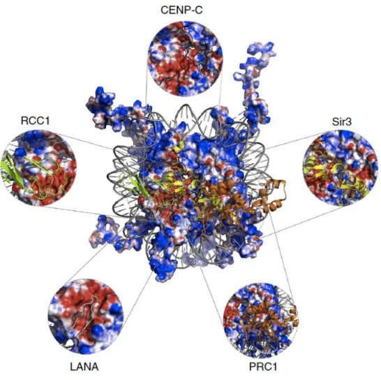
With the exception of the ATP dependent remodeling family of proteins identified interacting with the nucleosome, RNAPII, and the SOX family, the majority of NBPs focus on the acidic patch as their binding site, as seen in Figure 5.
Figure 5: Acidic patch is a docking hub for NBPs.
Figure 5: Acidic patch is a docking hub for NBPs. Overall charge view of the nucleosome, in blue positive and red negative. The acidic patch is highlighted and NBPs are overlaid. Adapted from (Cabral, Machado et al. 2016)
2.2. The acidic patch and NBPeps
As addressed in the previous section, the acidic patch can work as a docking hub for several NBPs, thus having an important role in gene regulation. This idiosyncratic region in the nucleosome surface is composed of 8 acidic amino acids between the histones H2A and H2B (E56, E61, E64, D90, E91, E92 of H2A and E102, E110 of H2B).
In 2014, in an attempt to displace LANA from the acidic patch and treat the latency aspect associated with KSVH, a group led by Keye, screened over 350,000 small molecules and all failed to do it. The authors concluded with the suggestion that more complex molecules, such as peptides,
might be a better option to displace the LANA from the nucleosome (Beauchemin, Moerke et al. 2014).
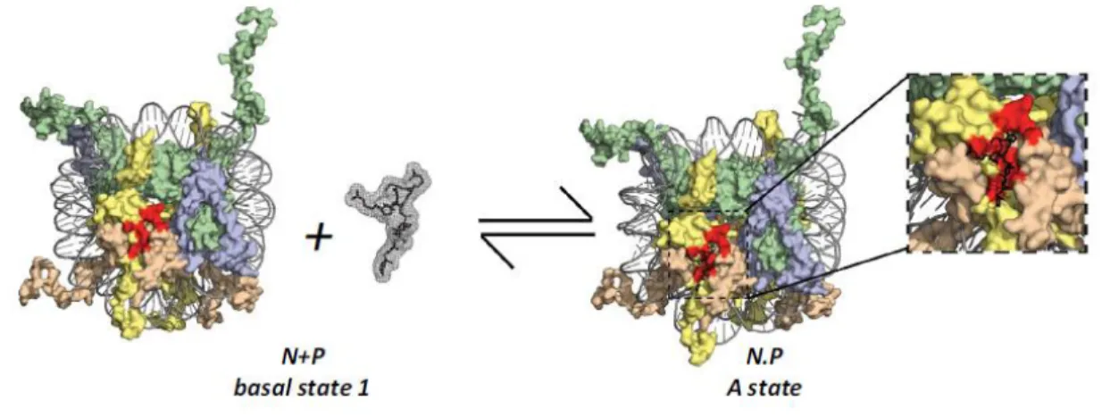
Due to the regulatory nature of the acidic patch, and the findings of Beachemin et al., Dr. Santos suggested that the nucleosome surface, and in specific the acidic patch, could be a potential pharmacological target using peptides for it (Silva, de Oliveira et al. 2015). In Dr. Santos’s paper, it is theorized that the binding of Nucleosome Binding Peptides (NBPeps) can induce specific outcomes in the chromatin and modulate cell function, as seen in Figure 6. In a similar manner of other epidrugs, NBPeps would modulate the chromatin architecture in a non-specific way. However, in this case changes would be direct to the chromatin, and not mediating chromatin remodeler enzymes. Furthermore, occupying the binding site for several NBPs can prevent the binding of several of these proteins, thus modulating chromatin architecture.
Figure 6: Kinetics of drug receptor for the nucleosome.
Figure 6: Kinetics of drug receptor for the nucleosome. The nucleosome and LANA peptide from Protein Data Bank (PDB) 1ZLA) are represented. H3 in green; H4 in blue; H2A in yellow; H2B in wheat; DNA in gray; acidic patch in red. The dynamic equilibrium between the nucleosome and NBPep, resulting in two states, bound or unbound to the nucleosome. Adapted from (Silva, de Oliveira et al. 2015).
In a novel work led by Dr. Luger, they developed binuclear ruthenium compounds that bind to the acidic patch, inducing aberrant chromatin condensation and alterations in the cell’s cycle, with potential applications in drug development and as tools for chromatin research (Davey, Adhireksan et al. 2017). It demonstrated that chromatin can in fact be modulated by exogenous
molecules.
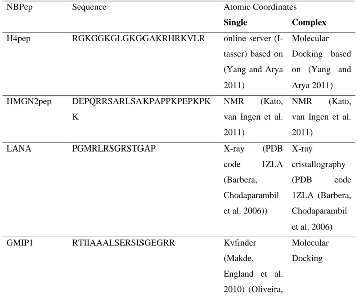
For the development of this work, four NBPeps were used, with GMIP1(Genetic Modified Inducible Peptide 1) being designed in silico and LANA, HMGN2pep, and H4pep based on the MBM (Minimum Binding Motif ) of these proteins that were already known to bind to the nucleosome, generating new peptides. Table 1 shows the sequence and mode of creation of these peptides.
Table 1: List of all NBPeps used in this work, the amino acid sequence, and method of resolution alone and in complex with the nucleosome.
Table 1: List of all NBPeps used in this work.
NBPep Sequence Atomic Coordinates
Single Complex
H4pep RGKGGKGLGKGGAKRHRKVLR online server
(I-tasser) based on (Yang and Arya 2011) Molecular Docking based on (Yang and Arya 2011) HMGN2pep DEPQRRSARLSAKPAPPKPEPKPK K NMR (Kato,
van Ingen et al. 2011)
NMR (Kato,
van Ingen et al. 2011)
LANA PGMRLRSGRSTGAP X-ray (PDB
code 1ZLA (Barbera, Chodaparambil et al. 2006)) X-ray cristallography (PDB code 1ZLA (Barbera, Chodaparambil et al. 2006)
GMIP1 RTIIAAALSERSISGEGRR Kvfinder
(Makde,
England et al. 2010) (Oliveira,
Molecular Docking
Ferraz et al. 2014)
The design of GMIP1 was based on the mode of interaction of RCC1 using the software KVFinder (Oliveira, Ferraz et al. 2014). It was simulated the mode of interaction of four proteins, RCC1 having the best fit (Teles, Fernandes et al. 2020). The binding mode of this protein, as previously stated, consists of one-part binding to the acidic patch and other to the DNA. The MBM of these two regions were connected by a three-alanine bridge, as seen in Figure 7.
Figure 7: Mode of binding of RCC1 and GMIP1 design.
Figure 7: Mode of binding of RCC1 and GMIP1 design. The MBM of RCC1 with the two regions of interaction with the nucleosome highlighted and the triple alanine bridge region connecting the two distinct regions. Images extracted and modeled using PDB (3MVD) from (Makde, England et al. 2010) and PyMol software.
In this work, I performed in vitro, cell-based assays, and in vivo assays in order to very if NBPeps (Nucleosome Binding Peptides) designed by Dr. Santos’ group have therapeutic potential
purposes and if targeting the nucleosome in order to module cell phenotypes is a feasible option to module cell phsyiology. The atomistic characterization of how NBPeps would affect the nucleosome was done by Fernandes. V, using in silico strategies at LBTC.
3. AIMS PART I (NBPeps)
3.1. PRIMARY AIM
This work’s goal is to understand how NBPeps that present distinct nucleosome binding sites affect the nucleosome and chromatin structure, thereby modulating chromatin status and influencing phenotypic outcomes.
3.2. SECONDARY AIMS
• Verify NBPeps binding to the nucleosome in vitro; • Verify cellular uptake of NBPeps;
• Verify the toxicity of NBPeps in different cell lineages; • Verify the toxicity of NBPeps in zebrafish embryos;
• Verify the distribution of NBPeps in tissues of zebrafish larvae; • Verify if NBPeps can reach the nucleus of cells in vivo.
4. METHODS
NBPeps: All Peptides were bought from Biomatik with 95%> purity and diluted in MiliQ H2O.
Fluorescent peptides were bought with TAMRA -(559/583nm) in the N-terminus. The concentration was determined by spectrophotometric method as described in (Murphy and Kies 1960). All peptides are described in Table 1.
In vitro nucleosome reconstitution: Histone octamers (HO) were purified from chicken
erythrocyte nuclei as described in Huynh, V. A. T., P. J. J. Robinson, and D. Rhodes, 2005. 601 DNA Widom with 167 base pairs (bp) was used to reconstitute mononucleosomes, using the slow salt dialysis method as described in (Huynh, Robinson et al. 2005).
The analyses of the reconstitution were verified by electrophoresis in native bis-acrylamide gels (6%).
Mononucleosome precipitation: Freshly reconstituted mononucleosomes (115nM
mononucleosome, Tris 10mM pH 7.4, EDTA 1.5mM NaCl 15mM) were incubated with the specified concentration of NBPeps for 30 minutes at room temperature. The samples were centrifuged (Sigma centrifuge-2K15) at 15493 x g for 20 minutes at 25 °C. The supernatant was transferred to another microcentrifuge tube and the pellet was resuspended in the same buffer as the mononucleosome. The samples were analyzed by electrophoresis in native 6% bis-acrylamide gel carried out with 0.5× TBE buffer at 15 mA. Densitometry was performed using ImageJ (National Institute of Health, Bethesda, MD, USA) version 1.49.
DNA binding assay: Widom 601 DNA fragments containing 167bp (30nM DNA, 10mM Tris pH 7.4, 135mM NaCl) were incubated with specified concentrations of GMIP1 for 2 hours at 37 ◦C and 100 RPM. The analysis was done in 0.8% agarose gel in TBE 0.5X. Samples were loaded with 30% glycerol, to avoid interaction caused by phenol blue and GMIP1.
Nucleosome binding assay: Freshly reconstituted mononucleosomes (115nM mononucleosome, Tris 10mM pH 7.4, EDTA 1.5mM NaCl 15mM) were incubated with the specified concentration of fluorescent NBPeps for 120 minutes at room temperature. Then samples were analyzed by electrophoresis in native 6% bis-acrylamide gel carried out with 0.5× TBE buffer at 15 mA. Gels
were analyzed using Amersham Imager 600 (GE) with the RGB laser kit detection for 520nm, to visualize the peptide, following incubation in ethidium bromide bath and analyzed with UV for ethidium bromide detection. For Kd determination, band densitometry was performed in the gel reveled with 520nm laser, using ImageJ (National Institute of Health, Bethesda, MD, USA) version 1.49, followed by analysis in Prism 6 Graphpad software using Binding - saturation binding to total and non-specific template.
MTT: For MTT (3-(4,5-dimethylthiazol-2-yl)-2,5-diphenyltetrazolium bromide) assays, 8000 Hela cells or ccd10595k cells were plated in 96-well culture plates and maintained at 37◦C and 5%
CO2 in DMEM medium with 10% fetal bovine serum, penicillin (100U/mL) and streptomycin
(100ug/mL) for 24 hours. Next, wells were washed 3 times with PBS 1X and filled with 100uL of DMEM medium as described above containing the specified amount of NBPeps and incubated for 24 hours in the same conditions. The MTT at 5mg/mL was added to the wells (10uL) and incubated
for 4 hours at 37◦C and 5% CO2. The wells were drained, and the formazan crystals were
solubilized in 100uL of acidic isopropanol solution (52uL of HCl 37% to 12 mL of isopropanol) and agitated for 30 minutes at room temperature. Absorbance at 570nm was determined with a plate spectrophotometer (DTX 800 Multimode Detector - Beckman Coulter) at 570 nm.
Flow cytometry: 70.000 Hela cells were plated in 12-wells culture plates for 16 hours and
maintained at 37◦C and 5% CO2 in DMEM medium with 10% fetal bovine serum, penicillin
(100U/mL) and streptomycin (100ug/mL). Prior to treatment with NBPeps, cells were incubated for 1 hour at 37 or 4 ◦C. Next, cells were washed with 1X PBS and filled with DMEM medium with the specified amount of fluorescent NBPeps and incubated at 37 or 4 ◦C for 1 or 3 hours. Wells were washed three times with ice cold 1X PBS and filled with 500uL 1X PBS, cells were harvested with a cell scraper and analyzed by flow cytometry on FACSCalibur (BD biosciences). Hela cells were gated to isolate the main population of living cells from cell debris. Data analysis was done using flowjo 8.7 software.
Zebrafish husbandry and embryo collection: Zebrafish (Danio rerio) were raised in an aquatic facility (ZebTec - Tecniplast, Italy) with a photoperiod cycle of 12:12 h (light:dark) at the University of Brasilia (Brazil). The water parameters were: temperature was maintained at 27.0 ±
1 °C, conductivity at 650 ± 100 μS/cm, pH at 7.0 ± 0.5 and dissolved oxygen≥95% saturation. Zebrafish embryos were collected immediately after natural mating, rinsed in water, and checked under a stereomicroscope (Stereoscopic Zoom Microscope – Stemi 2000, Zeiss, Germany). The unfertilized eggs and those showing cleavage irregularities or injuries were discarded (OECD 2013).
Fish embryo toxicity (FET): FET was adapted from Morash et al (Morash, Douglas et al. 2011). Briefly, Zebrafish embryos at 4, 28 and 52 hours post fertilization (hpf) were used to evaluate the toxicity of NBPeps in 96-well plates. Each peptide was tested at 0.1, 1, 10 and 100uM in 100uL of water from the aquarium system; pH in all conditions was tested using pH strips (92120 – MACHEREY-NAGEL). Embryos were stored at 27 ◦C with 14 hours light 10 hours dark cycle and evaluated Stemi 508 (Carl Zeiss) microscope with 1 and 24 hours of treatment. Embryos were assessed for pigmentation, development, hatching and lethality. 10 embryos were used for each condition, if the control group showed any alteration, the plate was discarded, alterations >10% were considered significant and were documented using Axiocam Erc 5s (Carl Zeiss) and ZEN software (Carl Zeiss).
Fluorescence fish embryo: Zebrafish larvae with 80 hpf were incubated with fluorescent NBPeps with specified concentration for 3 hours in 100uL in a 96-plate, larvae were washed 3 times in 100mL to remove the excess of NBPep, imaging was done using Axioskop 2 (Carl Zeiss) with HBO 100 lamps, Axiocam Erc 5s (Carl Zeiss) and ZEN software (Carl Zeiss) with appropriate laser filter for TAMRA (filter 4).
Fluorescence blood smear: Adults Zebrafish at 2 years old were injected in the abdomen with 50uL, 1mM of fluorescent NBPeps, and kept protected from light at 27 ◦C for 18 hours. Blood was extracted from the fins using a pipet tip and heparin 250 IU to make the blood smear in a microscope slide. Images were acquired with Axioskop 2 (Carl Zeiss) with HBO 100 lamps, Axiocam Erc 5s (Carl Zeiss) and ZEN software (Carl Zeiss) with appropriate laser filter for TAMRA (filter 4).
NMR: All NMR experiments were carried out on a Bruker advance III HD 600MHz. NMR spectra were processed in Bruker TopSpin (Delaglio, Grzesiek et al. 1995) and analyzed using Sparky
(Lee, Tonelli et al. 2015). Dimer samples of [C13,N15]H2A-H2B at 100uM in 5%D2O/95%H2O;
25mM NaPi + 100mM NaCl pH6.2 + 0,01% NaN3 + 1mM 2-Mercaptoethano + PIC (complete EDTA-free Protease Inhibitor Cocktail (Roche)) were titrated against GMIP1 using 600MHz
Lamour frequency at 308K. HSQC spectra were measured for free [C13,N15]H2A-H2B and after
the addition of GMIP1 at 308K. Titration consisting of 4 points in the range of 1:4.3 molar ratio
([C13,N15]H2A-H2B:GMIP1) was performed.
Circular dichroism: Measurement of secondary structure of NBPeps was performed in Jasco j-815 spectropolarimeter in a 0,1cm quartz cuvette in the range of 190-250nm. Samples were diluted in MiliQ water in the concentration of 0.125mg/mL for GMIP1, LANA, HMGN2pep and H4pep at 0.107mg/mL at 25 ◦C. Data were plotted using BestSel data base (available at: http://bestsel.elte.hu/).
5. RESULTS AND DISCUSSION (NBPeps)
5.1. NBPeps secondary structure characterization
All NBPeps, except for GMIP1, have been well characterized structurally (Luger, Mader et al. 1997, Barbera, Chodaparambil et al. 2006, Kato, van Ingen et al. 2011). Despite GMIP1 being based in the structure of RCC1, the triple alanine bridge connecting the two epitopes created a new structure. For this reason, I performed CD (Circular Dichroism) analysis, as seen in Figure 8.
Figure 8: Circular dichroism of NBPeps for determination of secondary structure.
Figure 8: Circular dichroism of NBPeps for determination of secondary structure. All NBPeps show low ellipticity above 210 and negative bands near 195nm, characterizing predominance of random coil structure.
All NBPeps that were developed have no well-defined secondary structure, result which is in agreement with NMR experiments based on the absence of medium or long-range NOEs (Nuclear Overhauser Effect) and random coil 13C chemical shifts for GMIP1 (unpublished data).
5.2. GMIP1 binding to the nucleosome in vitro
All NBPeps used, with exception of GMIP1, have their mode of interaction with the nucleosome surface already established at an atomic level, with two distinct modes of binding, (i), LANA and H4pep, that binds exclusively to the acidic patch, and (ii) HMGN2pep and GMIP1 that interacts with nucleosomal DNA and the acidic patch as well. To confirm the epitope of GMIP1, I performed NRM experiments, using isotope labeled dimers of H2A/H2B from Xenopus Laevis, as seen in Figure 9a. the binding of GMIP1 should induce changes in the electromagnetic environment of specifics residues in the NRM spectra, causing a shift in the peaks, that was not observed in the dimers even in a molar excess of 4.3 times, also GMIP1 interacted with DNA at a Kd of 50μM or weaker, which is probably in the range of non-specific binding of a charged peptide to DNA, Figure 9b.
Figure 9: NMR: HQSC spectra of [C13,N15]H2A-H2B dimers (black) and with GMIP1 (red).
Figure 9: NMR: HSQC spectra of [C13,N15]H2A-H2B dimers (black) and with GMIP1 (red). a) Nucleosomal DNA binding to GMIP1: b) Titration of GMIP1 on the DNA widom 601 (167 bp) analyzed in agarose gel 0,8% in TBE 0.5X. The assay was performed at least 3 times, and the representative gel was presented.
We aimed to create a novel NBPep that could bind to and present specificity for the nucleosome, with high dependence to DNA, that in future projects could be engineered to recognize specific sequences, in a similar manner that was done for transcriptional factors (Desai, Rodionov et al. 2009), giving more specificity to NBPeps with multiple contact sites. However, the biochemical data shows that GMIP1 has low nucleosome binding affinity. It is important to emphasize that all experiments performed were done with the Widom 601 DNA sequence, which is an artificial sequence with high specificity to the octamer.
5.3 NBPeps binding to the nucleosome
Despite most of the NBPeps having its sequences directly derived from NBPs, I wanted to evaluate if it would bind to the nucleosome in vitro. In this experiment, I reconstitute nucleosome
in vitro, with histones from chicken erythrocytes and DNA Widom 601 with 167bp and incubated
with the Tagged NBPeps, following analysis in polyacrylamide gel, the gel was then revealed at 510nm so I could visualize the fluorescence from the peptides, the gel was then stained with ethidium bromide and revealed, to show nucleosomal DNA, the bands from the NBPep and the nucleosome were compared to verify if they were at the same height. Peptides were also titrated against a fixed amount of nucleosome, its intensity used to determine the Kd of each NBPep, as seen in Figure 10.
Figure 10: NBPeps interaction assay.
Figure 10: NBPeps interaction assay. a) Nucleosome binding assay with fluorescent NBPeps, nucleosome is incubated with LANA at 0, 10, 20, 30, 40, 50 60, 70 80,90 u M, with HMGN2pep at 0, 20, 40, 60, 80 u M or with H4pep at 0, 200, 400, 600, 800, 1000, 1200, 1400, 1600 nM. It was then analyzed in acrylamide gel, following by detection of the fluorescent NBPep and subsequently detection of DNA. b) Densitometry of NBPeps bands Kd is represented by a vertical line in the densitometry graphs. These assays were performed at least 3 times, and the representative gel was presented.
The NBPeps testes showed a clear band for the peptides at the same height as the nucleosome, furthermore, all presented a dose-response behavior, this data suggests that the NBPeps are binding to the nucleosome in vitro with a Kd of 0.6, 8 and 35uM for H4pep, LANA
and HGMN2pep respectively, GMIP1 induced nucleosome aggregation even at low concentration, as seen in Figure 11, which difficulted to determine the binding affinity constant. HMGN2pep promoted an electrophoretic mobility shift, suggesting that it is binding at more than one site on the nucleosome surface, further investigations will be needed to explore this finding.
Figure 11: GMIP1 binding assay with nucleosome.
Figure 11: GMIP1 binding assay with nucleosome a) GMIP1 incubated with nucleosome at 0, 50, 100 and 150uM, gel on the left staining with EtBr, to the right gel visualizing the tagged peptide. The assay was performed at least 3 times, and the representative gel was presented.
5.4. NBPeps induce precipitation of the nucleosome in vitro
In order to observe if the fluorescent tag had the impact of the binding of NBPeps to the nucleosome and acquire more information about nucleosome interaction with NBPeps, I performed nucleosome precipitation assay. NBPeps were incubated at different concentrations with freshly reconstituted nucleosomes and samples were centrifuged. The supernatant was transferred to another micro-centrifuge tube, the pellet was resuspended and analyzed in polyacrylamide gel, as seen in Figure 12.
Figure 12: Nucleosome precipitation assay with NBPeps.
Figure 12: Nucleosome precipitation assay with NBPeps. Nucleosome without NBPeps stays in the supernatant (SN). The addition of 50uM GMIP1, 10uM LANA, 10uM HMGN2pep or 500nM H4pep induce precipitation and Pellet (P) formation. Non-centrifuged (NC) samples were used as control. Densitometry analysis allows a better quantification, with H4pep having the greatest impact.
It was observed that the nucleosome does not precipitate without NBPeps, although all peptides testes induced precipitation at different rates, suggesting binding to the acidic patch and charge neutralization (de Frutos, Raspaud et al. 2001). Notable, H4pep induced precipitation at 500 nM, corroborating to the data from the previous binding assay with fluorescent tagged NBPep.
5.5. NBPeps uptake by cells
In order to bind to the nucleosome, NBPeps must first penetrate the cell to reach the nucleus, one of the issues to use peptides as drugs is that peptides have very low permeability (Shaji and Patole 2008), although there is a class of peptide, known as CPP (Cell Penetrating Peptides) that can cross the cell wall (Prochiantz 2000). CPPs have an overall positive charge and
vary between 5-30 amino acids (Derakhshankhah and Jafari 2018). All NBPeps designed by Dr. Santos’ group have these characteristics (see Table 3). Using fluorescent tagged NBPep, I performed flow cytometry analysis in order to quantify and analyze if NBPeps can penetrate cells, and if this is done by passive or active mechanism. See Figure 13.
Figure 13: NBPeps cell penetration.
Figure 13: NBPeps cell penetration. Flow cytometry profile of Hela cells uptake of fluorescent NBPeps (TAMRA), in histogram view with 1 or 3 hours exposure and at 37 or 4˚C. The assay was performed at least 3 times, and the histogram was presented.
All NBPeps tested penetrated Hela cells, with GMIP1 having the highest uptake. Also by varying the temperature and incubating the cells at 4˚C the active mechanisms of endocytosis in the cell are inhibited (Fernando, Kandel et al. 2010). With the exception of H4pep, all NBPeps are uptake by active mechanisms, with H4pep penetrating both actively and passively.
5.6. NBPeps induce cytotoxicity in a specific manner
In order to verify the cytotoxicity of NBPeps, I performed MTT (3-(4,5-dimethylthiazol-2-yl)-2,5-diphenyltetrazolium bromide) analysis in Hela and ccd10595k cells. These two types of cells vary significantly, with Hela being an immortalized cell from an aggressive cervical cancer and ccd10595k a primary culture from fibroblasts (Rahbari, Sheahan et al. 2009). As seen in Figure
14, NBPeps have highly distinct cytotoxicity over these two cell lineages.
Figure 14: Cytotoxicity evaluation of NBPeps.
Figure 14: Cytotoxicity evaluation of NBPeps. Relative cell viability compared to control over 24 hours exposure to NBPeps in HeLa and CCD 10595K. HeLa cells showed a greater decreased in cell viability than CCD 10595k for every NBPep tested with the exception of GMIP1. Data is shown as mean ± SD. * represent significant statistical difference (one-way ANOVA test) between the control and treated groups with = p < 0.05 and n = 2.
For Hela cells, all NBPeps, with the exception of GMIP1 and LANA, showed cytotoxic effects, with a decrease in cell viability greater than 30%. When tested in ccd10595k there were a reduction in cell viability only for H4pep and HGMN2pep, with all other having little to no effect over cell viability. Testing NBPeps in other cell types might help elucidate if cytotoxicity is more prevalent in oncogenic cell lineages or not.
5.7. NBPeps can penetrate cell nucleus in vivo
For NBPeps to bind to the nucleosome in vivo, it is required to penetrate the cell and reach the nucleus. I evaluated the capability of NBPeps to reach the nucleus in vivo using Zebrafish (Danio rerio) as a model. The cellular uptake analysis described in section 4.4. could lead to false positives because the NBPeps could be accumulating in the cell membrane or in the cytoplasm. Therefore, I injected adult zebrafish with fluorescent tagged NBPeps, harvested the blood, performed a blood smear, and observed under the fluorescent microscope as seen in Figure 15.
Figure 15: NBPeps distribution in vivo.
Figure 15: NBPeps distribution in vivo. Injection of florescent NBPeps accumulate in the nucleus erythrocytes of adult zebrafish. At the left panel, HBO field, at right panel, visualization using 520 nm laser. The assay was performed at least 3 times, and the representative picture was presented.
It is possible to distinguish very clearly the accumulation of NBPeps in the nucleus, analyzing the formation of a thin halo (cytoplasmic content) around the concentrated red nucleus. This data suggests that NBPeps can reach and accumulate in the nuclear environment in vivo.
5.8. NBPeps penetrated different tissues of the zebrafish larvae
NBPeps were designed to bind to every cell with a nucleus, therefore I investigated how NBPeps would be distributed over zebrafish larvae, as seen in Figure 16.
Figure 16: NBPeps distribution in zebrafish larvae.
Figure 16: NBPeps distribution in zebrafish larvae with 80hpf. Fluorescent NBPeps incubated for 3h distributes heterogeneously over zebrafish larvae. At the left panel, HBO field, at right panel, visualization using 520 nm laser. The assay was performed at least 3 times, and the representative picture was presented.
The accumulation of NBPeps at the zebrafish larvae penetrated different tissues of the larvae. This result is in agreement with the cell uptake analysis and the blood smear, suggesting that NBPeps can penetrate cells.
5.9. NBPeps induces abnormalities in Zebrafish embryos development
To better understand the effects in vivo of NBPeps, I conducted a modified Fish Embryo Toxicity (FET) assay (Morash, Douglas et al. 2011). In my analysis, Zebrafish embryos were incubated with crescent concentrations of NBPeps reaching up to 100uM for 24 hours, at different stages of development, and evaluated for morphological modifications with 1 hour and 24 hours of exposure, see Table 2.
Table 2: Fish Embryo Toxicity assay. Zebrafish embryos at 4, 28 and 52hpf were exposed to NBPeps for 24 hours. The embryos were evaluated for alterations in the development with 1 hour and 24 hours of exposure. H4pep was the only NBPep that induce embryos mortality with 1hour exposure.
The outcome of embryo exposure to the NBPeps was highly dependent on the stage of development, with the first 28 hours having a higher impact. The hatching rate of zebrafish was largely affected by NBPeps, with GMIP1 having the most pronounced effect, GMIP1 also created defects in the pigmentation in 26% of the embryos and had no impact over mortality. Intriguingly LANA induced delay in the development of 100% on the embryos when they were exposed for 24h with 4hpf and at later stages of development, however, LANA caused no delay in the hatching processes. HMGN2pep affected 16% of the embryos in the development of melanocytes and had a low death rate when compared to H4pep, which was the only one that showed acute toxicity ( death with 1houre exposure), also inducing 100% mortality with 24hours of expousere for embryos with 4hpf. This data suggests that NBPeps overall have little toxicity to this animal model, see Figure 17, with increased mortality only at the early stages of development, corroborating the data of MTT in ccd10595 cells.
Figure 17: Fish Embryo Toxicity (FET) with NBPeps.
Figure 17: Fish Embryo Toxicity (FET) with NBPeps. Zebrafish embryos at 4, 28 and 52 hours post fertilization (hpf) were incubated with NBPeps or vehicle for 24h. Images are representative of three separate experiments.
interesting to observe that despite the similar binding region of these NBPeps, the effect in vivo remains distinct.
These results are not enough to provide a direct correlation between NBPeps binding sites and phenotypic outcomes. Furthermore, only H4pep presented high specificity, with the other three being non-specific nucleosomal interactors, raising the question of whether the NBPeps are not interacting with other chromatin machinery. In fact, Kim and collaborators showed that the tail of histone H4 can be used as a molecular tool to maintain the active state of p53 target genes via interaction with HDAC1 as a novel anticancer therapy (Heo, Kim et al. 2013). Here, I suggest that H4pep could affect tumoral cells, instead of acting only at the modulation of p53 activity, but thought direct nucleosome binding, since it showed higher affinity for the nucleosome
6. CONCLUSION
In conclusion, it was observed that NBPeps can affect the nucleosome structure in multiple ways, despite having a similar target, NBPeps had different effects over cell physiology, which might be due to the non-specificty in targeting the nucleosome surface. However, further experimentation should be performed to be able to correlate the effects of NBPeps binding sites with the physiological outcome. Despite the pioneering work done here, not all aspects that cover the modulation of chromatin via NBPeps were elucidated.
Nevertheless, considering that there are several pharmacological agents, such as DNA intercalators, with great relevance to the clinical practice, the fact that NBPeps are not specific would not preclude their potential as therapeutic agents. Taken all together, I believe that NBPeps open novel opportunities to design hybrid molecules with higher specificity to regulate a plethora of cellular disorders.


![Figure 9: NMR: HQSC spectra of [C13,N15]H2A-H2B dimers (black) and with GMIP1 (red).](https://thumb-eu.123doks.com/thumbv2/123dok_br/15206086.1018823/40.918.110.795.568.903/figure-nmr-hqsc-spectra-dimers-black-gmip-red.webp)
