655
Recebido para publicação: Outubro de 2008 • Aceite para publicação: Março de 2009 Received for publication: October 2008 • Accepted for publication: March 2009
ABSTRACT
Effects of stimulation and blockade of the autonomic nervous system on atrial refractoriness in patients with lone paroxysmal atrial fibrillation
Heterogeneous shortening of the atrial effective refractory period (AERP) and
increased dispersion of refractoriness (disp_A) predispose to recurrent episodes of atrial fibrillation (AF).
Aim: To evaluate the effects of stimulation and blockade of the autonomic nervous system (ANS) on atrial refractoriness in patients with ≥1 year clinical history of lone paroxysmal AF (PAF).
Methods: Ten patients (6 men, aged 55±14 years) underwent electrophysiological study while off medication. AERP was assessed at 5 sites – right atrial appendage (RAA) and low lateral right atrium (LRA), high interatrial septum (IAS), proximal (pCS) and distal (dCS) coronary sinus in basal conditions, during handgrip (HG) and carotid sinus massage (CSM), and after ANS blockade (ANSB) (atropine 0.04 mg/kg + propranolol 0.15 mg/kg). The AERP was taken as the longest S1-S2 interval that failed to initiate a response. Disp_A was calculated as the difference between the longest and shortest
RESUMO
A diminuição dos períodos refractários (PRE) e o aumento da dispersão da refractariedade (Disp_A) auriculares são marcadores de vulnerabilidade para recorrência de fibrilhação auricular (FA). Objectivo: Estudar os efeitos da estimulação e bloqueio do Sistema Nervoso Autónomo (SNA) nos PRE e Disp_A em doentes (D) com ≥1 ano de evolução clínica de FA paroxística idiopática (FAp). Métodos e Resultados: 10D (6 homens, 55±14 anos), submetidos a estudo electrofisológico sem sedação, após suspensão de antiarrítmicos. Os PRE foram avaliados em cinco locais (apêndice auricular direito - AAD -, aurícula direita lateral baixa - AD -, seio coronário proximal e distal - SCp e SCd - e septo interauricular alto - SIA -) durante pacing com ciclo básico de 600 ms e analisados em condições basais, com massagem do seio carotídeo (MSC) e handgrip (HG), e após bloqueio autonómico (BSNA) (atropina 0.04 mg/kg + propranolol 0.15 mg/kg). Os intervalos RR foram de 853±68 ms, 724±73 ms, 928±131 ms e 856±81 ms, respectivamente em basal, HG, MSC e após BSNA (basal versus HG, p<0,05). A pressão arterial (PA) sistólica aumentou durante HG (126±8 mmHg versus 135±10 mmHg, p<0,05).
Influência da Estimulação e Bloqueio
do Sistema Nervoso Autónomo
na Refractariedade Auricular em Doentes
com Fibrilhação Auricular Paroxística
Idiopática
[51]
MÁRIO MARTINS OLIVEIRA, NOGUEIRADA SILVA, JOANA FELICIANO, ANA TIMÓTEO, FERNANDO MARQUES, SOFIA SANTOS,
ISABEL ROCHA, LUÍS SILVA-CARVALHO, RUI FERREIRA Serviço de Cardiologia, Hospital de Santa Marta, Lisboa, Portugal
Unidade de Sistema Nervoso Autónomo, Instituto de Fisiologia, Faculdade de Medicina de Lisboa, Lisboa, Portugal
656
Vol. 28 Junho 09 / June 09
INTRODUCTION
A
trial fibrillation (AF) is the most common sustained cardiac arrhythmia in clinical practice. Its prevalence ranges between 1% in the general population and 5% in those aged over 65 years, and is increasing with aging populations(1-3). Recognition of the negative impact of AF on quality of life, morbidity and mortality(4,5) has led to increasing efforts to improve understanding of the mechanisms under lying the pathophysiology, of this arrythmia.INTRODUÇÃO
A
fibrilhação auricular (FA) é a arritmia cardía-ca mantida mais comum na práticardía-ca clínicardía-ca. A sua prevalência varia entre aproximadamente 1% na população geral e 5% acima dos 65 anos, vindo a aumentar com o envelhecimento da população(1-3). O reconhecimento do impacto desfavorável que representa para a qualidade de vida, morbilidade e mortalidade(4,5), combinado com o esforço crescente no desenvolvimento da compreensão dos mecanismos subjacentes Não se registaram diferenças significativas daPA durante MSC e BSNA. Os PRE foram de 208±15 ms, 212±22 ms, 252±43 ms, 256±37 ms e 246±31 ms, no AAD, AD, SIA, SCp e SCd, respectivamente (AAD versus SIA e SCp, p<0,05). Com MSC, os PRE diminuiram na AD e, após BSNA, houve aumento significativo no SCd. A Disp_A variou entre 70±39 ms em basal, 71±34 ms com HG, 75±46 ms com MSC e 54±37 ms após BSNA (p<0,05 para BSNA vs basal, HG e MSC). Os D com indução de FA tinham maior disp_A (70±15 ms versus 44±20 ms, p<0,05) e maior diminuição do PRE no AAD durante HG (11±9% versus 2±4%, p=0,02), sem diferenças relativamente aos PRE basais. Conclusões: Em D com FAp ocorrem alterações dos PRE durante estimulação do SNA, enquanto o BSNA aumenta a refractariedade no SCd e diminui a Disp_A. No grupo com FA indutível, a Disp_A é maior e os PRE são mais curtos no AAD durante estimulação simpática. Estes dados reforçam a complexidade da influência autonómica nas alterações da refractariedade relacionadas com vulnerabilidade para FA.
Palavras-Chave Fibrilhação auricular paroxística; Bloqueio autonómico;
Estimulação autonómica; Refractariedade auricular.
AERP. Results: RR intervals were 853±68 ms, 724±73 ms, 928±131 ms and 856±81 ms in basal conditions, HG, MSC and ANSB respectively (p<0.05 for basal vs. HG). Systolic blood pressure (BP) increased significantly during HG (from 126±8 mmHg to 135±10 mmHg, p<0.05), but there were no significant differences in BP values during CSM and ANSB. The AERPs were 208±15 ms, 212±22 ms, 252±43 ms, 256±37 ms and 246±31 ms, in RAA, LRA, IAS, pCS and dCS respectively (RAA vs. IAS and pCS, p<0.05). AERPs decreased significantly in LRA during CSM, and increased in dCS after ANSB. Disp_A was 70±39 ms in basal conditions, 71±34 ms during HG, 75±46 ms with CSM, and 54±37 ms after ANSB (p<0.05 for ANSB vs. all others). Patients with inducible AF had greater disp_A (70±15 ms vs. 44±20 ms, p<0.05) and a larger reduction of AERP in RAA during HG (11±9% vs. 2±4%, p=0.02), with no significant differences in basal AERP. Conclusion: In patients with PAF, ANS stimulation alters AERP, whereas ANSB increases AERP in dCS and decreases disp_A. Patients with inducible AF show greater disp_A and shorter AERP in RAA during sympathetic stimulation. These findings highlight the complexity of the influence of the ANS on alterations in refractoriness related to vulnerability to AF.
Key words
Paroxysmal atrial fibrillation; Autonomic blockade; Autonomic stimulation; Atrial refractoriness.
657
Paroxysmal AF (defined as lasting less than seven days, and usually terminating spontaneously) is considered idiopathic (lone) in around half of cases(6). Despite limitations in understanding of the sequence of phenomena responsible for recurrence of AF, it has been suggested that the autonomic nervous system (ANS) may play an important role in the genesis, perpetuation and termination of episodes of paroxysmal, particularly lone, AF(7-10). Evidence that atrial effective refractory periods (AERPs) and increased spatial dispersion of refractoriness provide an electrophysiological substrate for multiple reentry wavelets has aroused interest in studying these electrical properties and their importance in episodes of AF. Vagal stimulation leads to heterogeneous shortening of atrial refractoriness, while sympathetic stimulation acts as a trigger mechanism, by promoting repeated rapid depolarizations and reducing atrial refra c-toriness in a uniform manner(7-9,11). Fluctuations in autonomic tone may thus be a determining factor in paroxysmal AF. The electrical remodeling resulting from the exposure of atrial tissue to rapid frequencies also contributes to shortening of AERPs, with increased spatial dispersion of refractoriness, which leads to greater vulnerability to recurrence and perpetuation of AF(12,13). The influence of the ANS on atrial refractoriness in humans with a long clinical history of recurrent AF remains to be clarified. This study set out to evaluate the effects of stimulation and blockade of the ANS on AERPs and dispersion of refractoriness in patients with ≥1 year clinical history of lone paroxysmal AF.
METHODS
Ten patients (6 men and 4 women), mean age 55±14 years(25-66), were assessed following referral for electrophysiological study (EPS) and ablation therapy due to ≥1 year clinical history of recurrent episodes of paroxysmal AF, documented by electrocardiography and/ or Holter monitoring, despite antiarrhythmic drug therapy. None of the patients showed evidence of underlying heart disease on physical examination, chest X-ray, myocardial ischemia testing or echocardiography. Patients with hypertension, sinus node disease, permanent pacemaker, obstructive pulmonary disease, à fisiopatologia desta condição clínica tem
constituído um desafio clínico considerável. A FA paroxística (caracterizada por ter duração <7 dias e ser, habitualmente, de terminação espontânea) é considerada idiopática em cerca de metade dos casos(6). Apesar das limitações associadas ao completo esclarecimento da sequência de fenómenos responsáveis pela recorrência de FA, tem sido sugerido que, em particular na FA idiopática, o sistema nervoso autónomo (SNA) possa desempenhar um importante papel como modulador na génese, manutenção e interrupção de episódios de FA paroxística(7-10). A demonstração de que os períodos refractários efectivos auriculares (PRE) e o aumento da sua dispersão espacial proporcionam um substrato electrofisiológico para a reentrada de múltiplas ondas de propagação (wavelets), tem contribuído para o interesse do estudo destas propriedades eléctricas e sua importância nos episódios de FA. A refractariedade auricular encurta de modo heterogéneo com a estimulação vagal, enquanto a estimulação simpática aumenta a actividade trigger, facilitando a ocorrência de despolari za-ções rápidas repetitivas, e reduzindo a refracta-rie dade auricular de modo uniforme(7-9,11). Deste modo, as flutuações do tónus autonómico podem ser determinantes na ocorrência FA paroxística. A remodelagem eléctrica, resultante da exposição do tecido auricular a frequências rápidas, contribui também para o encurtamento dos PRE, com aumento da dispersão espacial da refractariedade, condicionado uma maior vulnerabilidade para recorrência e perpetuação de FA(12-13). Permanece por esclarecer a influência do SNA na refractariedade auricular em humanos com história clínica prolongada de episódios recorrentes de FA. No presente estudo, avaliámos os efeitos da estimulação e bloqueio do SNA nos PRE e dispersão da refractariedade auricular em doentes com FA paroxística idiopática, com evolução clínica superior a 1 ano.
POPULAÇÃO E MÉTODOS
Foram estudados 10 doentes (6 homens e 4 mulheres) com idade média de 55±14 anos (entre 25 e 66 anos), referenciados para estudo electrofisiológico (EEF) e terapêutica ablativa de FA, por episódios recorrentes de FA paroxística com ≥1 ano de evolução clínica, documentada
658
Vol. 28 Junho 09 / June 09
sleep apnea, diabetes, or thyroid dysfunction were excluded, as were those with AF during in-hospital electrocardiographic monitoring prior to EPS.
Their clinical history of paroxysmal AF varied between 1 and 5 years (2.6±2.0), with use of different antiarrhythmics (2±0.7/patient). Antiarrhythmics were suspended five half-lives before the procedure. Amiodarone therapy was suspended two months prior to EPS and replaced by a drug with a shorter half-life. No patient was taking any other cardiovascular medication. The study protocol was carried out in accordance with the Declaration of Helsinki and approved by the Ethics Committee of Hospital de Santa Marta. Informed consent was obtained before EPS was performed.
Electrophysiological study
EPS was performed after 6 hours fasting in a temperature-controlled environment (21º C) and without sedation. No serum electrolyte abnormalities were detected. For recording and electrical stimulation, 6F multipolar catheters with 2 mm interelectrode spacing were used (Daig Corp.), introduced percutaneously via the femoral and internal jugular veins. A quadripolar catheter was placed in the right atrial appendage (RAA) and, during the protocol, was positioned in the low lateral right atrium (LRA), the high interatrial septum (IAS) or the right ventricular apex. A second quadripolar catheter was used for recording in the para-Hisian position, while a decapolar catheter (Daig Corp.) was placed along the coronary sinus (CS) up to the most distal position. The electrograms and ECGs were recorded on a 32-channel polygraph (Bard LabSystem) with a frequency response of 50 to 500 Hz and saved onto optical disc.
The EPS included assessment of basal con-duction intervals and sinus and atrioventricular node function, exclusion of accessory pathways, and inducibility of supraventricular arrhythmias that could degenerate into AF, using the standard methodology as described in the literature(14-16).
AERPs were assessed at five different sites: RAA, LRA, IAS, and proximal and distal CS in basal conditions, during ANS stimulation maneuvers and after pharmacological ANS blockade (atropine 0.04 mg/kg + propranolol 0.15 mg/kg). Sympathetic stimulation was em electrocardiogramas e/ou registo de Holter,
apesar de terapêutica farmacológica antiarrí-tmica. Nenhum dos doentes apresentava evidência de cardiopatia subjacente após exame objectivo clínico, radiografia do tórax, prova de isquemia miocárdica e ecocardiograma. Foram excluídos casos com hipertensão arterial, doença do nódulo sinusal, portadores de pacemaker definitivo, doença pulmonar obstrutiva, apneia do sono, diabetes mellitus, disfunção tiróideia ou com FA durante o período de monitorização electrocardiográfica intrahospitalar que precedeu o EEF.
A história clínica de FA paroxística tinha uma duração variando entre 1 e 5 anos (2.6±2.0 anos), com recurso prévio a diferentes antiarrítmicos (2±0.7/doente). A medicação anti ar rítmica foi suspensa cinco semi-vidas antes do procedimento. No caso da amiodarona, interrompeu-se a terapêutica dois meses antes, substituindo-se por fármaco de semi-vida mais curta. Não havia utilização de outra medicação do foro cardiovascular. O protocolo do estudo foi efectuado de acordo com as recomendações da Declaração de Helsínquia e aprovado pela Comissão de Ética do Hospital de Santa Marta. O EEF foi realizado após autorização obtida em termo de consentimento informado.
Estudo electrofisológico
O EEF foi efectuado após jejum de seis horas, em ambiente com controlo de temperatura (21º C) e sem sedação. Não se detectaram anomalias do ionograma sérico. Para registo de electrogramas e estimulação eléctrica, utilizámos electrocateteres multipolares 6F (pólos com intervalos de 2 mm; Daig Co), introduzidos por via percutânea através das veias femural e jugular interna. Um cateter quadripolar foi colocado no apêndice auricular direito (AAD) e, durante o protocolo, posicionado na aurícula direita lateral-baixa (AD lateral), no septo interauricular alto (SIA) ou apex do ventrículo direito. Um segundo cateter quadripolar foi utilizado para registo em posição hisiana e um cateter decapolar (Daig Co) foi colocado ao longo do seio coronário (SC), até à posição mais distal. Os electrogramas e o ECG foram registados num polígrafo de 32 canais (Bard Lab System), com frequência de resposta de 50 a 500 Hz e gravados em sistema de disco óptico.
659
achieved by 3 minutes of static, intermittent handgrip (HG) of submaximal intensity until fatigue set in, and vagal activity was induced by right carotid sinus massage (CSM), with pressure applied at the point of strongest pulse at the level of the cricoid cartilage (for 10 s at 10-s intervals in 3-min periods). In the absence of a response, CSM was repeated on the left side. Continuous ECG and blood pressure (BP) monitoring, together with spectral analysis of RR intervals in the frequency domain (Task Force Monitor 3040i; CNSystems), were used to confirm ANS stimulation or blockade. The frequency spectrum was divided into three components: very low frequency (VLF) (0-0.04 Hz), low frequency (LF) (0.04-0.15 Hz) and high frequency (HF) (0.15-0.4 Hz). HF values, attributed to vagal modulation, are affected by mechanical stimulation of the carotid sinus, while LF values mainly reflect sympathetic activity and increase during HG (Figure 1). Intravenous administration of propranolol and atropine resulted in total suppression of HF and LF activity, thus enabling assessment of the intrinsic electrophysiological properties (Figure 1).
As etapas do EEF incluíram a avaliação dos intervalos de condução basais, da função dos nódulos sinusal e aurículo-ventricular, exclusão de via(s) acessória(s) e inducibilidade de arritmias supraventriculares com possibilidade de degenerar em FA, segundo metodologia habitual, descrita em detalhe na literatura(14-16).
Os PRE foram avaliados em cinco locais diferentes: AAD, AD lateral, SIA, SC proximal e SC distal em condições basais, durante manobras provocativas da actividade do SNA e após obtenção de bloqueio autonómico farmacológico (atropina 0.04 mg/kg + propranolol 0.15 mg/ kg). A estimulação da actividade simpática foi efectuada com recurso a três minutos de hand-grip (HG) estático, intermitente, de intensidade submáxima até à fadiga e a actividade vagal evocada através de massagem do seio carotídeo (MSC) direito, com pressão mantida no ponto de maior percepção do pulso carotídeo, ao nível da cartilagem cricóide (durante 10s, alternando com intervalos de 10s, em períodos de três minutos). Na ausência de resposta, a MSC era repetida à esquerda. Para confirmação da estimulação e bloqueio da actividade autonómica, utilizámos monitorização contínua do ECG e pressão
¡
HG¡
MSC | CSM
Atropina + propanolol Propanolol + atropine
Figura 1. Imagem do registo parcial da monitorização contínua da variabilidade dos intervalos RR obtida por análise espectral (Task Force Monitor, CNSystems) nas fases pré e pós-bloqueio autonómico farmacológico. As setas assinalam as manobras de hand-grip (HG), massagem do seio carotídeo (MSC) e a administração de propanolol e atropina. LF=baixa frequência (0.04-0.15 Hz), HF=alta-frequência (0.15-0.40 Hz).
Figure 1. Part of a recording during continuous monitoring of RR interval variability obtained by spectral analysis (Task Force Monitor, CNSystems) before and after autonomic blockade. The arrows indicate handgrip (HG), carotid sinus massage (CSM) and administration of propanolol and atropine. LF: low frequency (0.04-0.15 Hz), HF: high frequency (0.15-0.40 Hz).
660
Vol. 28 Junho 09 / June 09
In stable conditions, an extrastimulus (2 ms at twice the amplitude of the capture threshold) was introduced during continuous pacing with a cycle length of 600 ms. The initial coupling interval was 100 ms shorter than the basal pacing cycle, and was then reduced in 10-ms steps until the AERP was reached. To assess vulnerability to AF induction, programmed atrial stimulation (600 ms cycle, delivering up to 2 extrastimuli) and incremental pacing (continuous incremental pacing, 600-300 ms, for 5 s) were then performed using the distal bipole situated in the RAA and distal CS. The longest coupling interval not followed by a propagation response was taken as the AERP at that site. Dispersion of atrial refractoriness was calculated as the difference between the longest and shortest AERP at the different sites assessed. Atrial vulnerability was defined as the ability to induce AF with a duration of ≥10 s(17).
Statistical analysis
Continuous variables were expressed as means ± standard deviation and compared in absolute terms or as a percentage variation from basal values, with categorical variables expressed as frequencies and percentages. Comparisons between groups were made using the unpaired Student’s t test and ANOVA followed by Dunnett’s test for continuous variables. The Mann-Whitney test was used to compare con-tinuous variables with a non-normal distribution, and the chi-square test with Yates correction was used for categorical variables. Results with p<0.05 were considered significant. The statistical package used was GraphPad version 3.05 (GraphPad Software Inc., California, USA).
RESULTS
Values of basal conduction intervals and for sinus and atrioventricular node function are shown in Table I. Mild disturbance of atrioventricular supra-Hisian conduction or slight prolongation of infra-Hisian conduction were documented in two patients. The presence of accessory pathways was excluded in all cases. A self-limited counterclockwise cavo-tricuspid isthmus-dependent atrial flutter was induced in one patient and was successfully ablated the arterial e análise espectral dos intervalos RR
no domínio da frequência (Task Force Monitor 3040i;CNSystems). O espectro das frequências foi dividido em três componentes básicos: muito baixa frequência (VLF) – de 0 a 0,04 Hz, baixa frequência (LF) – de 0,04 a 0,15 Hz e alta frequência (HF) – de 0,15 a 0,4 Hz. Os valores superiores a 0,15 Hz, atribuídos à modulação pelo vago, são influenciados pela estimulação mecânica do seio carotídeo, enquanto a banda LF, se relaciona, sobretudo, com a actividade simpática aumentando durante a manobra de HG (Figura 1). Após administração e.v. de propranolol e atropina registou-se abolição total da actividade dos componentes HF e LF permitindo o estudo de propriedades electrofisiológicas intrínsecas (Figura1).
Em condições estáveis, introduziu-se um extra-estímulo (2 ms de duração com o dobro da amplitude do limiar de captura) durante pacing contínuo com ciclo de 600 ms. O intervalo de acoplamento inicial foi 100 ms inferior ao do ciclo basal de pacing, decrescendo em intervalos de 10 ms até atingir o PRE. Para avaliação da vulnerabilidade para indução de FA, procedemos a estimulação auricular programada (ciclo de 600 ms até dois extra- -estímulos) e pacing incremental (contínuo, 600 a 300 ms, durante 5 s), a partir do bipolo distal, situado no AAD e SC distal. O maior intervalo de acoplamento não seguido de propagação do impulso foi considerado como o PRE naquele ponto. A dispersão da refractariedade auricular foi calculada como a diferença entre o PRE mais longo e o mais curto obtidos nos diferentes locais avaliados. A vulnerabilidade auricular foi definida como a possibilidade de induzir FA com duração ≥10 s(17).
Análise estatística
As variáveis contínuas foram expressas sob a forma de média±desvio padrão, em termos absolutos ou como percentagem de variação em relação aos valores basais, e as variáveis categóricas em frequências e percentagens. As comparações intergrupos foram efectuadas pelo teste t de Student (não emparelhado) e análise de variância ANOVA seguida pelo teste de Dunnet para as variáveis contínuas. Para a comparação de variáveis contínuas sem distribuição normal usámos o teste de Mann-Whitney. O teste
661
electrophysiological study protocol.
The RR intervals were 853±68 ms, 724±73 ms, 928±131 ms and 856±81 ms respectively in basal conditions, during HG and CSM, and after pharmacological ANS blockade (basal vs. HG, p<0.05) (Figure 2A). Systolic BP increased with HG and CSM, reaching statistical significance during HG (from 126±8 mmHg to 135±10 mmHg, p<0.05), with no differences observed after ANS blockade (Figure 2B). There were no statistically significant changes in diastolic BP during ANS stimulation or blockade compared to basal values. Double product (heart rate x blood pressure) increased significantly during HG, showing highly significant differences between the various stages of the protocol (CSM vs. HG, p<0.001; CSM and HG vs. ANS blockade, p<0.01). No patient presented significant ventricular pauses or falls in BP of ≥50 mmHg during CSM. Supraventricular ectopic beats, isolated or in salvos, were recorded in 70% of patients (spontaneous in 5, during HG in 3, and during CSM in 2), without triggering AF.
de χ2, com correcção de Yates, foi utilizado para as variáveis categóricas. Considerámos estatisticamente significativos os resultados com p<0,05. O programa estatístico utilizado foi o GraphPAD na versão 3.05 (GraphPad Software,Inc.,California, USA).
RESULTADOS
Os valores dos intervalos de condução basais e da avaliação da função dos nódulos sinusal ou aurículo-ventricular estão descritos na Tabela I. Documentou-se ligeira perturbação da condução aurículo-ventricular suprahisiana ou prolongamento ligeiro da condução infrahisiana em dois doentes. Excluiu-se a presença de via acessória em todos os casos. Num doente, foi induzido flutter auricular anti-horário istmo cavo-tricúspide dependente auto-limitado, submetido a ablação com sucesso no final da avaliação electrofisiológica.
Tabela I. Características demográficas, intervalos basais e avaliação da função do nódulo sinusal e nódulo
aurículo-ventricular.
Doente idade sexo AH H HV TRNSc PW PRENAV intraA interA
1 52 M 98 20 40 256 350 320 28 78 2 75 M 94 20 55 258 375 240 50 108 3 57 F 90 20 65 258 360 250 40 92 4 69 F 124 18 45 368 560 550 54 88 5 43 M 78 19 45 0 340 360 24 103 6 54 M 70 18 45 240 300 320 22 72 7 23 M 56 14 40 242 260 300 60 66 8 70 F 100 19 45 440 420 440 20 118 9 60 M 118 15 44 440 400 390 36 56 10 55 F 48 13 55 172 280 280 38 67
M=masculino; F=feminino; AH=condução suprahisiana; H=condução intrahisiana; HV=condução infrahisiana; TRNSc=tempo de recuperação do nódulo sinusal corrigido; PW=ponto de Wenckbach; PRENAV=período refractário efectivo do nódulo aurículo-ventricular (ciclo base de 600 ms); ntraA=condução intraauricular em ritmo sinusal; interA=condução interauricular em ritmo sinusal (idade expressa em anos, restantes dados em milisegundos)
Table I. Demographic characteristics, basal intervals, and sinus and atrioventricular node function.
Patient Age Gender Supra-His Intra-His Infra-His cSNRT WP AVNERP IntraA InterA
1 52 M 98 20 40 256 350 320 28 78 2 75 M 94 20 55 258 375 240 50 108 3 57 F 90 20 65 258 360 250 40 92 4 69 F 124 18 45 368 560 550 54 88 5 43 M 78 19 45 0 340 360 24 103 6 54 M 70 18 45 240 300 320 22 72 7 23 M 56 14 40 242 260 300 60 66 8 70 F 100 19 45 440 420 440 20 118 9 60 M 118 15 44 440 400 390 36 56 10 55 F 48 13 55 172 280 280 38 67
AVNERP: atrioventricular node effective refractory period (at pacing cycle length of 600 ms); cSNRT: corrected sinus node recovery time; Infra-His: infra-Hisian conduction; InterA: interatrial conduction in sinus rhythm; IntraA: intra-atrial conduction in sinus rhythm; Intra-His: intra-Hisian conduction; Supra-His: supra-Hisian conduction; WP: Wenckebach point. Age expressed in years, other data in milliseconds.
662
Vol. 28 Junho 09 / June 09
Atrial refractoriness
In basal conditions, AERP increased progressively from RAA and LRA to IAS and proximal and distal CS (RAA vs. IAS and proximal CS, p=0.01) (Table II). HG was not associated with significant changes in AERP (Table II). During CSM, AERP decreased significantly in LRA compared to basal values (212±22 ms vs. 188±21 ms, p<0.05) (Table II). A significant increase of AERP in the distal CS was recorded after ANS blockade (246±31 ms vs. 265±37 ms, p<0.05) (Table II). Dispersion of refractoriness varied between 70±39 ms in basal conditions, 71±34 ms with HG, 74±46 ms with CSM, and 54±37 ms after ANS blockade (ANS blockade vs. basal, HG and MSC, p<0.05) (Figure 3A).
Vulnerability to atrial fibrillation induction
AF was induced in 50% of the patients with this protocol. There were no statistically signi-ficant differences between patients with and Os intervalos RR foram de 853±68 ms,
724±73 ms, 928±131 ms e 856±81 ms, respec-tivamente em basal, durante HG, MSC e após bloqueio autonómico farmacológico (basal versus HG, p<0,05) (Figura 2A). Ocorreu variação da pressão arterial sistólica com as manobras de HG e MSC, atingindo significado estatístico durante HG (126±8 mmHg versus 135±10 mmHg, p<0,05), sem registo de diferenças após blo-queio autonómico (Figura 2B). A pressão arterial diastólica não teve diferenças estatisticamente significativas durante estimulação ou bloqueio do SNA quando comparada com o basal. O duplo- -produto (frequência cardíaca x pressão arterial) aumentou significativamente com a manobra de HG e mostrou diferenças muito significativas na comparação entre as diferentes fases do protocolo (MSC versus HG, p<0,001; MSC e HG versus bloqueio autonómico, p<0,01). Nenhum doente apresentou pausas ventriculares significativas ou queda da pressão arterial ≥50 mmHg durante MSC. Documentaram-se ectopias supraventriculares, isoladas ou em salvas, em 70% dos doentes (5 com ectopias
Figura 2. Intervalos RR (2A), pressão arterial sistólica (2B) e duplo-produto (2C) médios registados durante o protocolo electrofisiológico.
basal=após posicionamento dos catetéres; HG=hand-grip; MSC=massagem do seio carotídeo; BSNA=bloqueio autonómico farmacológico;*=p<0.05 em comparação com os valores basais; §=p<0.01 na comparação dos valores com HG vs MSC; φ p<0.05 na comparação dos valores com HG vs BSNA.
Figure 2. Mean RR intervals (2A), systolic blood pressure (2B) and double product (2C) recorded during the electrophysiological protocol.
Basal: after positioning of catheters; HG: handgrip; CSM: carotid sinus massage; ANSB: autonomic nervous system blockade. * p<0.05 compared to basal values; § p<0.01 comparing HG vs. CSM; φp<0.05 comparing HG vs. ANSB.
2A ms Basal 853±68 928±131 856±81 724±73*§φ HG MSC CSM BSNA ANSB 2B mmhg Basal 126±8 121±10 125±9 135±10*§φ HG MSC CSM BSNA ANSB 2C Basal 8665±697 7816±575§φ 9843±900§φ 12435±1060*§φ HG MSC CSM BSNA ANSB mmhg x bpm
663
espontâneas, 3 durante HG e 2 durante MSC), sem desencadearem episódios de FA.
Refractariedade auricular
Em condições basais, os PRE aumentaram progressivamente do AAD e AD lateral para o SIA, SC proximal e distal (AAD versus SIA e SC proximal, p=0,01) (Tabela II). A manobra de HG não se associou a modificação significativa dos PRE (Tabela II). Durante a MSC, os PRE diminuiram significativamente na AD lateral, quando comparado com os valores basais (212±22 ms versus 188±21 ms, p<0,05) (Tabela II). Após bloqueio autonómico, registou-se aumento significativo dos PRE no SC distal (246±31 ms versus 265±37 ms, p<0,05) (Tabela II). A dispersão da refractariedade variou entre 70±39 ms no registo basal, 71±34 ms com HG, 74±46 ms com MSC e 54±37 ms após bloqueio autonómico (bloqueio do SNA versus basal, HG e MSC, p<0,05) (Figura 3A).
Vulnerabilidade para indução de fibrilhação auricular
Com o protocolo utilizado, induziu-se FA em 50% dos doentes. Não havia diferenças estatísticamente significativas entre os grupos relativamente às características clínicas (Tabela III), nem nos valores de PRE obtidos nos cinco locais avaliados (Tabela IV). No entanto, com a manobra de HG obteve-se uma maior redução dos PRE nos doentes com vulnerabilidade para indução de FA (11±9% versus 2±4%, p=0,02). O grupo com FA indutível tinha valores de dispersão da refractariedade auricular em condições basais significativamente superiores aos do grupo sem indução de FA (70±15 ms versus
Tabela II. Períodos refractários efectivos avaliados em cinco localizações auriculares em condições basais, durante
hand-grip (HG), massagem do seio carotídeo (MSC) e após bloqueio autonómico farmacológico (BSNA).
PRE (ms) Basal HG MSC BSNA
AAD 208±15 206±34 205±16 213±17
AD lateral-baixa 212±22 213±23 188±21* 212±15
SIA alto §252±43 234±40 232±48 241±41
SCp §256±37 238±28 243±28 240±20
SCd 246±31 242±32 247±43 §264±35*
PRE=período refractário efectivo; AAD=apêndice auricular direito; AD=aurícula direita; SIA=septo interauricular; SCp=seio coronário proximal; SCd=seio coronário distal (valores expressos em milissegundos, média±desvio padrão). * p<0.05 na comparação com o PRE basal; § p<0.05 na comparação do PRE na mesma condição.
Table II. Effective refractory periods assessed at 5 atrial sites in basal conditions, during handgrip (HG) and carotid sinus massage (CSM) and after autonomic nervous system blockade (ANSB).
AERP (ms) Basal HG CSM ANSB
RAA 208±15 206±34 205±16 213±17
Low lateral RA 212±22 213±23 188±21* 212±15
High IAS §252±43 234±40 232±48 241±41
pCS §256±37 238±28 243±28 240±20
dCS 246±31 242±32 247±43 §264±35*
AERP: atrial effective refractory period; dCS: distal coronary sinus; IAS: interatrial septum; pCS: proximal coronary sinus; RAA: right atrial appendage; RA: right atrium. Values expressed in milliseconds, mean ± standard deviation. * p<0.05 compared to basal AERP; § p<0.05 comparing AERPs under the same conditions.
Basal 70±39 ms 71±34 ms 75±46 ms * 54±37 ms HG MSC CSM BSNA ANSB 3A 3B Basal HG MSC CSM BSNA ANSB 70±15 ** ** NS NS 45±20 78±28 44±34 58±32 48±25 50±20 34±20 ms
Figura 3. Dispersão da refractariedade auricular em condições basais, durante hand-grip (HG), massagem do seio carotídeo (MSC) e após bloqueio autonómico farmacológico (BSNA) . 3A – valores obtidos no total dos doentes estudados (n=10). 3B – comparação entre o grupo com indução de fibrilhação auricular (linha vermelha) e o grupo sem indução de fibrilhação auricular (linha preta).
(valores expressos em milissegundos, média±desvio padrão. As linhas representam a média, valores máximos e mínimos em cada fase do protocolo). *=p<0.05 na comparação com basal, HG e MSC; **=p<0.05 na comparação entre os 2 grupos; ns=não significativo.
Figure 3. Dispersion of atrial refractoriness in basal conditions, during handgrip exercise (HG) and carotid sinus massage (CSM), and after autonomic nervous system blockade (ANSB). 3A: Values for all patients studied (n=10). 3B: Comparison between the group with (red line) and the group without atrial fibrillation induction (black line).
Values expressed in milliseconds, mean ± standard deviation. The lines represent mean, maximum and minimum values at each stage of the protocol. * p<0.05 compared to basal conditions, HG and CSM; ** p<0.05 comparing the two groups; NS: non-significant
664
Vol. 28 Junho 09 / June 09
without AF induction in clinical characteristics (Table III) or in AERP values at the five sites assessed (Table IV). However, there was a greater reduction in AERP during HG in patients with vulnerability to AF induction (11±9% vs. 2±4%, p=0.02). Those with inducible AF had significantly higher values for dispersion of atrial refractoriness in basal conditions than those without (70±15 ms vs. 45±20 ms, p<0.05). The differences in dispersion remained during stimulation with HG, but failed to reach statistical significance with CSM or after ANS blockade (Figure 3B). Dispersion of refractoriness was >40 ms in 40% of the patients without vulnerability to AF induction and in 100% of those with inducible AF (p=0.08).
DISCUSSION
The aim of this study was to evaluate the effects of autonomic stimulation through HG and CSM maneuvers, and of pharmacological ANS blockade on AERP and dispersion of refractoriness, in patients with a long clinical 45±20 ms; p<0,05). A diferença na dispersão
dos PRE manteve-se durante a estimulação com HG, perdendo o significado estatístico com a MSC e após bloqueio autonómico (Figura 3B). A dispersão da refractariedade auricular foi >40 ms em 40% dos doentes sem vulnerabilidade para indução de FA e em 100% dos doentes com FA indutível (p=0,08).
DISCUSSÃO
Este estudo foi efectuado para avaliar o impacto da estimulação autonómica, através de manobras provocativas como a MSC e o HG, e do bloqueio farmacológico dos receptores do SNA nos valores dos PRE e dispersão da
refractariedade, em doentes com história clínica de longa duração consistindo em episódios recorrentes de FA paroxística. Estudámos ainda a possibilidade da vulnerabilidade para indução de FA, durante o EEF, se associar a respostas diferentes nas modificações da refractariedade auricular relacionadas com o SNA. Os resultados
Tabela III. Características clínicas e tamanho da auricula esquerda (ecocardiograma modo M, incidência paraesternal) nos doentes com e sem vulnerabilidade para indução de fibrilhação auricular (FA) durante o protocolo do estudo electrofisiológico. com indução de FA (n=5) sem indução de FA (n=5) p idade (anos) 56±12,1 52,5±17,6 ns sexo masculino 4(80%) 3(60%) ns AE (modo M;mm) 42,8±2,2 40,6±3,1 ns AE >22mm/m2 3(60%) 1(20%) ns duração da FA (anos) 2.3±2.0 2.0±1.9 ns nº prévio de antiarrítmicos 1.7±0.9 1.6±0.8 ns
AE=auricula esquerda; ns=não significativo.
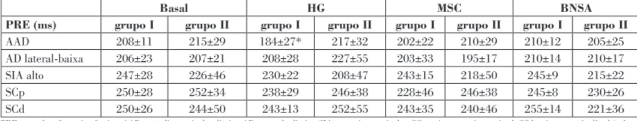
Tabela IV. Comparação dos períodos refractários efectivos avaliados em cinco localizações auriculares em condições basais, durante hand-grip (HG), massagem do seio carotídeo (MSC) e após bloqueio autonómico farmacológico (BSNA) nos doentes com (grupo I, n=5) e sem (grupo II, n=5) vulnerabilidade para indução de fibrilhação auricular.
Basal HG MSC BNSA
PRE (ms) grupo I grupo II grupo I grupo II grupo I grupo II grupo I grupo II
AAD 208±11 215±29 184±27* 217±32 202±22 210±29 210±12 205±25
AD lateral-baixa 206±23 207±21 208±28 227±55 203±33 195±17 210±14 210±17
SIA alto 247±28 226±46 230±22 208±47 243±15 218±50 245±9 215±22
SCp 250±28 252±34 238±29 246±38 228±46 246±38 245±8 230±26
SCd 250±26 244±50 243±13 252±55 243±35 240±46 255±14 221±36
PRE=período refractário efectivo; AAD=apêndice auricular direito; AD=aurícula direita; SIA=septo interauricular; SCp=seio coronário proximal; SCd=seio coronário distal (valores expressos em milissegundos, média±desvio padrão).
* p=0.05 na comparação com o PRE do grupo II e correspondendo a uma diminuição significativa do PRE durante hand-grip (12±9% no grupo I vs 2±4% no grupo II, p=0.02).
Table III. Clinical characteristics and left atrial size (assessed by M-mode echocardiography in parasternal view) of patients with and without vulnerability to AF induction during electrophysiological study.
with AF induction (n=5) without AF induction (n=5) p Age (years) 56±12.1 52.5±17.6 NS Male 4 (80%) 3 (60%) NS LA (M-mode; mm) 42.8±2.2 40.6±3.1 NS LA >22 mm/m2 3 (60%) 1 (20%) NS Duration of AF (years) 2.3±2.0 2.0±1.9 NS No. of previous antiarrhythmics 1.7±0.9 1.6±0.8 NS
665
history of recurrent paroxysmal AF. We also assessed whether vulnerability to AF induction during EPS was associated with different ANS-related responses in terms of atrial refractoriness. The results show that vagal stimulation was associated with shortening of AERPs in the lateral RA, with no significant impact at the other sites assessed or in dispersion of refractoriness. On the other hand, increased sympathetic tone during HG was not associated with significant change in AERPs, whereas ANS blockade not only significantly increased AERPs at the distal CS, reflecting the refractoriness of left atrial tissue according to Chen et al. (18), but also markedly decreased dispersion of refractoriness compared to basal conditions. These findings highlight the role of the ANS as a modulator of dynamic variations in atrial refractoriness.
Autonomic influence on atrial electro-physiology
It is known that ANS activity can affect atrial electrophysiological properties and cause changes in AERP, conduction velocity and automatism(7). Although the sequence of mechanisms underlying episodes of paroxysmal AF has yet to be fully clarified, it has been suggested that nonuniformity of AERPs at different sites is one of the most important factors in vulnerability to AF, since it is consistently associated with spontaneous or induced AF (19-21). In addition, atrial refractoriness heterogeneity shows a stronger correlation with vulnerability to AF than wavelength, which depends on AERP and conduction velocity(20). Thus, nonuniform spatial dispersion of atrial refractoriness may be a key factor in the pathophysiology of AF.
Various authors have suggested that auto-nomic activity, both sympathetic and para-mostram que, nestas condições, a estimulação
vagal se associa a uma diminuição dos PRE na AD lateral, sem impacto significativo noutros locais auriculares avaliados ou na dispersão da refractaridade. Por outro lado, o aumento do tónus simpático durante HG não se associou a modificação significativa dos PRE e o bloqueio autonómico, não só aumentou significativamente os PRE a nível do SC distal, reflectindo, de acordo com Chen et al, a refractariedade no tecido auricular esquerdo(18), como diminuiu, de forma marcada, a dispersão da refractariedade em comparação com os valores basais. Estes da - dos reforçam o papel do SNA como factor modu-lador das variações dinâmicas da refractariedade auricular.
Influência autonómica na electrofisiologia das aurículas
Tem sido reconhecido que a actividade do SNA pode condicionar alterações nas propriedades electrofisiológicas das aurículas envolvendo os PRE, a velocidade de condução e o automatismo(7). Em doentes com FA paroxística, apesar da sequência de mecanismos subjacentes aos episódios desta arritmia permanecer por esclarecer, a não uniformidade dos PRE em diferentes locais é apontada como um dos aspectos mais importantes da vulnerabilidade para FA, associando-se, de forma consistente, à ocorrência de FA espontânea ou induzida(19-21). Além disso, a heterogeneidade da refractariedade auricular apresenta maior correlação com a vulnerabilidade para FA do que o comprimento de onda (wavelenght), que depende do PRE e da velocidade de condução(20). Deste modo, o conceito de dispersão da refractariedade auricular baseada na distribuição espacial não uniforme dos PRE pode representar um factor
Table IV. Comparison of effective refractory periods assessed at 5 atrial sites in basal conditions, during handgrip (HG) and carotid sinus massage (CSM) and after autonomic nervous system blockade (ASNB) in patients with (group I, n=5) and without (group II, n=5) vulnerability to AF induction.
Basal HG CSM ANSB
AERP (ms) group I group II group I group II group I group II group I group II
RAA 208±11 215±29 184±27* 217±32 202±22 210±29 210±12 205±25
Low lateral RA 206±23 207±21 208±28 227±55 203±33 195±17 210±14 210±17
High IAS 247±28 226±46 230±22 208±47 243±15 218±50 245±9 215±22
pCS 250±28 252±34 238±29 246±38 228±46 246±38 245±8 230±26
dCS 250±26 244±50 243±13 252±55 243±35 240±46 255±14 221±36
AERP: atrial effective refractory period; dCS: distal coronary sinus; IAS: interatrial septum; pCS: proximal coronary sinus; RAA: right atrial appendage; RA: right atrium. Values expressed in milliseconds, mean ± standard deviation.
666
Vol. 28 Junho 09 / June 09
sympathetic, may play an important role in modulating recurrence of paroxysmal AF(7-9,22). Vagal stimulation significantly reduces atrial refractoriness in a heterogeneous manner, creating the conditions for the appearance of reentrant atrial arrhythmias and AF(7-9), while sympathetic activity shortens AERP, with no significant effect on heterogeneity(7-9,23). Studies based on analysis of heart rate variability have shown that significant changes in the sympatho-vagal balance precede the onset of spontaneous AF(22,24,25). Thus, fluctuations in autonomic tone may be crucial in characterizing the electrophysiological substrate for episodes of AF.
Atrial effective refractory periods and dispersion of refractoriness
As reported by other authors(26), the AERP increased progressively from right to left at the different sites assessed in our study, with lower values in the RAA, LRA and IAS than those recorded in the proximal and distal CS, possibly due to the nonuniform distribution of vagal nerve terminals, which appear to affect AERP more in the RAA than in the left atrium(27).
The maneuvers used for ANS stimulation were handgrip, which is considered a simple, reproducible test with high sensitivity and specificity for sympathetic stimulation(28), and carotid sinus massage, which is frequently used in neuroautonomic assessment since it produces a parasympathetic reflex response(29). Although both maneuvers produced slight changes in heart rate and systolic BP (with statistical significance in the case of HG), there were considerable differences in double product (HG vs. MSC, p<0.001), and assessment of RR variability by spectral analysis in the frequency domain confirmed the effect on autonomic activity of these maneuvers and of pharmacological blockade with propranolol and atropine (Figure 1).
In patients with ≥1 year clinical history of paroxysmal AF, alterations in electrophysiological properties resulting from atrial remodeling are to be expected, particularly heterogeneous shortening of AERPs, which contributes to perpetuation of the condition(30,31). Even so, vagal stimulation significantly reduced AERPs. However, only ANS blockade had a significant impact on refractoriness in the distal CS, with a marked decrease in dispersion. These results chave na fisiopatologia da FA.
Diferentes autores têm sugerido que a actividade autonómica, quer simpática quer parassimpática, pode desempenhar um papel importante como modulador na recorrência de episódios de FA paroxística(7-9,22). A estimulação vagal reduz significativamente a refractarie-dade auricular, de modo heterogéneo, criando condições para o aparecimento de arritmias auriculares de reentrada e FA(7-9), enquanto a actividade simpá tica reduz os PRE, sem efeito significativo nos índices de heterogeneidade(7-9,23). Estudos com base na análise da variabilidade da frequência cardíaca têm mostrado que modi-ficações significativas no balanço simpático- -vagal precedem o início de FA espontânea(22,24,25). Portanto, as flutuações do tónus autonómico podem ser determinantes na caracterização do substrato electrofisiológico para a ocorrência de episódios de FA.
Períodos refractários efectivos auriculares e dispersão da refractariedade
No nosso estudo, à semelhança do descrito por outros autores(26), o PRE aumentou gradualmente nos diferentes locais analisados da direita para a esquerda, com valores mais baixos no AAD, AD lateral e SIA quando comparados com as determinações no SC proximal e distal. Facto possivelmente causado pela distribuição não uniforme das terminações vagais nervosas, que parecem influenciar de forma mais acentuada o PRE no AAD que na aurícula esquerda(27).
As manobras provocativas do SNA utilizadas envolveram o HG, considerado como um teste simples, facilmente reprodutível, com elevada sensibilidade e especificidade para estimulação da actividade simpática(28) e a MSC, aplicada frequentemente como método de avaliação neuro-autonómica capaz de evocar uma res-pos ta reflexa parassimpática(29). Apesar de condicionarem alterações ligeiras da frequência cardíaca e pressão arterial sistólica (com signi-ficado estatístico para o HG), associaram-se a uma grande amplitude na variação dos valores do duplo-produto (HG versus MSC, p<0.001) e a monitorização da variabilidade RR no gráfico da análise espectral no domínio da frequência permitiu confirmar os efeitos destas manobras e do bloqueio farmacológico com propranolol e atropina na actividade autonómica (Figura 1).
667
may indicate the protective effect of drugs with sympatholytic and vagolytic action. Thus, in patients with a long clinical history of recurrent paroxysmal AF, autonomic stimulation, parti-cularly of vagal activity, appears to play an important role in reducing atrial refractoriness. A recent study by Lu et al. demonstrated that the intrinsic cardiac autonomic nervous system plays a critical role in acute atrial electrical remodeling induced by rapid atrial pacing, and that it is possible to prevent AERP reduction and AF induction with ANS blockade(32). In the present study, autonomic blockade enabled changes in AERP and dispersion of refractoriness to be documented that may influence vulnerability to AF in patients with a long clinical history of such episodes.
Atrial refractoriness and vulnerability to atrial fibrillation induction
Our results show that AERP heterogeneity is a determining factor in atrial vulnerability, and that AF induction is associated with greater basal dispersion of refractoriness, despite similar absolute AERP values at the different sites assessed. This finding is in agreement with the study by Fareh et al., in which vulnerability to AF correlated with AERP heterogeneity but not with AERP or wavelength(20). The present study found a tendency for the left atrium to be larger in patients with AF induction, but there were no statistically significant differences in the clinical characteristics of the two groups. The larger reduction in AERP observed at the RAA in the group vulnerable to AF induction, together with increased dispersion of refractoriness in response to sympathetic stimulation, may reflect greater susceptibility to arrhythmias following ectopic beats or extrastimuli with shorter coupling intervals, which demonstrates the possible role of this electrophysiological phenomenon as a substrate for AF inducibility.
STUDY LIMITATIONS AND CONCLUSIONS
The size of the sample did not allow for subgroup analysis based on the presence of spontaneous supraventricular ectopic beats or on the strength of response to autonomic Num grupo de doentes com FA paroxística e
evolução clínica ≥1 ano são esperadas alterações das propriedades electrofisiológicas, como con - se quência do fenómeno de remodelagem auri-cular, associando-se, em partiauri-cular, a uma diminuição heterogénea dos PRE que contribui para perpetuar a situação clínica(30,31). Ainda assim, a estimulação vagal permitiu obter uma redução significativa dos PRE na AD. No entanto, apenas o bloqueio autonómico influenciou de modo significativo o valor da refractariedade ao nível do SC distal, associando-se a uma marcada diminuição da dispersão da refractariedade. Estes resultados podem traduzir um efeito protector da utilização de fármacos com acção simpaticolítica e vagolítica, face ao aumento da refractariedade em tecido auricular esquerdo combinado com a redução da variabilidade dos PRE. Assim, em situações de FA paroxística com clínica recorrente de longa duração, a estimulação autonómica, em particular da actividade vagal, parece manter um papel importante na redução da refractariedade auricular. Num estudo re-cente, Lu et al demonstraram que a inervação cardíaca autonómica tem um impacto crucial na fase aguda da remodelagem eléctrica induzida por pacing auricular de alta frequência, sendo possível prevenir a redução dos PRE e a indução de FA com bloqueio do SNA(32). No presente trabalho, o bloqueio autonómico permitiu evi-denciar modificações nos PRE e dispersão da refractariedade que podem ter impacto na vul-nerabilidade para episódios de FA em situa ções de evolução clínica de longo prazo.
Refractariedade auricular e vulnerabilidade para indução de fibrilhação auricular
Os resultados mostram que a heterogeneidade dos PRE é um factor determinante da vulnera-bilidade auricular, associando-se a indução de FA à presença de valores basais mais altos da dispersão da refractariedade, apesar de valores absolutos semelhantes para os PRE nos diferen-tes locais analizados. Esdiferen-tes dados estão de acordo com o estudo de Fareh, onde a a vulnerabilidade para FA se correlacionava com a heterogenei-dade dos PRE auriculares mas não com o PRE ou wavelenght(20). No presente trabalho, verificou--se uma tendência para o tamanho da aurícula esquerda ser maior nos doentes com indução de FA mas não houve diferenças estatisticamente
668
Vol. 28 Junho 09 / June 09
maneuvers. Furthermore, the fact that patients with paroxysmal AF of recent onset were not included means that the study’s conclusions can only be applied to cases with a long clinical history of the condition. Nevertheless, this group of patients with lone paroxysmal AF undergoing a clinical and electrophysiological protocol as the initial selection stage of an AF, ablation program, showed similar behavior in terms of the effect of autonomic stimulation and blockade on the aspects of atrial refractoriness studied. Another limitation is that pulmonary vein refractoriness was not studied, and thus no information is available to compare AERPs at the five sites assessed with those of the pulmonary veins, which are known to be shorter than those of the left atrium(33,34). These points should be taken into consideration in any future study in order to better understand the electrophysiological phenomena involved in the onset, perpetuation and termination of AF episodes. Finally, although the effects of HG and CSM were confirmed on the basis of heart rate, blood pressure and spectral analysis of frequency variations, use of other maneuvers to stimulate autonomic activity, such as cold pressor test, deep breathing or Valsalva maneuver, would have improved the results of stimulation of sympathetic and/or parasympathetic tone.
In conclusion, patients with a long clinical history of lone paroxysmal AF show slight changes in atrial refractoriness during ANS stimulation, but maintain significant shortening of AERP in the right atrium with vagal stimulation. Increased AERP in the distal CS, associated with decreased dispersion of refractoriness during autonomic blockade, suggests a possible role for pharmacological or non-pharmacological intervention in changes to the electrophysiological substrate related to vulnerability to AF.
significativas nas características clínicas dos dois grupos. A documentação duma redução mais acentuada dos PRE no AAD, combinada com a maior dispersão da refractariedade em res posta à estimulação simpática no grupo com vulnerabilidade para indução de FA, pode repre sentar uma maior susceptibilidade para a ocorrência de arritmias na sequência de ectopias ou extra-estímulos com intervalos de acoplamento mais curtos, traduzindo assim um papel potencial deste fenómeno electrofisioló gi-co gi-como substrato para a inducibilidade de FA.
LIMITAÇÕES DO ESTUDO E CONCLUSÕES
A dimensão da amostra não permitiu a análise estatística em subgrupos tendo em conta a presença de ectopias supraventriculares espontâneas ou o grau de intensidade da resposta às manobras autonómicas. Além disso, a não inclusão de doentes com FA paroxística de início recente limita as conclusões do estudo a situações com história clínica de longa duração. No entanto, trata-se dum grupo restrito de doentes com FA paroxística idiopática, incluído em protocolo de avaliação clínica e electrofisiológica de selecção numa fase inicial do programa de ablação de FA, que mostrou um comportamento homogéneo no que concerne à influência da activação e bloqueio autonómico nos aspectos avaliados da refractariedade auricular. Outra limitação está relacionada com a não inclusão do estudo da refractariedade das veias pulmonares, não dispondo assim de informação completa para comparação dos PRE dos cinco locais avaliados com os das veias pulmonares, aceites como mais curtos que os da aurícula esquerda(33,34). No futuro, a importância destes aspectos deverá ser considerada para melhor compreensão dos fenómenos electrofisiológicos envolvidos no início, manutenção e interrupção dos episódios de FA. Finalmente, apesar da confirmação dos efeitos do HG e MSC com base na frequência cardíaca, pressão arterial e análise espectral da variabilidade da frequência, o recurso a outras manobras provocativas da actividade autonómica, como o teste do frio, a respiração profunda (deep-breathing) ou a manobra de Valsalva teriam contribuído para melhorar os resultados da estimulação do tónus simpático e/ ou parassimpático.
669
Pedido de separatas: Address for reprints: MÁRIO OLIVEIRA Cardiology Department Santa Marta Hospital Rua Santa Marta
1169-024 Lisboa - Portugal Fax: 351 213 560 368 Phone: 351 213 594 311
e-mail: m.martinsoliveira@gmail.com Em conclusão, nos doentes com FA
paroxísti-ca idiopátiparoxísti-ca de longa evolução clíniparoxísti-ca ocorrem ligeiras alterações da refractariedade auricular du rante estimulação do SNA, mantendo encur-tamento significativo dos PRE na AD com esti-mulação vagal. O aumento dos PRE no SC dis tal associado à menor dispersão da refractarie dade resultante do bloqueio autonómico sugere um papel potencial para medidas terapêuticas de âmbito farmacológico ou não farmacológico nas alterações do substrato electrofisiológico relaci-onado com vulnerabilidade para FA.
BIBLIOGRAFIA / REFERENCES
[1] Camm AJ. Preface. In: Murgatroyd, FD, Camm, AJ, editors. Nonpharmacological Treatment of Atrial Fibrillation. Armonk, NY: Futura; 1997.
[2] Feinberg WM, Blackshear JL, Laupacis A, et al. Prevalence, age distributions, and gender of patients with atrial fibrillation: analysis and implications. Arch Intern Med 1995;155:469–73. [3] Go AS, Hylek EM, Philips KA, et al. Prevalence of diagnosed atrial fibrillation in adults: national implications for rhythm management and stroke prevention: the AnTicoagulation and Risk Factors in Atrial Fibrillation (ATRIA) Study. JAMA 2001; 285:2370-2375.
[4] Benjamin EJ, Wolf PA, D’Agostino RB, et al. Impact of atrial fibrillation on the risk of death. The Framingham Heart Study. Circulation 1998;98: 946–52.
[5] Van Den Berg MP, Hassink RJ, Tuinenburg AE, Van Sonderen EF, Lefrandt JD, et al. Quality of life in patients with paroxysmal atrial fibrillation and its predictors: importance of the autonomic nervous system. Eur Heart J 2001;22:247–53. [6] Murgatroyd FD, Camm AJ. Atrial arrhythmias. Lancet, 1993, 341:1317-1322.
[7] Chen P, Tan AY. Autonomic nerve activity and atrial fibrillation. Heart Rhythm. 2007 March; 4(3 Suppl):S61-64. [8] Coumel P. Autonomic influences in atrial tachyarrhythmias. J Cardiovasc Electrophysiol, October 1996; vol. 7(10): 999-1007.
[9] Tai CT. Role of autonomic influences in the initiation and perpetuation of focal atrial fibrillation. J Cardiovasc Electrophysiol. 2001 Mar;12(3):292-3.
[10] Elosua R, Arquer A, Mont L, et al. Sport practice and the risk of lone atrial fibrillation: a case-control study. Int J Cardiol, 2006, 108:332-337.
[11] Liu L, Nattel S. Differing sympathetic and vagal effects on atrial fibrillation in dogs: role of refractoriness heterogeneity. Am J Physiol 1997; 273:H805-H816.
[12] Wijfeels MC, Kirchoff CJ, Dorland R, Allessie MA. Atrial fibrillation begets atrial fibrillation. A study in awake chronically instrumented goats. Circulation, 1995, 92:1954-1968. [13] Soylu M, Demir AD, Özdemir Ö, Soylu O, et al. Increased Dispersion of Refractoriness in Patients with Atrial Fibrillation in the Early Postoperative Period after Coronary Artery Bypass Grafting. J Cardiovasc Electrophysiol, January 2003, vol. 14, pp. 28-31.
[14] Miller JM. Diagnosis of Cardiac Arrhythmias. In: Libby P, Bonow RO, Mann DL, Zipes DP, eds. Braunwald’s Heart Disease: A Textbook of Cardiovascular Medicine, 8th ed. St. Louis, Mo: WB Saunders; 2007: chap. 32.
[15] Olgin JE, Zipes P. Specific Arrhythmias: Diagnosis and Treatment. In: Libby P, Bonow RO, Mann DL, Zipes DP, eds. Braunwald’s Heart Disease: A Textbook of Cardiovascular Medicine, 8th ed. St. Louis, Mo: WB Saunders; 2007: chap. 35 [16] Fogoros, RN. The Electrophysiology Study in the Evaluation and Therapy of Cardiac Arrhythmias. In: Fogoros, RN. Electrophysiologic Testing. Blackwell Publishing: Malden, MA, 2006: 35-157.
[17] Oliveira M, Silva N, Timóteo AT, et al. Dispersão da Refractariedade Auricular como Substrato Electrofisiológico da Vulnerabilidade Auricular em Doentes com Fibrilhação Auricular Paroxística. Rev Port Cardiol 2007; 26 (7-8):1-12.
670
Vol. 28 Junho 09 / June 09
[26] Ishimatsu T, Hayano M, Hirata T, Iliev II, et al. Electrophysiological properties of the left atrium evaluated by coronary sinus pacing in patients with atrial fibrillation. Pacing Clin Electrophysiol 1999 Dec; 22(12):1739-46.
[27] Zipes DP, Mihalick MJ, Robbins GT. Effects of selective vagal and stellate ganglion stimulation on atrial refractoriness. Cardiovasc Res 1974;8:647-55.
[28] Khurana RK, Setty A. The value of the isometric hand-grip test–studies in various autonomic disorders. Clin Auton Res. 1996;6:211–218.
[29] Tea SH, Mansourati J, L’Heveder G, Mabin D, Blanc JJ. New insights into the pathophysiology of carotid sinus syndrome. Circulation 1996; 93:1411–6.
[30] Daoud EG, Bogum F, Goyal R, Harvey M, Man KC, Strickberger SA, Morady F. Effect of atrial fibrillation on atrial refractoriness in humans. Circulation 1996;94:1600–6. [31] Goette A, Honeycutt C, Langberg JJ. Electrical remodeling in atrial fibrillation: time course and mechanisms. Circulation 1996;94:2968–74.
[32] Lu Z, Scherlag B, Lin J, et al. Atrial Fibrillation Begets Atrial Fibrillation. Autonomic Mechanisms for Atrial Electrical Remodeling Induced by Short-Term Rapid Atrial Pacing. Circulation 2008;1:184-192.
[33] Adragão P, Santos K, Aguiar C, Neves J, Abecassis M, et al. Atrial fibrillation and effective refractory period of the pulmonary vein ostia. Rev Port Cardiol 2002 Oct;21(10):1125-34.
[34] Jais P, Hocini M, Macle L, Choi KJ, Deisenhofer I, Weerasooriya R, et al. Distinctive electrophysiological properties of pulmonary veins in patients with atrial fibrillation. Circulation. 2002 Nov 5;106(19):2479-85.
[18] Chen M, Guo GB, Chang HW. Atrial electrophysiological properties evaluated by right and left atrial pacing in patients with or without atrial fibrillation. Jpn Heart J 2002; 43:231-240.
[19] Zhen L, Hertervig E, Carlson J, Camilla J, Bertil O, Yuan S. Dispersion of refractoriness in patients with paroxysmal atrial fibrillation. Evaluation with simultaneous endocardial recordings from both atria. Journal of Electrocardiology 2002; Vol. 35 No. 3:227-34.
[20] Fareh S, Villemaire C, Nattel S. Importance of refractoriness heterogeneity in the enhanced vulnerability to atrial fibrillation induction caused by tachycardia-induced atrial electrical remodeling. Circulation 1998; 98: 2202–2209.
[21] Oliveira M, da Silva MN, Timoteo AT, Feliciano J, Sousa L, Santos S, Silva-Carvalho L, Ferreira R. Inducibility of atrial fibrillation during electrophysiologic evaluation is associated with increased dispersion of atrial refractoriness. Int J Cardiol. 2008 Aug 1doi:10.1016/j.ijcard.2008.04.097.
[22] Tomita T, Takei M, Saikawa Y, et al. Role of autonomic tone in initiation and termination of paroxysmal atrial fibrillation in patients without structural heart disease. J Cardiovasc Electrophysiol, 2003 June, vol. 14:559-64.
[23] Liu L, Nattel S. Differing sympathetic and vagal effects on atrial fibrillation in dogs: role of refractoriness heterogeneity. Am J Physiol Heart Circ Physiol 1997, 273: H805-H816. [24] Klingenheben T, Gronefeld GC, Li YG. Heart rate variability to assess changes in cardiac vagal modulation before the onset of paroxysmal atrial fibrillation in patients with and without structural heart disease. Ann Noninvasive Electrocardiol 1999;4:19–26.
[25] Bettoni M, Zimmermann M. Autonomic tone variations before the onset of paroxysmal atrial fibrillation. Circulation. 2002;105:2753–2759.