Universidade de Lisboa
Faculdade de Medicina de Lisboa
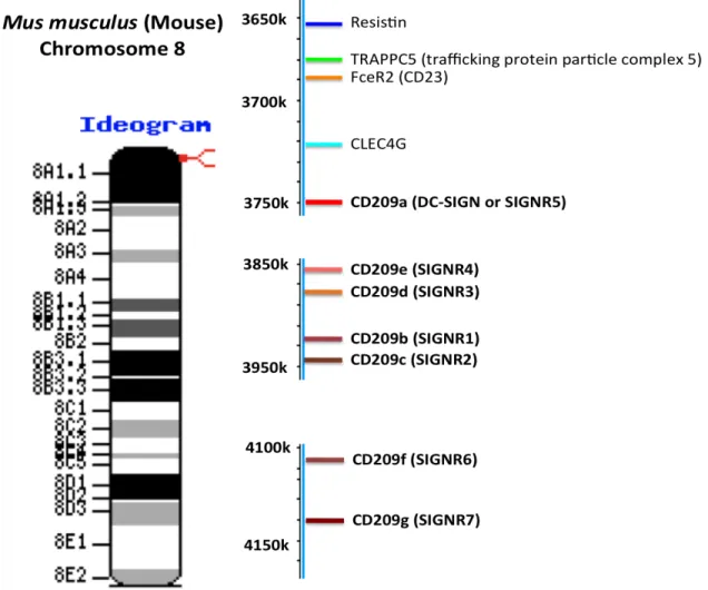
The mouse C-type lectin DC-SIGN/CD209a is
a marker for monocyte derived DCs and
reveals a new pathway of DC differentiation
upon bacterial stimulation
Inês Grazina de Matos
Doutoramento em Ciências Biomédicas
Especialidade de Ciências Biopatológicas
Universidade de Lisboa
Faculdade de Medicina de Lisboa
The mouse C-type lectin DC-SIGN/CD209a is a marker
for monocyte derived DCs and reveals a new pathway of
DC differentiation upon bacterial stimulation
Inês Grazina de Matos
Tese orientada por:
Professor Doutor Ralph M. Steinman, Laboratory of Cellular
Physiology and Immunology, Rockefeller University
Co-Orientador:
Professor Doutor Luís Graça, Faculdade de Medicina da
Universidade de Lisboa
Doutoramento em Ciências Biomédicas
Especialidade de Ciências Biopatológicas
Todas as afirmações apresentadas no presente documento são da
exclusiva responsabilidade do seu autor, não cabendo qualquer
responsabilidade á Faculdade de Medicina de Lisboa pelos
conteúdos nele apresentados.
A impressão desta dissertação foi aprovada pelo
Conselho Científico da Faculdade de Medicina de
Lisboa em reunião de 20 de Julho de 2011.
! ! !
"!
Prefácio
Nesta dissertação apresentam-se os resultados do trabalho desenvolvido entre Setembro de 2007 e Maio de 2011, no Laboratório de Fisiologia Celular e Imunologia (Rockefeller University, New York, New York, USA) sob a orientação do Professor Doutor Ralph M. Steinman (supervisor internacional), e tendo como supervisor nacional o Professor Doutor Luís Graca.
Este trabalho teve como principal objectivo a produção de anticorpos monoclonais contra a letina DC-SIGN/CD209a do rato. Este reagente foi importante para a identificação e caracterização de um novo subtipo de células dendriticas que se diferenciam a partir de monócitos do sangue em resposta ao LPS (lipossacarídeo) um constituinte da parede celular de bactérias gram-negativas.
A presente dissertação encontra-se dividida em 7 capítulos: Introduction, onde se encontram resumidos os conhecimentos até à data acerca das células dendriticas, suas funções, principais subtipos e receptores que expressam para reconhecimento de patógenes e onde focamos a abordagem na lectina do tipo C denominada DC-SIGN/CD209a; Experimental procedures, com a descrição dos métodos e reagentes utilizados durante a investigação levada a cabo neste estudo; Results, onde são apresentados todos os resultados originais obtidos neste estudo, parte dos quais são apresentados também na forma de artigos no último capítulo. Discussion, onde são discutidos detalhadamente os resultados obtidos, Conclusion and Future Perspectives com as conclusões a que permitiu chegar este estudo e onde são também apresentadas perspectivas de investigação futura; References, onde são enumeradas as publicações consultadas durante a elaboração desta tese. Finalmente, Publications, onde constam os artigos publicados em revistas científicas durante o período de execução da presente dissertação.
! ! !
#!
De acordo com o disposto no artigo 40° do Regulamento de Estudos Pós-Graduados da Universidade de Lisboa, Deliberação n°961/2003, publicada no Diário da República – IIa Série, n° 153 de 5 de Julho de 2003, foram incluídos nesta tese resultados dos artigos abaixo indicados:
Manuscripto 1. Cheong C, Matos I, Choi JH, Schauer JD, Dandamudi DB, Shrestha E, Makeyeva JA, Li X, Li P, Steinman RM, Park CG. New monoclonal
anti-mouse DC-SIGN antibodies reactive with acetone-fixed cells. 2010 Aug 31. J
Immunol Methods. 360(1-2):66-75.
Manuscripto 2. Cheong C*, Matos I*, Choi JH, Dandamudi DB, Shrestha E, Longhi MP, Jeffrey KL, Anthony RM, Kluger C, Nchinda G, Koh H, Rodriguez A, Idoyaga J, Pack M, Velinzon K, Park CG, Steinman RM.. Microbial stimulation
fully differentiates monocytes to DC-SIGN/CD209(+) dendritic cells for immune T cell areas. 2010 Oct 29. Cell. 143(3):416-29. *NOTE: Equal contribution.
No cumprimento do disposto na referida deliberação, a autora esclarece serem da sua inteira responsabilidade, excepto quando referido em contrário, a execução das experiências que levaram aos resultados apresentados nesta tese assim como a interpretação e discussão dos mesmos. Resultados obtidos por outros autores foram tambem incluidos com autorização dos mesmos, de forma a facilitar a compreensão dos trabalhos e estão devidamente assinalados nas respectivas figuras.
! ! !
$!
Resumo
As células dendríticas (DCs) desempenham um papel fundamental na regulação da resposta imunitária pela sua eficiência na apresentação de
antigénios. Sabe-se que as células dendriticas derivam de precursores da medula óssea, mas o seu potencial para se desenvolverem directamente a partir de monócitos em circulação no sangue nunca foi devidamente
demonstrado in vivo.
Neste estudo provamos que a diferenciação de DCs pode ocorrer a partir de monócitos (DC, de “monocyte-derived DCs”) no murganho. Estas Mo-DCs caracterizam-se pela expressão de CD209a, também designada como DC-SIGN, uma proteína da família das lectinas do tipo C. Células que expressam esta proteína são encontradas nos nódulos linfáticos após a injecção de lipossacarídeo (LPS) purificado ou de bactérias gram-negativas. Mostrámos ainda que a mobilização destas Mo-DCs requer a presença dos receptores TLR4 e CD14 e da proteína intracelular Trif.
As células Mo-DCs descritas neste trabalho apresentam uma morfologia típica de DCs e localizam-se nos nódulos linfáticos na área das células T. Esta localização está de acordo com sua função na apresentação de antigénios, que inclui a apresentação cruzada de antigénios associados a moléculas de MHC classe I. Foi ainda demonstrado que a mobilização das células Mo-DCs in vivo depende da expressão da molécula de adesão L-selectina e da quimoquina CCR7.
Em resumo, este trabalho mostra pela primeira vez que monócitos presentes no sangue se podem diferenciar em células dendríticas. Este processo de diferenciação ocorre em resposta à presença de LPS, um componente da parede celular de bactérias Gram-negativas, e involve a
aquisição de diversas características tipicamente associadas a DCs. Esta nova sub-polulação de DCs é também caracterizada pela expressão da proteína CD209a.
! ! !
! ! !
&!
Abstract
Dendritic cells (DCs), critical antigen-presenting cells for immune control, normally derive from bone marrow precursors distinct from monocytes. It is not yet established if the large reservoir of monocytes can develop into cells with critical features of DCs in vivo. We now show that fully differentiated monocyte-derived DCs (Mo-DCs) can develop in mice and are marked by the C-type lectin DC-SIGN/CD209a. Here, we report that Mo-DCs are recruited from blood monocytes into lymph nodes by lipopolysaccharide (LPS) and live or dead gram-negative bacteria. These Mo-DCs mobilization requires TLR4 and its CD14 coreceptor via a Trif dependent signaling pathway. When tested for antigen-presenting function, Mo-DCs are as active as conventional DCs, including cross-presentation of antigens of proteins or live gram-negative bacteria on MHC I. Fully differentiated Mo-DCs acquire DC morphology and localize to T cell areas via the adhesion molecule L-selectin and the chemokine CCR7. Thus the large reservoir of blood monocyte can become the dominant presenting cell in response to select microbes, acquire the expression of DC-SIGN and other critical features of DCs.
! ! !
! ! ! (! Prefácio!!!!!!!!!!!!!!!!!!!!!!!!!!!!!!!!!!!!!!!!!!!!!!!!!!!!!!!!!!!!!!!!!!!!!!!!!!!!!!!!!!!!!!!!!!!!!!!!!!!!!!!!!!!!!!!!!!!!!!!!!!!!!!!!!!!!!! "! Resumo!!!!!!!!!!!!!!!!!!!!!!!!!!!!!!!!!!!!!!!!!!!!!!!!!!!!!!!!!!!!!!!!!!!!!!!!!!!!!!!!!!!!!!!!!!!!!!!!!!!!!!!!!!!!!!!!!!!!!!!!!!!!!!!!!!!!!! #! Abstract!!!!!!!!!!!!!!!!!!!!!!!!!!!!!!!!!!!!!!!!!!!!!!!!!!!!!!!!!!!!!!!!!!!!!!!!!!!!!!!!!!!!!!!!!!!!!!!!!!!!!!!!!!!!!!!!!!!!!!!!!!!!!!!!!!!!!! $! List of abbreviations!!!!!!!!!!!!!!!!!!!!!!!!!!!!!!!!!!!!!!!!!!!!!!!!!!!!!!!!!!!!!!!!!!!!!!!!!!!!!!!!!!!!!!!!!!!!!!!!!!!!!!!!!!!!! %! Acknowledgements!!!!!!!!!!!!!!!!!!!!!!!!!!!!!!!!!!!!!!!!!!!!!!!!!!!!!!!!!!!!!!!!!!!!!!!!!!!!!!!!!!!!!!!!!!!!!!!!!!!!!!!!!!!! ""! Chapter 1. Introduction!!!!!!!!!!!!!!!!!!!!!!!!!!!!!!!!!!!!!!!!!!!!!!!!!!!!!!!!!!!!!!!!!!!!!!!!!!!!!!!!!!!!!!!!!!!!!!!!!!!!! "$! A. The Immune System!!!!!!!!!!!!!!!!!!!!!!!!!!!!!!!!!!!!!!!!!!!!!!!!!!!!!!!!!!!!!!!!!!!!!!!!!!!!!!!!!!!!!!!!!!!!!!!!!!! "&! B. Dendritic Cells!!!!!!!!!!!!!!!!!!!!!!!!!!!!!!!!!!!!!!!!!!!!!!!!!!!!!!!!!!!!!!!!!!!!!!!!!!!!!!!!!!!!!!!!!!!!!!!!!!!!!!!!!!!!!! "'!
1. DC features!!!!!!!!!!!!!!!!!!!!!!!!!!!!!!!!!!!!!!!!!!!!!!!!!!!!!!!!!!!!!!!!!!!!!!!!!!!!!!!!!!!!!!!!!!!!!!!!!!!!!!!!!!!!!!!!!!!!!!!!!!!!!!!!! "(!
1.1. Potent antigen presenting cells))))))))))))))))))))))))))))))))))))))))))))))))))))))))))))))))))))))))))))))))))))))))))))))))))"*!
1.2. DC migration))))))))))))))))))))))))))))))))))))))))))))))))))))))))))))))))))))))))))))))))))))))))))))))))))))))))))))))))))))))))))))))))))))))))"+!
1.3. DC Maturation as a Control Point for Initiating Immunity))))))))))))))))))))))))))))))))))))))))))))))#"!
2. DC subsets!!!!!!!!!!!!!!!!!!!!!!!!!!!!!!!!!!!!!!!!!!!!!!!!!!!!!!!!!!!!!!!!!!!!!!!!!!!!!!!!!!!!!!!!!!!!!!!!!!!!!!!!!!!!!!!!!!!!!!!!!!!!!!!!!! )*!
2.1. Classification criterion)))))))))))))))))))))))))))))))))))))))))))))))))))))))))))))))))))))))))))))))))))))))))))))))))))))))))))))))))))))#%!
2.2. Phenotype of mouse DCs subsets in peripheral lymphoid organs (Table 1))))))#&!
3. Origin of DCs in lymphoid tissues!!!!!!!!!!!!!!!!!!!!!!!!!!!!!!!!!!!!!!!!!!!!!!!!!!!!!!!!!!!!!!!!!!!!!!!!!!!!!!!!!!!! )'!
3.1. In steady state conditions))))))))))))))))))))))))))))))))))))))))))))))))))))))))))))))))))))))))))))))))))))))))))))))))))))))))))))))#(!
3.2. Under inflammatory conditions)))))))))))))))))))))))))))))))))))))))))))))))))))))))))))))))))))))))))))))))))))))))))))))))))))$#!
4. Different DC subsets are involved in different types of immune responses.!! #*!
5. C-type lectins on DCs are key modulators of the immune responses!!!!!!!!!!!!!!! #&!
5.1. C-type lectins receptors are differentially expressed on different DC subsets))$(!
5.2. Therapeutic potential of C-Type lectins)))))))))))))))))))))))))))))))))))))))))))))))))))))))))))))))))))))))))))))))))$+!
6. The DC-SIGN Receptor!!!!!!!!!!!!!!!!!!!!!!!!!!!!!!!!!!!!!!!!!!!!!!!!!!!!!!!!!!!!!!!!!!!!!!!!!!!!!!!!!!!!!!!!!!!!!!!!!!!!!!!!!! *+!
6.1. Structure of DC-SIGN))))))))))))))))))))))))))))))))))))))))))))))))))))))))))))))))))))))))))))))))))))))))))))))))))))))))))))))))))))))%,!
6.2. Expression of DC-SIGN)))))))))))))))))))))))))))))))))))))))))))))))))))))))))))))))))))))))))))))))))))))))))))))))))))))))))))))))))%#!
6.3. Immunological functions of DC-SIGN)))))))))))))))))))))))))))))))))))))))))))))))))))))))))))))))))))))))))))))))))))))%&!
C. Aims of the project!!!!!!!!!!!!!!!!!!!!!!!!!!!!!!!!!!!!!!!!!!!!!!!!!!!!!!!!!!!!!!!!!!!!!!!!!!!!!!!!!!!!!!!!!!!!!!!!!!!!!! *&!
Chapter 2. Experimental Procedures!!!!!!!!!!!!!!!!!!!!!!!!!!!!!!!!!!!!!!!!!!!!!!!!!!!!!!!!!!!!!!!!!!!!!!!!!! *'!
2.1. Cell lines, mAbs production and labelling!!!!!!!!!!!!!!!!!!!!!!!!!!!!!!!!!!!!!!!!!!!!!!!!!!!!!!!!!!!!!!!!! *(!
2.2. Animals!!!!!!!!!!!!!!!!!!!!!!!!!!!!!!!!!!!!!!!!!!!!!!!!!!!!!!!!!!!!!!!!!!!!!!!!!!!!!!!!!!!!!!!!!!!!!!!!!!!!!!!!!!!!!!!!!!!!!!!!!!!!!!!!!!!!! *(!
2.3. Antibodies!!!!!!!!!!!!!!!!!!!!!!!!!!!!!!!!!!!!!!!!!!!!!!!!!!!!!!!!!!!!!!!!!!!!!!!!!!!!!!!!!!!!!!!!!!!!!!!!!!!!!!!!!!!!!!!!!!!!!!!!!!!!!!!! *(!
2.4. Lipopolysaccharide and Bacteria!!!!!!!!!!!!!!!!!!!!!!!!!!!!!!!!!!!!!!!!!!!!!!!!!!!!!!!!!!!!!!!!!!!!!!!!!!!!!!!!!! *%!
2.5. Generation of DCs from Bone marrow progenitors!!!!!!!!!!!!!!!!!!!!!!!!!!!!!!!!!!!!!!!!!!!!!!! $+!
2.6. Monocyte and Bone Marrow Transfer!!!!!!!!!!!!!!!!!!!!!!!!!!!!!!!!!!!!!!!!!!!!!!!!!!!!!!!!!!!!!!!!!!!!!!!!!! $"!
2.7. BrdU labeling!!!!!!!!!!!!!!!!!!!!!!!!!!!!!!!!!!!!!!!!!!!!!!!!!!!!!!!!!!!!!!!!!!!!!!!!!!!!!!!!!!!!!!!!!!!!!!!!!!!!!!!!!!!!!!!!!!!!!!!!!! $"!
2.8. Lymph node and spleen cell suspensions!!!!!!!!!!!!!!!!!!!!!!!!!!!!!!!!!!!!!!!!!!!!!!!!!!!!!!!!!!!!!!!!! $"!
2.9. Microscopy!!!!!!!!!!!!!!!!!!!!!!!!!!!!!!!!!!!!!!!!!!!!!!!!!!!!!!!!!!!!!!!!!!!!!!!!!!!!!!!!!!!!!!!!!!!!!!!!!!!!!!!!!!!!!!!!!!!!!!!!!!!!!! $)!
2.10. Antigen Presentation!!!!!!!!!!!!!!!!!!!!!!!!!!!!!!!!!!!!!!!!!!!!!!!!!!!!!!!!!!!!!!!!!!!!!!!!!!!!!!!!!!!!!!!!!!!!!!!!!!!!!!!! $)!
2.11. Quantitative PCR for TLR and CD14 Expression by Monocytes and Mo-DCs !!!!!!!!!!!!!!!!!!!!!!!!!!!!!!!!!!!!!!!!!!!!!!!!!!!!!!!!!!!!!!!!!!!!!!!!!!!!!!!!!!!!!!!!!!!!!!!!!!!!!!!!!!!!!!!!!!!!!!!!!!!!!!!!!!!!!!!!!!!!!!!!!!!!!!!!!!!!!! $#!
2.12. Microarray analysis!!!!!!!!!!!!!!!!!!!!!!!!!!!!!!!!!!!!!!!!!!!!!!!!!!!!!!!!!!!!!!!!!!!!!!!!!!!!!!!!!!!!!!!!!!!!!!!!!!!!!!!!!!! $#!
! ! !
*! Chapter 3. Results!!!!!!!!!!!!!!!!!!!!!!!!!!!!!!!!!!!!!!!!!!!!!!!!!!!!!!!!!!!!!!!!!!!!!!!!!!!!!!!!!!!!!!!!!!!!!!!!!!!!!!!!!!!!!!! $$!
3.1. Production of mouse DC-SIGN/CD209a monoclonal antibody!!!!!!!!!!!!!!!!!!!!!!!!!! $&!
3.2. Comparison of different monoclonal antibodies available against the mouse DC-SIGN/CD209a extracellular domain!!!!!!!!!!!!!!!!!!!!!!!!!!!!!!!!!!!!!!!!!!!!!!!!!!!!!!!!!!!!!!!!!!!!!!!!!!!!!!!! $&!
3.3. DC-SIGN/CD209a is a specific marker for Bone Marrow derived Mo-DCs!!!! $(!
3.4. TLR4 Agonists Rapidly Recruit DC-SIGN+ Cells to the T Cell Area of LNs!!! &*! 3.5. Mo-DCs Can Be Selectively Labeled with Injected Anti-DC-SIGN/CD209a Antibody and Isolated from Classical DCs in LPS treated Skin draining Lymph Nodes!!!!!!!!!!!!!!!!!!!!!!!!!!!!!!!!!!!!!!!!!!!!!!!!!!!!!!!!!!!!!!!!!!!!!!!!!!!!!!!!!!!!!!!!!!!!!!!!!!!!!!!!!!!!!!!!!!!!!!!!!!!!!!!!!!!!!!!!!!!!!!!!! &&!
3.6. DC-SIGN+ cells in LPS-Stimulated Lymph Nodes Derive from Monocytes!!! '+! 3.7. L-Selectin and CCR7 Are Required for LPS to Generate Mo-DCs!!!!!!!!!!!!!!!!!!!! '$!
3.8. DC-SIGN+ Mo-DCs Efficiently Present Proteins Captured In Vivo to T Cells'%! 3.9. Mo-DCs Selectively Express CD14, a Needed Coreceptor for Trif-Dependent LPS Signaling!!!!!!!!!!!!!!!!!!!!!!!!!!!!!!!!!!!!!!!!!!!!!!!!!!!!!!!!!!!!!!!!!!!!!!!!!!!!!!!!!!!!!!!!!!!!!!!!!!!!!!!!!!!!!!!!!!!!!!!!!!!!!!!!! ("!
3.10. DC-SIGN is not required during Leishmania major infection!!!!!!!!!!!!!!!!!!!!!!!!!! (&! Chapter 4. Discussion!!!!!!!!!!!!!!!!!!!!!!!!!!!!!!!!!!!!!!!!!!!!!!!!!!!!!!!!!!!!!!!!!!!!!!!!!!!!!!!!!!!!!!!!!!!!!!!!!!!!!!! (%!
Chapter 5. Conclusions and Future perspectives!!!!!!!!!!!!!!!!!!!!!!!!!!!!!!!!!!!!!!!!!!!!!!!!! %%!
Chapter 6. References!!!!!!!!!!!!!!!!!!!!!!!!!!!!!!!!!!!!!!!!!!!!!!!!!!!!!!!!!!!!!!!!!!!!!!!!!!!!!!!!!!!!!!!!!!!!!!!!!!!!!"+#!
! ! ! +! List of abbreviations 2-!ME / ME – 2-!-Mercaptoethanol APC – antigen presenting cell APC – Allophycocyanin BM – bone marrow
BMDC – bone marrow-derived dendritic cell CD – Cluster of Differentiation
cDC – conventional or classical dendritic cell CDP – common Dendritic cell precursor CDP – Common Dendritic cell Progenitor CFU – colony forming unit
CHO – Chinese hamster ovary CHS – contact hypersensitivity
CLA – Cutaneous Leucocyte-associated Antigen CLEC-1 – C-type lectin receptor 1
CLP – common lymphoid precursor CLR – C- type lectins
CMP – common myeloid precursor CSP – circumsporozoite protein CTL – cytotoxic T lymphocyte DC – dendritic cell
DC-SIGN – Dendritic Cell-Specific Intercellular adhesion molecule-3-Grabbing Non-integrin
DC-SIGN – dendritic-cell specific ICAM-3 grabbing non-integrin DCIR – dendritic cell immunoreceptor
DLEC – dendritic cell lectin
DMEM – Dulbecco's modified Eagle's medium DIC – differential interference contrast
DT – diphtheria toxin
DTR – diphtheria toxin receptor
EDTA – Ethylenediaminetetraacetic acid ELISA – Enzyme-linked immunosorbent assay FACS – Fluorescence-activated cell sorting FBS – Fetal Bovine serum
FITC – Fluorescein isothiocyanate
Flt3L – fms-related tyrosin kinase 3 Ligand G-CSF – granulocyte colony-stimulating factor GFP – Green fluorescent protein
GM-CSF – granulocyte macrophage colony-stimulating factor HRP – Horseradish peroxidase
i.p. – intraperitoneal i.v. – intravenous
! ! ! ",! IFN – interferon IgG – Immunoglobulin G IL – Interleukin
iNOS – inductible Nitric Oxide Synthase
ITAM – immunoreceptor tyrosine-based activation motif ITIM – immunoreceptor tyrosine-based inhibitory motif KO – knock out
LC – Langerhans cell LN – lymph node
LPS – Lipopolysaccharide
M-CSF – macrophage colony-stimulating factor mAb – Monoclonal Antibody
MDP – Macrophage Dendritic cell Progenitor MHC I – Major histocompatibility class I MHC II – Major histocompatibility class II MLR – Mixed Leucocyte Reaction
MMR – macrophage mannose receptor Mo-DC – Monocyte-derived dendritic cell
MyD88 – Myeloid differentiation primary response gene (88) NK – natural killer
ON – Overnight OVA – Ovalbumin
PBS – Phosphate buffered saline PCR – Polymerase chain reaction PDC – plasmacytoid dendritic cell PE – phycoerythrin
PRR – Pattern Recognition Receptor RAG – Recombination-activating genes
RPMI – Roswell Park Memorial Institute medium RT – Room temperature
RT-PCR – Reverse-transcription polymerase Chain reaction SD – Standard Deviation
SDS-PAGE – Sodium dodecyl sulfate polyacrylamide gel electrophoresis TBP – TATA-binding protein
TCR – T cell receptor TG – transgenic
TGF – Transforming growth factor
Tip-DC – TNF-!/iNOS producing dendritic cell TLR – toll like receptor
TNF – Tumor necrosis factor
TRIF – TIR-domain-containing adapter-inducing interferon-" UV – ultraviolet
WB – Western blot WT – wild type
! ! !
""!
Acknowledgements
The study of dendritic cells is an exciting and interesting area of immunology and I’m really happy that I had the chance not only to develop my PhD research on this topic but also for having done so in one of the best labs of the world in the field. I can’t thank enough Dr Ralph Steinman for having accepted me as his student and welcomed me into his lab. He has been an amazing advisor and mentor in science. I am very grateful to him for being so involved in my research at all times. He was always available, extremely reliable and provided the most insightful comments and suggestions. His very high standards in science were a great challenge to me. I hope that during the course of these four years I was able to live up to his high performance and intellectual reasoning. He is someone I deeply admire and respect for his intelligence, creativity, high-energy levels and determination. There aren’t enough words to compliment Dr Steinman but I just want to add that he is a model to me and I can only wish that one day I will be like him.
I want to thank the Portuguese Foundation for Science and Technology (Fundação para a Ciência e Tecnologia) for having awarded me with a PhD fellowship, one that gave me all the freedom and support to do my PhD in the United States.
Even tough my PhD fellowship allowed me to do my research in Steinman’s Lab I am a graduate student at the Medical School of the University of Lisbon and I would like to thank Professor Luis Graca, my advisor at the Medical School.
I want to thank Professor Antonio Coutinho, my first PhD advisor, for believing in me and very much stimulated my will to pursue a scientific career and to do a PhD in Immunology and in the USA.
I would also like to thank Dr. Christophe Gregoire and Dr. Elsa Seixas, who supervised my first steps in the laboratory at Instituto Gulbekian de Ciencia for her guidance, understanding and friendship.
I am very grateful to the members of the Steinman lab for their guidance, stimulating discussions and determination. I’m very thankful to Juliana Idoyaga, for being there for me since the beginning, both as a friend and a colleague. For all the good moments we shared and for her relentless support in and outside
! ! !
"#!
work, indispensable to help me get through the inevitable frustrations and disappointments. For all the scientific and non-scientific discussions we had throughout these years and for always being straightforward. I want to say “thanks so much” to Courtney Kluger, who is now in Medical school, and worked with me in the first years of my PhD for being such a good colleague, always ready to help with anything and always ready to give. A special thank you to Olga Mizenina for technical help and support. I’m also very grateful to both past and present members of the Steinman lab for creating such a great dynamics for science learning, especially to Gaelle, Christine, Leonia, Paula, Marina, Maggi, Chae Guy, Cheolho, Anthony, Niro and Durga. Finally, I would like to thank to Marguerite and Jackie, for all their friendship, support and advice, and for always being ready to help.
Distance does not change a thing between some people. I was fortunate enough to feel this with some of my Portuguese friends. The Atlantic was not enough to separate me from them. I am so grateful for the companionship, sweetness, phone calls, and above all the ability to understand the non-linear sides of life of Ines Cardoso, Irene Sempere, Ana Almeida, Sara Pinela and Paulo Rodrigues. I would like to leave a note of gratitude for Daniel Ferreira and Sonia Lourenco, two amazing friends I lost during these 4 years, for being so inspiring to me and someone I will never forget.
During the course of my PhD a lot of people arrived and left New York. Some are now my friends for life because we shared a piece of it and it worked so fine between us. I want to thank Sara Marques and Rosa Silva for sharing the good and the bad moments during the first years of my PhD and for making my adaptation to New York so much easier. Also to Florian, Ana Magalhaes, Tony, Joana Silva and Irina Franco.
A very special thanks for my every day friends. They have brought a lot of joy to my life, Maria Frias, Rossana Henriques and Miguel Amado, Joao Dias, Sergio Simoes, Ana Domingos, Irina Matos and Christian Zierhut.
Finally, I thank my family, especially my parents, José e Maria Matos, to whom I dedicate this thesis, for their unconditional support and understanding. My heartfelt gratitude and affection go to my boyfriend, Sandro Pereira, for his ceaseless moral and emotional support. I thank him for standing side by side every step on the way.
! ! ! "$! !
To my parents,
Maria e José Matos
! ! ! ! ! ! ! ! ! ! ! ! ! !
! ! !
! ! !
"&!
Chapter 1.
Introduction
! "'!
A. The Immune System
The vertebrate body is constantly challenged by a variety of pathogens, such as viruses, bacteria, fungi and parasites. In order to respond to this constant threat, vertebrates have evolved an elaborate immune system that, in many cases, confers protective immunity to disease-causing microorganisms.
The immune system is commonly divided into two major branches: innate and adaptive immunity (Janeway, 2006).
At the primary stage of any infection, our immune system initially relies on the quick but unspecific defenses that constitute innate immunity. Innate immune responses involve both soluble and germline-encoded cell surface receptors that recognize a finite set of molecular patterns associated with tissue damage and certain pathogens (Janeway and Medzhitov, 2002).
Dendritic cells (DCs) form an integral part of this innate immune system, supported by the activity of other bone marrow derived non-specific immune cells – such as mast cells, natural killer cells, granulocytes and other phagocytes like monocytes or macrophages – and various resident tissue cells, including epithelial cells. These innate immune cells respond rapidly to invading microorganisms in the mucosa and other exposed tissues, releasing inflammatory cytokines that recruits effector cells and initiate antimicrobial activity.
Innate immunity is still considered to lack specific memory, which is a primarily feature of the adaptive immune system (Janeway, 2006) and therefore, repeated exposure to the same pathogen does not substantially alter the nature of the ensuing response.
Adaptive immunity takes longer to develop and is conveyed by two main
types of lymphocytes: B cells, which mature in the bone marrow and are responsible for humoral-mediated immune responses, and T cells, which mature in the thymus and are involved in cell-mediated immunity.
Adaptive immune responses can provide long-lasting protection against reinfection and are highly specific for a particular pathogen. Once activated lymphocytes must first undergo clonal expansion before they differentiate into
! "(! effector cells and be able to clear the infection. Memory B and T cells mount strong immune responses that are qualitatively and/or quantitatively enhanced at a second encounter with a certain pathogen. Moreover, the versatility of adaptive responses is almost unlimited with regard to antigen specificity. A principal mechanism generating this diversity is the random recombination of variable, diversity and joining gene segments (VDJ) during lymphocyte development, which depends on the synergistic activity of proteins encoded by recombination-activating gene 1 (Rag1) and Rag2 and gives rise to millions of naive T cells and B cells, each with a unique antigenic specificity. This complex combinatorial diversity allows the human immune system to make potentially 1011 different
receptors expressed on B cells and 1018 different T cells (Janeway, 2006),
although the actual number of distinct receptors present in the body at any given time is far less than this. Adaptive immune responses finally rely on the presence of these rare B and T cells specific for a certain invading pathogen.
B. Dendritic Cells
Until 1973, the year in which dendritic cells were discovered, the link between innate and adaptive immune system was not accurately understood. At the time, the induction of immune responses in the lymphoid organs was believed to require both lymphocytes and a yet uncertain type of “accessory cells” though to be typical macrophages (Mosier, 1967).
Steinman and Cohn were working on macrophages as a model to study immune responses initiation. They found that these cells could not keep whole antigens on the cell surface but simply degraded it and were therefore unable to present them to lymphocytes. Steinman decided then to leave peritoneal macrophages beyond and look at mouse splenocytes, since previous work supported the presence of a pool of splenic accessory cells required for the generation of in vitro primary antibody responses (Mishell and Dutton, 1966; Mosier, 1967). When looking at these accessory cells in the culture dish and using phase-contrast light microscopy, the investigators found a small number of
! "*! extensively branched, motile and mitochondria-rich cells mixed in and different from any other immune cell so far described. Because of their unique morphology with the tree-like processes, Steinman and Cohn named them “dendritic cells” (DCs) from the greek word dendron, or tree (Steinman and Cohn, 1973).
1. DC features
With the development of new protocols to isolate and purify DCs from the mouse spleen (comprising ~1% of the total cells in this organ), it was possible to begin the functional studies (Steinman et al., 1975; Steinman and Cohn, 1973; Steinman and Cohn, 1974; Steinman et al., 1979; Steinman et al., 1974; Steinman and Witmer, 1978). Although current understanding of the biology of DCs is still expanding, in the following sections I will talk about some important features so far ascribed for these cells.
1.1. Potent antigen presenting cells
Steinman and colleagues soon noticed that this new type of cells expresses very high levels of the surface antigen MHC class II (Steinman et al., 1979). This observation led them to test if DCs could then induce a so-called mixed leukocyte reaction (MLR) (Steinman and Witmer, 1978), a clinical model for graft rejection to identify the degree of compatibility of tissue transplants between donors and recipients. In the MLR, leukocytes from one individual, the potential transplant donor, are mixed with T cells from the responder or graft recipient. If donor and recipient are mismatched at the MHC, the T cells begin to proliferate whereas those from compatible individuals will not. Before the discovery of DCs, the assay was known as the mixed “lymphocyte” reaction, because it was presumed that the B lymphocytes were presenting MHC products from the organ transplant donor to the recipient’s T cells (Lonai and McDevitt, 1974). However, the results obtained by Steinman and colleagues showed that purified DCs were an impressive 100-fold better at activating T cells in the mix compared with
! "+! macrophages or B cells, and were more potent than bulk spleen cells. In this regard, only 1 DC can turn on 100-3.000 T cells whereas activation of T cells by Macrophages and B cells is weak even at a 1:1 cell ratio (Steinman and Witmer, 1978).
Since these early years of DC research, several studies have proved that DCs are the most specialized antigen-presenting cell (APC) and are unique among all APCs because they have the capacity to initiate cellular immunity.
But before DCs can perform their major function—to initiate the immune response—two events typically need to take place: migration and maturation.
1.2. DC migration
Most DCs circulate in the body in an "immature" state and lack many features that lead to T-cell activation. Nonetheless, immature DCs act as “immunological sentinels”, continuously circulating through peripheral and lymphoid tissues where they capture microbes and other sources of antigens. Thus, they are stationed at surfaces where antigens gain access to the body. For example, they can be found in the epithelial of the skin where they are termed Langerhans cells (Schuler and Steinman, 1985). DCs are also located in the afferent lymphatic vessels, which allow cells to move from peripheral tissues to lymphoid organs. There they can encounter naïve lymphocytes, selecting those cells that specifically recognize the antigens being carried by the DCs (Randolph et al., 2005). At this point the immune response begins.
The “migration” process occurs at a basal rate in the steady-state (Henri et al., 2001; Kissenpfennig et al., 2005) but is enhanced during inflammation (Cumberbatch et al., 1997; Jakubzick et al., 2008; Roake et al., 1995). On exposure to immune or inflammatory signals, DCs undergo functional maturation and reenter the circulatory system to home to the T cell areas of lymphoid organs. To do so, two routes are so far described, via the lymphatic vessels to reach the lymph nodes or through the blood to reach the spleen.
Cell migration involves the coordinate activation of two classes of effector molecules, adhesion molecules and chemotactic factors.
! #,! Adhesion molecules, such as CD62L, CD49d, CD44 and CLA, mediate binding to endothelial cells found lining the lymphatics and blood vessels, and at the same time these adhesion molecules regulate the interaction of the migrating cells with the extracellular matrix (Sozzani et al., 1999).
Chemokines are a much complex group of molecules. This protein superfamilly includes about 50 different members that can be classified into four groups according to the position of the N-terminal cysteines. Virtually all cell types produce chemokines under appropriate conditions of stimulation. The stimuli and the cellular context dictate the pattern of chemokine production. For instance, certain chemokines have different or divergent effects with regard to the target cell and the chemokine considered.
Chemokines evoke their biological functions by interacting with specific chemokine receptors expressed on leukocytes, which are named according to their ligands (CC, CXC, CX3C), plus R for receptor. These receptors are seven transmembrane-spanning receptors that signal through coupled heterotrimeric G proteins. Chemokine receptor binding induces a signal transduction cascade that leads to activation of protein kinases and an increase in intracellular Ca2+ (Horuk 2001). Within each subfamily, most chemokine receptors share multiple ligands (Cravens and Lipsky, 2002). Receptor expression is also a crucial determinant of the spectrum of action of chemokines. Leukocyte subpopulations (e.g., polarized Th1 and Th2 lymphocytes) or DCs at different stages of maturation show regulated receptor expression and responsiveness to chemokines. For example, expression of 'inflammatory' chemokine receptors, such as CCR2, CCR5, CCR6, CXCR1, and CXCR2 allows immature DC to respond to inflammatory chemokines produced at sites of inflammation by non-immune cells such as, for example, keratinocytes in the skin (Sallusto et al., 1999). Upon receiving appropriate chemokine stimuli, DC exits the tissues and enters the draining lymphatic vessel and move into the T-cell area of the lymph node. Migration of DC from the tissues into the lymphatics is accompanied by the down-regulation of chemokine receptors that bind inflammatory chemokines and an up-regulation of the chemokine receptor, CCR7, that binds to the chemokines, CCL21
! #"! expressed on the endothelial cells lining the lymphatic vessels and to CCL19 expressed in the T cell areas of the lymphoid tissue (Förster et al., 1999; Robbiani et al., 2000).
Although our understanding of DC migration in response to inflammatory stimuli has grown, DC trafficking from normal tissues into the lymphatics in the absence of inflammation is less understood and it is likely that the mechanisms that regulate DC trafficking in a healthy individual will be disrupted in inflammatory diseases.
1.3. DC Maturation as a Control Point for Initiating Immunity
Immature DCs have phagocytic and endocytic function. For these purposes, they express different groups of receptor families on their surface, such as Fc receptors for antigen-antibody complexes, and pattern recognition receptors (PRR), such as the Toll like receptors (TLRs) and C-type lectin receptors (CLRs) that mediateendocytosis and, at the same time, enable DCs to recognize a wide range of microbial stimuli (Geijtenbeek et al., 2004).Once DCs have captured antigens, they receive signals that induce further differentiation, called maturation. At this point, DCs form large vesicles to sample the foreign substances and then convert them into MHC-peptide complexes, while the cells migrate from peripheral sites to the T cell areas of the secondary lymphoid organs. The MHC class II–peptide complexes are then transported from the intracellular lysosomes to the cell surface. At the same time, DCs upregulate costimulatory molecules, such as CD80, CD86 and CD40. In addition, they increase MHC class I expression on their cell surface, and can activate a pathway called "presentation" (Heath and Carbone, 2001). Through cross-presentation, DCs can present nonreplicating exogenous antigens, e.g., from dying cells (Liu et al., 2002) (Luckashenak et al., 2008), noninfectious microbes, (Morón et al., 2003) and immune complexes (Regnault et al., 1999) to CD8+ cytotoxic T cells. Thus and although other cell types possess APC capacity, the ability of cross-presentation in context with the expression of costimulatory signals makes DCs superior in activating effector T cells. Other changes
! ##! occurring during the maturation process include production of proinflammatory cytokines by DCs and extension of their characteristic dendrites (Mellman and Steinman, 2001).
Finally the MHC-peptide complex expressed on the DC surface will be able to efficiently interact and activate naïve T cells expressing a specific T cell receptor (TCR). Strong adhesion between the cells is important for the crosstalk between DC and T cell, and is provided by integrin molecules that help form a dynamic structure described as “the immunological synapse”. Antigen recognition by the specific TCR leads to both clonal expansion and differentiation of CD4+ T cells, cytotoxic CD8+ T cells, or natural killer T cells that is targeted towards a specific pathogen. These ‘‘clonal expansion’’ phase occurs 7–10 days after the initiation of the immune response when the frequency of antigen specific T cell will markedly increase, as much as 100,000-fold, in the case of antigen-specific CD8+ T cells responding to viruses (Butz and Bevan, 1998; Murali-Krishna et al., 1998). Importantly, some of these T cells will be used to immediately fight the infection while others will be maintained by the host as memory cells. The pool of memory cells can be quickly re-expanded to provide a strong response against future challenges by the same pathogen, affording long-lasting immunity.
In the absence of a microbial stimulus, immature dendritic cells can also present self-antigens to self-reactive T cells, which in turn get deleted or silenced, i.e. tolerized or expanded in the form of regulatory T cells. The nature of these tolerogenic DCs is still not clearly understood (Bonifaz et al., 2002).
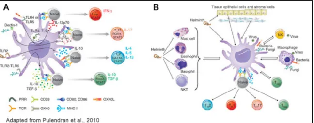
The type of the maturation stimuli that tweak the DCs is also a critical aspect influencing the development of CD4+ T helper (Th)-cells into Th1 or Th2 subsets or the activation of CD8+ Cytotoxic T cells or the class of antibodies secreted by B cells (Banchereau and Steinman, 1998; Iwasaki and Medzhitov, 2004). Entry of a microbe into the body can also activate a range of other cell types, such as NK cells, NK T cells, basophils, mast cells, myeloid suppressor cells, tissue epithelial cells and stromal cells, all of which can influence DC function. Thus, DCs can sense microbes directly but also indirectly, through factors secreted by other immune cells and the microenvironment, and integrate
! #$! this information to orchestrate the immune response (Figure 1.1) (Pulendran et al., 2010). In this way, the type of response is adapted to the type of invading pathogen and the infected tissue.
Naïve CD4+ T cells are particularly plastic and can differentiate into several functional effector classes, including:
- Th1 cells, which produce IFN-" and help the induction of CD8+ cytotoxic T cells (CTLs) are critical for the eradication of intracellular microorganisms such as
Listeria monocytogenes bacteria and Leishmania major protozoa (Szabo et al.,
2003);
- Th2 cells, producing interleukin-4 (IL-4), IL-5, IL-9, IL-13, and IL-25, are important for the elimination of extracellular parasites such as helminthes and nematodes (Pulendran et al., 2010);
- Th17 cells, a more recently recognized pathway of helper T cell development, are abundant at mucosal interfaces, where they respond to infections with pathogenic bacteria and fungi. These cells produce mainly IL-17 and IL-22 cytokines (Dong, 2008).
Figure 1.1 Dendritic-cell control of pathogen-driven T-cell polarization
(A) The classical view of how DCs polarize TH responses involves sensing microbial stimuli
directly through various innate immune receptors expressed by DCs and the stimulation of distinct signaling pathways that mediate the production of different cytokines and factors that control TH polarization. (B) A revised view places the classical picture in the context of the
cell-cell interactions that occur (for example, basophils help TH2 polarization), together with
conditioning from stromal cells and epithelial cells (Pulendran et al., 2008).
Besides effector cell lineages, CD4+ T cells can also differentiate into distinct regulatory lineages so-called T regulatory cells that suppress the
! #%! proliferation and differentiation of Th or cytotoxic T cells and serve to limit the potential immunopathology that might be caused by an overexuberant immune response.
A number of other factors can also influence the Th lineage commitment, including the type of activated DC presenting the antigenic peptide to the naïve T cell (Coquerelle and Moser, 2010). But first, the existence of different DC subsets needs to be introduced.
2. DC subsets
In the recent years it has become clear that DCs do not represent one homogeneous population but rather form a plethora of distinct subsets that can be found in vivo. These DC subsets differ not only in phenotype, but also in their genetic program, are distributed in different microenvironments within the body and equipped to sense different types of pathogens and to modulate distinct classes of immune responses.
2.1. Classification criterion
Classification of the DC subsets can be difficult and opinions vary on which criteria is the best for categorization of the different sub-populations.
For example, the classification can be development oriented. For instance, DCs in the mouse lymphoid organs can require one of the two hematopoietins, the Fms-like tyrosine kinase ligand (Flt-3L) or the Granulocyte/Macrophage Colony Stimulating Factor (GM-CSF) for their development. This classification is an active current area of research.
In terms of lineage origins, DCs were once divided into myeloid or lymphoid DCs. This division was proposed in 1992 when it was found that T cell precursors, which are lymphoid committed cells, could form thymic DC (Vremec et al., 1992). The expression of markers that are normally expressed by lymphocytes (i.e., CD8!) was taken as a hint that these cells derived from lymphoid committed cells (Ardavin et al., 1993). In the meantime, it was also recognized that DCs could arise from myeloid precursors. Until now it is still not
! #&! clear what is the function of this cell marker on DCs. Nonetheless, the terms myeloid and lymphoid DC subsets are still used by some authors in relation to their expression of myeloid vs. lymphoid markers (CD11b vs. CD8!) but most feel that this type of distinction is not accurate in developmental terms.
Another type of nomenclature divides DCs according to whether they are “resident” or migratory. All DCs are generated in the bone marrow and migrate as precursor cells to sites of potential entry of pathogens. “Resident DCs” move directly to lymphoid tissue from a blood precursor, while “migratory DCs” first enter peripheral tissues (e.g. skin) before migrating through the lymphatics to the draining lymphoid organs.
An alternative classification distinguished plasmacytoid (PDC) from conventional DC (cDC) based on different morphology, surface markers, and as a result of more recent studies, gene-expression profiling that clearly put these two subsets apart (Robbins et al., 2008).
DCs can also be categorized on whether they are present in the uninfected steady state or whether they arise with infection or inflammation.
Any cell of the DC family, at any one time, should fit one of the classifiers described above.
2.2. Phenotype of mouse DCs subsets in peripheral lymphoid
organs (Table 1)
Murine splenic DCs are certainly the most widely studied cells in DC biology. At steady state, splenic DCs constitutively express MHC class II and the integrin CD11c and can be subdivided into three major subsets:
(i) CD11chigh CD8#+ CD11b- DEC205+ DCs [so called “CD8#+ DCs”]. This subpopulation corresponds to the original “lymphoid DC subset”.
(ii) CD11chigh CD8#- CD11b+ DEC205- DCs [so called “CD8#- DCs”]. This subset can be further divided into two, based on the expression of CD4 or DCIR.
(iii) CD11cintermediate CD8#+/- CD11b-DEC205- B220+ DCs [PDCs].
Subsets (i) and (ii) correspond to the cDC for conventional or “classical” dendritic cells.
! #'! In addition to these subsets we can find at least two additional subpopulations of DCs in the mouse lymph nodes, particularly the ones draining the skin, also called skin-draining lymph nodes (sdLNs):
(i) CD11chigh CD8#low CD11b+ DEC205high Langerin+ [Langerhans cells (LCs)].
(ii) CD11chigh CD8#- CD11b+ DEC205+ [dermal DCs]. This subset can be further sub-divided in two, based on the expression or not of the CD103 integrin.
These two subsets found in the lymph nodes are commonly referred to as migratory DCs since, even in germ-free mice, they constantly travel from peripheral tissues (the epidermis or the dermis of the skin respectively), via the lymphatics, to the draining lymph node. In contrast, because the spleen lacks afferent lymphatic vessels, migratory DCs are distinctively absent from this organ.
Table 1.1. Major DC subsets in the mouse lymphoid organs.
! #(! The described DC subsets are found in the lymphoid organs under steady state conditions and represent just one aspect of the scope of the DC family. The complex DC system also includes many other intricacies associated with other non-lymphoid tissues such as the intestine and the lung where the different types of DCs are conditioned for important immune mechanism by the particular tissues, but that will not be explored in my thesis.
DC heterogeneity can be further extended to include monocyte-derived DCs generated under inflammatory conditions and that will be further discussed in this chapter.
DC subsets are currently well described on the basis of their surface phenotype. However, the origin and developmental relationship of these DC types is not yet entirely clear. In the next section, I will discuss some of proposed mechanisms concerning the origin and developmental relations existing between the different DC subsets in steady state or inflammatory conditions
3. Origin of DCs in lymphoid tissues
3.1. In steady state conditions
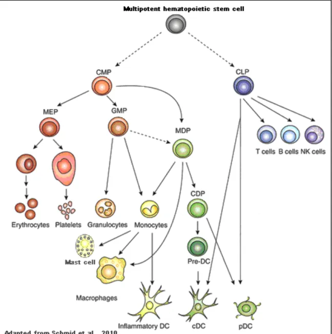
Lymphoid and myeloid lineage divergence is an early step in haematopoiesis. Monocytes, macrophages, granulocytes, megakaryocytes, and erythrocytes differentiate from a common myeloid progenitor (CMP), whereas B, T, and natural killer (NK) cells differentiate from a common lymphoid progenitor (CLP) (Figure 1.2.). As with all other leukocytes, DCs also develop from bone marrow-derived hematopoietic stem cells. In the lymphoid tissues, DCs are in a dynamic balance, with an estimated half-life of only a few days during steady state conditions (Liu et al., 2007). Although this rapid turnover mandates a continuous replacement of DCs by a precursor population, the identities of hematogenous DC precursors that contribute to steady-state DC populations remain a subject of controversy, and attempts to identify committed DC precursors have led to a range of results that I will summarize in the next paragraphs.
! #*!
Figure 1.2 Hematopoietic tree for dendritic cell development.
Hematopoiesis is initiated in bone marrow multipotent hematopoietic stem cells. Further downstream, lineage differentiation potential branches into progenitors committed to myeloid cells, common myeloid progenitors (CMPs), or lymphoid cells, common lymphoid progenitors (CLPs). CMPs then further differentiate to megakaryocyte/erythrocyte progenitors (MEPs) and granulocyte/macrophage progenitors (GMPs). Alternatively, CMPs can develop into macrophage-DC progenitors (MDPs) that give rise to monocytes, macrophages, classical macrophage-DCs (cmacrophage-DCs), and plasmacytoid DCs (pDCs). MDPs-derived monocytes can further differentiate into inflammatory DCs. MDPs lie upstream of the common DC progenitors (CDPs), which are DC-restricted, giving rise to pDCs and, via pre-DCs, to cDCs. Solid arrows show demonstrated pathways; dotted arrows show suggested pathways that have not been formally proven(Schmid et al., 2010).
The differentiation of hematopoietic stem cells requires the integration of environmental signals surrounding the cell. In their microenvironments, hematopoietic progenitors interact via receptors with ligands present in the
! #+! extracellular matrix. In these developmental niches, cytokines can act locally as secreted factors or in membrane-bound forms in direct cell-to-cell contacts. In addition, cytokines can act distantly from where they were produced by travelling along the bloodstream or lymph vessels (Kondo et al., 2003).
To maintain homeostasis in the DC compartment, two major players are involved, the GM-CSF and the Flt3L, that were previously mentioned in the context of DC classification criterion. The macrophage–colony stimulating factor (M-CSF) is also involved and controls the segregation of monocytes into fully differentiated macrophages. These are all growth factors know to be potent stimulators of hematopoietic progenitor cell expansion and mobilization (Robinson et al., 2000) (Figure 1.3).
The use of genetically deficient mice has shaped our current view on the identity of cell progenitors and the cytokine requirements for the development of different DC subsets. For example, GM-CSF-deficient mice or mice lacking the GM-CSF receptor common ! chain have only minor decreases in splenic cDCs compared with wildtype mice and a maximal threefold reduction in lymph node cDCs. In addition, transgenic mice overexpressing GM-CSF showed only a small increase in cDCs (Vremec et al., 1997), whereas monocytes are missing in mice lacking M-CSF receptor (c-fms or CD115) (Dai et al., 2002; Ginhoux et al., 2006). On the other hand, mice with a targeted gene deletion of Flt3L or Flt3 (the Flt3L receptor also known as CD135 or Flk2) have severely reduced numbers of cDCs and PDCs (D'Amico and Wu, 2003; Geissmann et al., 2010; Ginhoux et al., 2006; Maraskovsky et al., 2000; Waskow et al., 2008). Consistent with these observations, Flt3L injection induces selective expansion of cDCs, PDCs and myeloid cells but not B or T lymphocytes (Maraskovsky et al., 1996). Thus, the data suggest that most DCs found in the lymphoid organs in steady state are independent of monocytes. The exception seems to occur with DCs in peripheral organs like lung and intestine that do appear to arise from monocytes on an ongoing basis (Varol et al., 2007), raising the interesting possibility that the origin of DCs in non-lymphoid tissues is different from spleen and lymph nodes. In this thesis I will however concentrate on the mechanisms that determine DC
! $,! development from monocytes in the lymphoid tissues.
Figure 1.3. Model for cytokine-induced differentiation of DCs
MDPs and CDPs commitment is dependent on cytokine exposure. These two cell percursors express the receptors for Flt3L (green), GM-CSF (red), and M-CSF (yellow). MDPs have the potential to differentiate into macrophages, monocytes, and inflammatory DCs, and via CDPs to cDCs and PDCs. Different microenvironments with variations in the combination and concentration of the three cytokines influence the lineage commitment and differentiation of MDPs and CDPs into mature cells (Schmid et al., 2010).
The initial reports regarding the origin of DCs were the studies of Inaba et
al. reporting that granulocytes, macrophages and DCs could arise from a yet
unknown MHC class-II-negative common progenitor cell during in vitro cultures of mouse bone marrow cells. In this setting, the development of monocytes into DCs (called monocyte-derived DCs [Mo-DCs]) required the presence of the GM-CSF in the cultures (Inaba et al., 1993). Sallusto and Lanzavecchia, and Romani further proved this notion in a report indicating that human DCs can be differentiated in vitro from blood monocytes (Sallusto and Lanzavecchia, 1994) (Romani et al., 1994).
! $"! Traver et al. in 2000, showed that the DC potential is maintained along the two early branches of the hematopoietic system and both CMP or CLP could give rise to spleenic cDCs (Traver et al., 2000) (Figure 1.2). Therefore, the terms “lymphoid” and “myeloid” are not longer used in the context of DC development. In this same study, MDPs were identified. The name stands for Macrophage and DC progenitor, because of the potential to develop into DC or Macrophages depending on the microenvironment encountered at tissue sites e.g. cytokine signals (Traver et al., 2000) (Figure 1.2. and Figure 1.3.). Phenotypically, MDPs are defined by the absence of markers for lineage-committed precursors such as CD3 (a T cell marker), CD19 (a B cell marker) and CD11b (a marker of monocytes in the BM), but express both the CD117 (c-kit, the receptor for stem cell factor) or the chemokine receptor Cx3CR1. MDPs were shown to give rise to monocytes, to several subsets of macrophages, and to steady state splenic CD11c+CD8#+ and CD11c+CD8#- DCs (cDCs) in vivo. In contrast, the MDP is devoid of lymphoid, erythroid, and megakaryocytic potential, also lacking polymorphonuclear leukocytes differentiation potential and therefore do not give rise to any granulocytes including eosinophils, basophils and neutrophils (Traver et al., 2000) (Figure 1.2).
Some years later, Onai et al, identified the CDPs for common DC precursor, in the mouse bone marrow. CDPs give rise exclusively to cDCs and pDCs, but not to other cell lineages, both in vitro and in vivo. Phenotypically, CDPs lack specific lineage markers and are negative for CD11c and MHC-II. On the other hand, they express the receptor for the Flt3L (CD135), the receptor for M-CSF (CD115) and the receptor for stem cell factor (c-Kit) (Onai et al., 2007). With this study, the divergence between the monocyte and the DC lineages was found to occur in the bone marrow and between the MDP and the CDP stages of development (Figure 1.2). Thus, lymphoid tissue cDCs, PDCs, and monocytes share a common progenitor, the MDP, whereas the CDP is restricted to produce cDCs and PDCs under steady state conditions.
A more recent study by Liu et al, defined the point of divergence between multipotential CDP precursors and cDC-restricted progenitors in the bone
! $#! marrow. Taking advantage of the persistent expression of CD135 throughout pre-DCs development, it was shown that CDPs move into the blood to seed both lymphoid and non-lymphoid tissues forming CD11chi, MHC IIhi DCs (Liu et al., 2009).
3.2. Under inflammatory conditions
As previously denoted, monocytes can also develop many of the phenotypic features of DCs but they are not, in contrast with CDPs, precursors for lymphoid organ DCs in the steady state.
The question of how DCs can arise from monocyte precursors has proven to be a vexing issue and will be explored in more detail in the next paragraphs.
Monocytes are circulating cells of the mononuclear phagocyte system that were first studied as macrophage precursors, mainly in vitro (Johnson et al., 1977) (de Villiers et al., 1994). Monocytes were later recognized to have an added potential to develop into DCs and this facet was primarily suggested from the in vitro studies with mouse bone marrow monocytes or using human blood monocytes, as previously cited (Inaba et al., 1993) (Sallusto and Lanzavecchia, 1994) (Romani et al., 1994). These in vitro fully differentiated Mo-DCs acquire a typical dendritic cell probing morphology, while losing the capacity to phagocytose and to adhere to various tissue culture surfaces. At the same time,
in vitro derived Mo-DCs acquire strong capacities to initiate immunity including
enhanced capacity for antigen presentation and T cell stimulation. Hereof, Mo-DCs can immunize humans (Dhodapkar et al., 1999) (Schuler-Thurner et al., 2000) and home to the T cell areas of lymph nodes (LNs) (de Vries et al., 2003). In addition, monocytes are almost 20 times more abundant than DCs in blood and bone marrow, so the mobilization of this cell reservoir in vivo in order to generate potent antigen-presenting DCs has an important therapeutic potential but is not fully understood.
The first description of the differentiation of mouse monocytes into DCs in
vivo was reported in 1999 by Gwendalyn Randolph using an experimental model
! $$! with the purpose of inducing a local dermal inflammation to which monocytes could be recruited and allowing the tracking of the cells that had internalized them. Data from this study allowed for the conclusion that monocytes recruited to the inflamed dermis differentiated locally into macrophages, as well as DCs that subsequently migrated to the draining lymph nodes (Randolph et al., 1999). In line with these findings, but using a different experimental model that promotes the exit of Langerhans cells from the inflamed skin after exposure to UV, the group of Miriam Merad has established that monocytes recruited to the skin could differentiated into epidermal Langerhans cells and that this process was dependent on M-CSF (Ginhoux et al., 2006). However, in steady state conditions, epidermal langerhans cells derive from local non-monocytic precursors (Merad et al., 2002).
More recent reports, have also reveled that monocytes could be precursors of some DC subsets found in the lung and intestinal laminal propria under steady state conditions, but majors efforts has been taken in order to identify the role played by Mo-DCs during the immune response against clinically relevant pathogens. In this regard, several reports have begun to document in mice the differentiation of CD11c# and MHC II# blood monocytes into large numbers of CD11c+ MHC II+ Mo-DCs during different infection models, e.g., Leishmania
major infection via the skin (León et al., 2007), intravenous infection with Listeria monocytogenes (Serbina et al., 2003), influenza virus infection via the airway
(Nakano et al., 2009), Aspergillus fumigatus in the lung (Hohl et al., 2009), T cell-mediated colitis (Siddiqui et al., 2010), and injection of the adjuvant, alum (Kool et al., 2008). Although these studies support the view that monocytes could behave as DC precursors after recruitment to the inflammatory foci related to each one of these pathological conditions, research on Mo-DCs have been limited by the lack of specific markers to localize them in vivo. and to isolate them, in order to look for features that characterize dendritic cells such as their morphology, their location into the T cell areas, and their capacity to capture antigens for cross presentation on MHC I.
! $%! In summary, there is a certain type of flexibility in terms of DC subtype production, at the level of the early hemopoietic precursors. The points downstream of these precursors where the DC sublineages diverge and become more fixed, and the factors determining this divergence, need to be elucidated. However, different functions have been attributed to the different DC subsets. Some of these functional differences will be further discussed.
4. Different DC subsets are involved in different types of immune
responses.
Classically, knowledge on DC biology was concentrated on their capacity to initiate immune responses following encounter with different maturation stimuli. The field has expanded considerably with the studies showing the existence of different DC subsets expressing different receptors for antigen uptake and thus involved in different immune mechanisms. Therefore, for each DC subset previously described, specialized immune functions have been recognized. This specific DC-subset function has been also shown to correlated with the profile of pattern recognition receptors (PRR) expression, such as Toll Like Receptors and C-Type lectins, with the type of cytokines that each subset secrets, and with the microenvironmental location of a certain DC subtype within the lymphoid organs (Figure 1.1 and Table 1)
For example, CD11chigh CD8#+ DCs express high amounts of messenger RNA for TLR3 but do not express TLR7 and may be therefore specialized for the recognition of double stranded RNA viruses, known agonists for TLR3 (Edwards et al., 2003) (Schulz et al., 2005). More recent proteomics data also revealed the specific expression of TLR12 and TLR13 on CD8!+ DCs (Luber et al., 2010). This particular subset of DC localize in the T-cell rich areas of the spleen and lymph nodes (Vremec et al., 2000) and upon TLR activation secrete large amounts of IL-12 (p70) (Reis e Sousa et al., 1997). This cytokine is important for the formation of CD4+ Th1 cells (Maldonado-López et al., 1999) and cytotoxic CD8+ T cell priming (Felzmann et al., 2005). By contrast, CD8#- DCs do express
! $&! receptors, the RIG-I-like helicases RIG-I and MDA-5, important in the recognition of single stranded RNA viruses (Luber et al., 2010). CD8#- DC are localized in the red pulp and marginal zone of the spleen (Dudziak et al., 2007) where they can be marked using the 33D1 anti-DCIR2 antibody. But the study of this population in the lymph nodes has been limited by the lack of specific markers for staining. CD8#- DCs are a major source of IL-10 and IL-4 and have been shown to induce Th2 responses (Maldonado-López et al., 1999; Maldonado-López et al., 2001).
Another important subset in the DC biology, the PDCs are able to produce large amounts of type I interferons upon exposure to both live and inactivated viruses. This is because PDCs express Toll-like receptor 7 (TLR7) and TLR9 in the endossomes (Villadangos and Young, 2008).
With regard to cross-presentation of particulate or soluble protein antigens to CD8+ T cells, the CD8#+ DCs are more efficient comparing with the other DC subsets (den Haan et al., 2000) (Pooley et al., 2001). Furthermore, CD8#+ DCs
are preferentially able to endocytose dying cells in vivo and present cell-associated antigens to both CD4 and CD8 T cells (Iyoda et al., 2002). The CD8# -DCs are, in contrast, more efficient for presentation of antigens for the MHC-II pathway, irrespective of activation status or mode of Ag acquisition (Kamphorst et al., 2010) and this is consistent with the differential antigen-processing ability of CD8#+ versus CD8#- DC subsets (Dudziak et al., 2007). In contrast, activated PDCs express lower levels of CD11c, MHC class II and other T-cell costimulatory molecules and are therefore much less efficient at T-cell priming comparing with the other DC subsets (Villadangos and Young, 2008).
The other two additional subsets found in the lymph nodes, the Langerhans cells and the dermal DCs (or migratory DCs) are found in the periphery (e.g. skin) in an immature state. However, by the time they reach the draining lymph node, they are found in a mature state expressing high levels of MHCII and costimulatory molecules (Jakubzick et al., 2008; Wilson et al., 2003). In terms of T cell priming capacities, a recent study has shown that CD103+ dermal DCs were the migratory subset most efficient in the presentation of antigens of herpes
! $'! simplex virus type 1 to naive CD8+ T cells, but were comparable to all other DC subsets in the draining lymph node for presentation of these antigens to CD4+ T cells (Bedoui et al., 2009).
In order to find additional molecular differences between DC subsets, several genomic approaches have been taken. This led to the identification of a series of genes that encode lectin-like receptors, some of which are remarkably DC specific. These lectins are members of the C-type lectin receptor family (CLRs) and will be subject of detailed description in the next section.
5. C-type lectins on DCs are key modulators of the immune
responses
The term C-type lectin was introduced to distinguish between Ca2+ -dependent and Ca2+-independent carbohydrate-binding lectins. CLRs share at least one carbohydrate recognition domain (CRD), which is a compact structural module that contains conserved residue motifs and determines the carbohydrate specificity of the CLR (Zelensky and Gready, 2005). CLRs exist both as soluble and transmembrane proteins, but for the purpose of my thesis I will only discuss the transmembrane CLRs that function as PRRs.
As previously noted, immature DCs express an abundant variety of PRRs to interact with a broad spectrum of invading pathogens. The most abundant PRRs expressed on DCs include the archetypical TLRs, and CLRs. In contrast to TLRs, CLRs can recognize carbohydrate structures on pathogens. Thus, CLRs are particularly important for internalization of glycosylated antigens into intracellular compartments present on DCs enhancing presentation of the antigens on MHC I and II, without inducing DC maturation (Figdor et al., 2002). For this reason, CLRs are also called endocytic receptors or antigen-uptake receptors, and some of them do not have any known innate signaling function other than endocytosis.
Although TLRs and CLRs recognize different determinants and have distinct functions, various studies suggest that CLRs may also modulate immune reactions through cross-talk with other receptors and especially with TLRs. This indicates that the outcome of an immune response is determined by the balance
! $(! between triggering of the two receptor families (Lee and Kim, 2007).
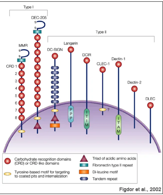
So far more than 15 CLRs have been identified on DCs and macrophages. These CLRs can be divided into two groups depending on the orientation of their amino (N) terminus (Figure 1.4.) (Figdor et al., 2002).
Group I – the N-terminus is pointing outwards from the cytoplasm of the cell, and include the Macrophage Mannose Receptor (MMR or CD206) and the DEC-205 (CD205). Lectins of this group contain several CRDs.
Group II - the N-terminus is pointing into the cytoplasm of the cell. Include, the Dendritic Cell-Specific Intercellular adhesion molecule 3-Grabbing Nonintegrin (DC-SIGN or CD209), the DC-associated C-type lectin 1 (Dectin-1; also known as CLEC7A) and DC immunoreceptor (DCIR; also known as CLEC4A). The type II surface lectins produced by DCs contain a single CRDs domain.
Depending on the amino acid sequence, the CRD bears specificity for mannose, galactose, or fucose structures. However, binding of these carbohydrate structures to the different CLRs is also dependent on carbohydrate branching, spacing, and multivalency (Zelensky and Gready, 2005).
5.1. C-type lectins receptors are differentially expressed on
different DC subsets
CLRs are differentially expressed by various subsets of DCs. For example, DEC-205 (CD205) is most abundant on CD8#+ DCs . In contrast, the DCIR2 is a marker for the CD8#- DCs subset especially in the spleen. MMR and
DC-SIGN are hallmarks of in vitro cultured monocyte derived DCs, while Langerin (CD207) expression is restricted to epidermal Langerhans cells and dermal DCs found in the skin and migrating to the skin draining lymph nodes. The CLR CLEC9A is expressed at high levels by the murine and human CD8#+
DCs (Geijtenbeek and Gringhuis, 2009).
A broad set of mAb against particular CLRs is currently available and used to study specific DC-subsets. In addition, it is becoming clear that these differences in expression reflect differences in antigen capture, developmental pathways and migration patterns attributed to each DC-subset.