Programa Doutoral em Saúde Pública
Maria Teresa Pegado Barroso Abilheira Monjardino
Effects of growth on childhood bone status:
A study in the Generation XXI birth cohort
Porto | 2019
Dissertação de candidatura ao grau de Doutor apresentada à Faculdade de
Medicina da Universidade do Porto
ii
Art.º 48º, § 3º - “A Faculdade não responde pelas doutrinas expendidas na dissertação.”
iii
Corpo Catedrático da Faculdade de Medicina do Porto
Professores Catedráticos Efetivos
Doutora Maria Amélia Duarte Ferreira Doutor José Agostinho Marques Lopes
Doutor Patrício Manuel Vieira Araújo Soares Silva Doutor Alberto Manuel Barros da Silva
Doutor José Henrique Dias Pinto de Barros
Doutora Maria Fátima Machado Henriques Carneiro Doutora Isabel Maria Amorim Pereira Ramos
Doutora Deolinda Maria Valente Alves Lima Teixeira Doutora Maria Dulce Cordeiro Madeira
Doutor Altamiro Manuel Rodrigues Costa Pereira Doutor Manuel Jesus Falcão Pestana Vasconcelos
Doutor João Francisco Montenegro Andrade Lima Bernardes Doutora Maria Leonor Martins Soares David
Doutor Rui Manuel Lopes Nunes
Doutor José Eduardo Torres Eckenroth Guimarães Doutor Francisco Fernando Rocha Gonçalves Doutor José Manuel Pereira Dias de Castro Lopes
Doutor António Albino Coelho Marques Abrantes Teixeira Doutor Joaquim Adelino Correia Ferreira Leite Moreira Doutora Raquel Ângela Silva Soares Lino
iv
Professores Jubilados ou Aposentados
Doutor Alexandre Alberto Guerra Sousa Pinto Doutor Álvaro Jerónimo Leal Machado de Aguiar Doutor António Augusto Lopes Vaz
Doutor António Carlos de Freitas Ribeiro Saraiva Doutor António Carvalho Almeida Coimbra
Doutor António Fernandes Oliveira Barbosa Ribeiro Braga Doutor António José Pacheco Palha
Doutor António Manuel Sampaio de Araújo Teixeira Doutor Belmiro Dos Santos Patrício
Doutor Cândido Alves Hipólito Reis
Doutor Carlos Rodrigo Magalhães Ramalhão Doutor Cassiano Pena de Abreu e Lima
Doutor Eduardo Jorge Cunha Rodrigues Pereira Doutor Fernando Tavarela Veloso
Doutor Henrique José Ferreira Gonçalves Lecour de Menezes Doutor Jorge Manuel Mergulhão Castro Tavares
Doutor José Carlos Neves da Cunha Areias Doutor José Carvalho de Oliveira
Doutor José Fernando Barros Castro Correia Doutor José Luís Medina Vieira
Doutor José Manuel Costa Mesquita Guimarães Doutor Levi Eugénio Ribeiro Guerra
Doutor Luís Alberto Martins Gomes de Almeida Doutor Manuel Alberto Coimbra Sobrinho Simões Doutor Manuel António Caldeira Pais Clemente Doutor Manuel Augusto Cardoso de Oliveira Doutor Manuel Machado Rodrigues Gomes Doutor Manuel Maria Paula Barbosa
Doutora Maria da Conceição Fernandes Marques Magalhães Doutora Maria Isabel Amorim de Azevedo
Doutor Ovídio António Pereira da Costa Doutor Rui Manuel Almeida Mota Cardoso Doutor Serafim Correia Pinto Guimarães
Doutor Valdemar Miguel Botelho dos Santos Cardoso Doutor Walter Friedrich Alfred Osswald
v
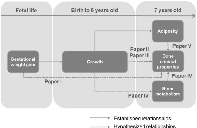
Ao abrigo do Art.º 4º, § 2º do Decreto-Lei n.º 707/2018, fazem parte desta dissertação as seguintes publicações:
I. Gestational Weight Gain and Offspring Bone Mass: Different Associations in Healthy Weight Versus Overweight Women
II. Early Childhood as a Sensitive Period for the Effect of Growth on Childhood Bone Mass: Evidence from the Generation XXI Birth Cohort
III. Weight Trajectories from Birth and Bone Mineralization at 7 Years of Age
IV. Bone Formation and Resorption Markers at 7 Years of Age: Relations with Growth and Bone Mineralization
V. When to Stop Looking at a Cause: an Application to Adiposity and Bone Physical Properties in Children
Ao longo da elaboração da presente dissertação, colaborei ativamente na definição e operacionalização das hipóteses em estudo e dos objetivos a responder em cada um dos artigos, bem como na análise estatística dos dados e interpretação dos resultados. Participei na recolha dos dados durante a reavaliação aos 7 anos de idade da Geração XXI. Fui responsável pela redação da versão inicial de todos os manuscritos e participei ativamente na preparação das suas versões finais.
vii
Esta investigação foi realizada no Instituto de Saúde Pública da Universidade do Porto e no Departamento de Ciências da Saúde Pública e Forenses e Educação Médica, da Faculdade de Medicina, Universidade do Porto sob orientação da Professora Doutora Raquel Lucas (Instituto de Saúde Pública e Faculdade de Medicina da Universidade do Porto), e co-orientação da Professora Doutora Ana Cristina Santos (Instituto de Saúde Pública e Faculdade de Medicina da Universidade do Porto) e da Professora Doutora Teresa Rodrigues (Centro Hospitalar de São João, Instituto de Saúde Pública e Faculdade de Medicina da Universidade do Porto).
Esta investigação foi financiada pelo Fundo Europeu de Desenvolvimento Regional (FEDER) através do Programa Operacional Competitividade e Internacionalização e por Fundos Nacionais através da FCT – Fundação para a Ciência e a Tecnologia (029087; 016838 e POCI-01-0145-FEDER-016837), no âmbito dos projetos “STEPACHE - Raízes pediátricas da resposta ampliada à dor: das influências contextuais à estratificação do risco” (Ref. FCT: PTDC/SAU-EPI/29087/2017); “BioAdversity: Como a adversidade social na infância condiciona a saúde: A biologia da adversidade social” (Ref. FCT: PTDC/DTP-EPI/1687/2014) e “PathMOB: Risco cardiometabólico na infância: desde o início da vida ao fim da infância” (Ref. FCT: PTDC/DTP-EPI/3306/2014). Este trabalho foi também financiado pela Unidade de Investigação em Epidemiologia - Instituto de Saúde Pública da Universidade do Porto (EPIUnit) (POCI-01-0145-FEDER-006862; Ref. UID/DTP/04750/2013) e pela bolsa de doutoramento SFRH/BD/92370/2013 (Teresa Monjardino), cofinanciada pela FCT e pelo Programa Operacional Potencial Humano (POPH/FSE). Este trabalho resulta também do projeto “DOCnet: Diabetes & obesity at the crossroads between Oncological and Cardiovascular diseases – a system analysis NETwork towards precision medicine” (NORTE-01-0145-FEDER-000003), cofinanciado pelo Programa Operacional Regional do Norte (NORTE 2020), através do Acordo de Parceria Portugal 2020 e dos Fundos FEDER.
ix
Júri da Prova de Doutoramento
Doutor José Henrique Dias Pinto de Barros (Presidente)
Faculdade de Medicina da Universidade do PortoDoutora Laura Howe
Population Health Science Institute, Bristol Medical School, University of Bristol
Doutora Katrine Strandberg-Larsen
Department of Public Health, University of Copenhagen
Doutora Carla Maria de Moura Lopes
Faculdade de Medicina da Universidade do PortoDoutora
Liane Maria Correia Rodrigues da Costa Nogueira Silva
Instituto de Ciências Biomédicas Abel Salazar da Universidade do PortoDoutora Raquel Lucas Calado Ferreira (Orientadora)
Faculdade de Medicina da Universidade do PortoDra. Maria Lúcia Carvalho Dias Costa
Centro Hospitalar São Joãoxi
Agradecimentos
À minha orientadora Prof. Raquel Lucas por ser a Melhor, à qual reconheço a enorme inteligência, capacidade de trabalho, humildade, sensatez e bom humor. Porque me faz perceber, sem nunca me rebaixar, que ainda tenho muito para percorrer até me tornar uma verdadeira epidemiologista. Esta tese tem a sua contribuição desde o primeiro rascunho do projeto até ao último ponto final. À minha amiga Raquel agradeço os momentos de risota, “cusquise”, partilha de roteiros gastronómico, conversa sobre as maravilhas e angústias da maternidade e devaneios da vida. O mais profundo e sincero obrigado é mesmo para ti.
Ao Prof. Henrique Barros que criou uma escola à qual me orgulho de pertencer. Agradeço as oportunidades que me tem dado de integrar tantos e tão diferentes projetos que me fazem crescer profissional e pessoalmente.
À Prof. Ana Cristina Santos o meu agradecimento por todos os momentos de partilha de conhecimento. Obrigada por me ter “assustado” tantas vezes pelas costas. Isto significava sempre o inicio de uma discussão muito frutífera para enriquecer o meu trabalho.
À Prof. Teresa Rodrigues agradeço as questões desafiadoras e a ajuda na reflexão das implicações práticas do trabalho.
Deixo também uma palavra de agradecimento às pessoas que desde 2005 têm feito parte da equipa de investigação Geração XXI. Aos participantes e respetivas famílias da Geração XXI agradeço profundamente por partilharem e confiarem tanta informação sobre as suas vidas.
Um especial agradecimento à Joana Amaro, Joana Araújo, Sara Soares, Samantha Morais pela leitura cuidada das versões finais da tese.
A todos os colegas que passaram pela sala 406 (Sílvia, Diogo, Margarida, Ana Raquel, Maja, Ofélia, Sara, Fábio, Poliana, Daniela) mas principalmente aos que agora a ocupam (Raquel, Sara, Vânia, Paula, Flávia, Armine, Ana, Joana, Makram), agradeço terem criado as condições ideais para que eu pudesse trabalhar e descontrair nos momentos certos. Nem toda a gente tem a sorte de escrever a tese até ao último dia numa sala com 10 pessoas que são tão trabalhadoras, respeitadoras e descontraídas. Os chocolates e bolos na mesa da entrada, os piqueniques, os “desentendimentos” com a porta, a disposição das mesa, janelas e ar condicionado, fazem parte da razão de todos os dias, eu acordar tão bem-disposta e com tanta vontade de vir trabalhar.
xii
Aos restantes coautores dos trabalhos (Ana Henriques, Carla Moreira, Maria João Fonseca, Hazel Inskip, Nicholas Harvey, Cyrus Cooper, Nuno Adubeiro, Luísa Nogueira, João Tiago Guimarães) agradeço as revisões cuidadas e sugestões mais que pertinentes que foram fundamentais para enriquecer os meus trabalhos
Ao grupo de amigas da faculdade (Diana, Mónica, Liliana, Susana, Manuela) que me inspiram pelo seu profissionalismo e me emocionam com todas as palavras amigas e motivadoras, em especial à Joana que me trouxe para o Departamento de Epidemiologia.
À minha grande amiga Francisca e restante grupo de amigas transmontanas de sempre (Teresa AL, Teresinha Janeira, Raquel, Pilar, Mariana) agradeço os momentos de “não doutoramento” que me trazem a alegria, ânimo, humor e coragem e me fazem lembrar do que realmente importa na vida.
Aos meus Pais, manas Pia e Matilde e Avó Zi, porque são o exemplo vivo de uma linhagem à qual me orgulho de pertencer, em especial o Avô Pedro e o tio Alexandre, que projetaram em mim o orgulho de ser estudante universitária. Juntamente com os tios Paula e Zé, Rita e Pedro e minhas queridas sobrinhas Benedita, Camila e Mafalda são a família que adoro e a quem tudo agradeço.
Ao Miguel, a quem não sei como agradecer pela vida maravilhosa que temos partilhado. Nesta fase em particular obrigada por teres sido o pai e mãe que não consegui ser, pelo otimismo e confiança, afinal “é só uma tese de doutoramento, Teresa”.
Aos meus pequeninos Teresinha e Zé, porque são a minha maior fonte de felicidade, inspiração e energia.
Table of contents
ABSTRACT ... 3
RESUMO ... 9
1.INTRODUCTION ... 15
1.1. The burden of musculoskeletal disorders ... 17
1.2. The population burden of fragility fractures ... 18
1.3. Bone fragility and fractures ... 24
1.4. Estimating bone fragility - Bone mass as a surrogate for bone strength ... 26
1.5. The life course approach to bone health ... 28
1.6. Early life growth ... 32
1.7. Early life bone development ... 33
1.8. Factors influencing childhood bone mass accrual ... 35
1.8.1. Intrauterine influences on childhood bone mass ... 36
1.8.2. Postnatal growth-related changes of the skeleton ... 37
1.8.3. Adiposity and childhood bone mass ... 42
2.STUDY OBJECTIVES ... 45
3.PARTICIPANTS AND METHODS ... 49
3.1. The Generation XXI cohort study ... 51
3.1.1. Participants ... 51
3.1.2. Ethical considerations ... 52
3.1.3. Data collection ... 52
3.1.4. Additional wave of assessment for musculoskeletal evaluation ... 56
4.RESULTS ... 61
4.1. Description of bone mineral properties in the Generation XXI birth cohort ... 63
4.2. Gestational Weight Gain and Offspring Bone Mass: Different Associations in Healthy Weight versus Overweight Women ... 79
4.3. Early Childhood as a Sensitive Period for the Effect of Growth on Childhood Bone Mass: Evidence from the Generation XXI Birth Cohort ... 101
4.4. Weight Trajectories from Birth and Bone Mineralization at 7 Years of Age ... 129
4.5. Bone Formation and Resorption Markers at 7 Years of Age: Relations with Growth and Bone Mineralization ... 141
4.6. When to Stop Looking at a Cause: an Application to Adiposity and Bone Physical Properties in Children ... 173
5.OVERALL DISCUSSION ... 199
5.1. Main findings ... 201
5.2. Implications for clinical practice and policy ... 202
5.3. Methodological considerations ... 203
5.3.1. Selection bias ... 203
5.3.2. Participation bias ... 204
5.3.3. Misclassification ... 205
5.3.4. Confounding ... 207
5.4. Interpretation of main findings ... 208
5.5. Future research ... 211
6.CONCLUSIONS ... 213
3
Abstract
Background
Fragility fractures constitute a major public health problem associated with considerable and growing individual, societal and economic burden. One of the main modifiable factors that determine fracture risk is bone strength that depends on bone size, morphology and material properties. As with many other chronic diseases, the approach currently used to explain the etiology of suboptimal bone strength is built on the premise that there is important tracking of this characteristic throughout the life course, modulated by gene-environment interactions. Body size is recognized as one of the major determinants of bone fragility and strong cross-sectional relations between body size and composition and bone properties have been clearly established since pediatric ages. However, important issues regarding longitudinal effects of early life growth on childhood mineralization remain to be clarified. Namely, there is still no clear understanding of the impact of maternal gestational weight gain (GWG) on offspring bone physical properties. Additionally, it is unknown whether knowledge of children’s growth trajectories since birth improves the statistical prediction of bone mineralization or turnover during the first decade of life. Finally, the potential for adiposity to specifically influence bone physical properties in children remains to be clarified.
Objectives
In the present work our objectives were:
1. To quantify the associations between gestational weight gain and bone mineralization
in offspring, testing early pregnancy body mass index as an effect modifier (Paper I)
2. To characterize the impact of early life growth on bone mineralization in childhood by:
a. Identifying sensitive periods for the effect of early growth on childhood bone mass
(Paper II)
b. Assessing whether different trajectories of weight gain since birth influence
childhood bone mineralization (Paper III)
3. To describe markers of bone turnover in generally healthy children and assess their
4
4. To test the potential for adiposity to specifically influence bone mass in children, using
common causes as a benchmark (Paper V)
Methods
We used data from the Generation XXI, the first Portuguese birth cohort study assembled in Porto. All women who delivered live-born children with gestational age above 23 weeks in one of the five public units in the metropolitan area of Porto between April 2005 and August 2006, and whose residence was in the units’ catchment area were eligible for recruitment. Seventy percent of eligible mothers were invited and, of these, 91.4% accepted to participate. All 8647 children enrolled at birth were invited to attend follow-up assessments at ages four (2009-2011) and seven (2012-2014) years (86.3% and 79.7% participation, respectively).
At baseline, in the first 24 to 72 hours after delivery, trained interviewers were responsible for presenting the study and inviting mothers. Subsequent follow-up evaluations of the cohort took place at our research center. In face-to-face interviews at baseline and follow-up evaluations, information was collected using structured questionnaires concerning the child’s and mother’s health. Family medical history and data on demographic, socioeconomic and psychosocial circumstances, and lifestyles were also collected. A physical examination including anthropometric assessment was performed to children and mothers. Routine primary care data regarding the child’s development were periodically extracted from the Child’s Health Book. Velocities of growth in weight and length/height, across different age periods, were obtained through multilevel linear spline models. Weight trajectories since birth up to the age of 6 years were identified through normal mixture modeling for model-based clustering.
In each evaluation, a child blood sample drawn from an antecubital vein, after overnight fasting, were collected and, at 7 years of age, a subsample of 400 participants was selected to measure the following bone metabolism markers: total alkaline phosphatase (tALP), osteocalcin (OC) and β-isomerized C-terminal telopeptides of type I collagen (β-CTx).
In the 7 years-old follow-up evaluation a consecutive subsample of children who attended the face-to-face interview was invited to undergo a whole body dual-energy X-ray absorptiometry (DXA). Scanning was successfully performed in 2408 children (79.9% of the eligible sample and 27.8% of the entire cohort). Bone mineral content (BMC) and areal bone mineral density (aBMD) were obtained from DXA scan and a measure of size-corrected BMC (scBMC) was calculated.
5
Results
Paper I
In 2167 mother-child pairs, 35.8% of the mothers were overweight or obese [body mass index (BMI) ≥ 25 kg/m2] at the beginning of pregnancy. According to Institute of Medicine (IOM)
recommendations for gestational weight gain, we found that 36.6% mothers gained excessive weight during pregnancy. We estimated heterogeneous associations between GWG and bone mineralization in the offspring at 7 years of age according to maternal BMI. Among under/normal weight mothers there was a positive association between GWG and offspring bone measures: per 5 kg of GWG, BMC: 0.07 standard deviation (SD) (95% confidence interval - 95% CI:, 0.01, 0.12); aBMD: 0.10 SD (95% CI: 0.05, 0.15), while in overweight/obese mothers no effect of GWG on bone was observed: BMC: 0.02 SD (95% CI: –0.04, 0.09); aBMD: 0.02 SD (95% CI: –0.04, 0.08). When GWG was analyzed using IOM recommended categories, there were no differences in offspring mean bone properties between mothers gaining excessive and adequate weight during pregnancy, for both early pregnancy BMI groups.
Paper II
In 1853 children, all the five weight velocities (“early neonatal”: 0-10 days, “early infancy”: 10 days-3 months, “late infancy”: 3-12 months, “early childhood”: 1-3 years, and “later childhood”: 3-6 years) and four length/height velocities (“early infancy”: 0-3 months, “late infancy”: 3-12 months, “early childhood”: 1-3 years, and “later childhood”: 3-6 years) were associated with increased bone mass and height at 7 years. Strongest associations were observed for growth in early childhood [(per 1 SD increase in weight velocity height-adjusted BMC z-score increased by 0.27 (95% CI: 0.22, 0.32) in girls and 0.24 (95% CI: 0.19, 0.29) in boys; per 1 SD increase in length/height velocity height-adjusted BMC z-score increased by 0.12 (95% CI: 0.07, 0.17) in girls and 0.11 (95% CI: 0.06, 0.16) in boys]. Estimates remained positive, albeit attenuated, after adjusting for preceding velocities of growth.
6
Paper III
In 1889 7-year-old children, 66.2% had a trajectory of growth characterized by “normal weight gain”, 8.4% had a “weight gain during infancy” trajectory, 14.6% had a “weight gain during childhood” trajectory and 10.9% had a “persistent weight gain” trajectory. Compared with the “normal weight gain” group, bone measures were greater in all the remaining trajectories. Children who followed a “persistent weight gain” trajectory since birth had clearly increased bone mass and density in comparison to “normal weight gain” children (girls [BMC: 674.0 vs. 559.8 g, aBMD: 0.677 vs. 0.588 g/cm2, scBMC: 640.7 vs. 577.4 g], boys [BMC: 689.4 vs. 580.8
g, aBMD: 0.682 vs. 0.611 g/cm2, scBMC: 633.0 vs. 595.6 g]). After adjustment for current
weight, girls with a “weight gain during childhood” trajectory had greater bone measures than those with a “normal weight gain” trajectory (BMC: 601.4 vs. 589.2 g, aBMD: 0.618 vs. 0.609 g/cm2).
Paper IV
In 395 children, reference intervals (2.5th- 97.5th percentile) for serum tALP was 159 - 439 U/l;
for β-CTx was 470 - 1690 ng/l and for OC was 52.5 - 137.7 µg/l in girls and 50.0 - 129.9 µg/l in boys. We found a moderate correlation between serum concentrations of bone-specific metabolism markers OC and β-CTx, likely representing the dynamic nature of bone turnover. Concentration of tALP increased slightly with height (Pearson partial correlations (rpartial)
controlled for sex=0.26, 95% CI: 0.17, 0.35), was higher in overweight than in healthy weight children, and in children who gained weight above average during infancy. No correlations were found between OC or β-CTx and growth. In girls, OC was slightly correlated with measures of total body less head BMC (rpartial=0.22, 95% CI: 0.08, 0.35), subtotal aBMD (rpartial=0.20, 95%
CI: 0.06, 0.33) and lumbar spine aBMD (rpartial=0.23, 95% CI: 0.09, 0.36). tALP and β-CTx were
not correlated with any of the DXA-derived bone measures. Therefore, bone formation and resorption markers seemed to have limited usefulness to describe overall anthropometric growth and bone mineralization status in generally healthy children.
7
Paper V
Using association between socioeconomic position (SEP) and fat mass as a benchmark, in a subsample of 2408 children, we found that among 10 SEP indicators associated with fat mass only educational level and occupational level of caregivers was associated with BMC: [educational level of the main (less than higher vs. higher education, standardized linear regression coefficient: 0.15, 95% CI: 0.07, 0.24) and secondary (0.13, 95% CI: 0.03, 0.22) caregivers; occupations of the main (blue and lower white vs. upper white collar: 0.12, 95% CI: 0.03, 0.20) and secondary (0.10, 95% CI: 0.01, 0.18) caregivers]. A similar pattern to that observed for BMC was found for lean mass and height. In summary, assuming that traits that are causally related share causes to a measurable extent, the observation that fat mass and BMC did not share socioeconomic causes, may mean that fat mass has a small specific effect on BMC in childhood beyond its contribution to body size.
Conclusions
In the present study, we found no beneficial effect of excessive maternal GWG on childhood bone mineralization. Therefore, adherence to current recommendations for pregnancy weight gain may contribute to optimize child’s skeletal health. Regarding early life growth, our findings suggested that second and third years of life might represent a sensitive period for the effect of growth in height and weight on childhood bone mass, partly through their effect on concurrent body size. Additionally, the shape of the overall trajectory of weight gain since birth up to 6 years was associated with DXA-derived bone measures, but not with bone turnover, at 7 years of age. Markers of bone turnover were also not particularly related to bone mineralization suggesting their limited usefulness in generally healthy children. Finally, we found that fat and bone mass did not share socioeconomic causes, from which we concluded that adiposity is likely to have a small specific effect on bone mass in childhood beyond its contribution to body size.
9
Resumo
Introdução
As fraturas de fragilidade estão associadas a elevada morbilidade e mortalidade e constituem um importante problema de saúde pública associado a custos substanciais para os doentes e para a sociedade. Um dos principais fatores modificáveis que determinam o risco de fratura é a resistência óssea que depende do tamanho, morfologia e propriedades materiais do osso. Como em muitas outras doenças crónicas, a abordagem teórica atualmente utilizada para explicar a etiologia do desenvolvimento de pior resistência óssea, e subsequente risco de fratura, baseia-se na premissa de que a qualidade óssea é em grande parte conservada desde as primeiras décadas de vida, e modulada por interações gene-ambiente. A corpulência é reconhecida como um dos principais determinantes da fragilidade óssea, e têm sido encontradas fortes relações transversais entre o tamanho e composição corporal e as propriedades do osso desde idades pediátricas. No entanto, permanecem por esclarecer algumas questões importantes sobre os efeitos longitudinais do crescimento no início da vida na mineralização óssea das crianças. Nomeadamente, não é claro o efeito do ganho de peso gestacional (GPG) da mãe nas propriedades físicas do osso da criança. Além disso, desconhece-se em que medida o conhecimento das trajetórias de crescimento desde o nascimento melhora a predição estatística das medidas de mineralização ou do turnover ósseo durante a primeira década de vida. Finalmente, permanece por esclarecer o potencial especifico da adiposidade para influenciar as propriedades físicas do osso em crianças.
Objetivos
Os objetivos do presente trabalho foram:
1. Quantificar as associações entre o ganho de peso gestacional e a mineralização óssea
da criança, testando o índice de massa corporal no início da gravidez como modificador de efeito (Artigo I)
2. Caracterizar o impacto do crescimento no início da vida na mineralização óssea na
10
a. Identificação de períodos sensíveis para o efeito do crescimento no início da
vida na mineralização óssea na infância (Artigo II)
b. Determinação do efeito de diferentes trajetórias de ganho de peso desde o
nascimento na mineralização óssea na infância (Artigo III)
3. Descrever marcadores de remodelação óssea em crianças globalmente saudáveis e
determinar as suas correlações com o crescimento antropométrico e a mineralização óssea (Artigo IV)
4. Testar o potencial específico da adiposidade para influenciar a massa óssea em
crianças, usando causas comuns como um benchmark (Artigo V)
Métodos
Foram utilizados dados do estudo Geração XXI, a primeira coorte de nascimentos portuguesa realizado no Porto. Foram consideradas elegíveis para o estudo, todas as mulheres que tiveram partos originando nados-vivos com idade gestacional superior a 23 semanas numa das cinco maternidades de nível III do Porto, entre abril de 2005 e agosto de 2006, e que residiam na área de referência das maternidades. Das mães elegíveis, 70% foram convidadas e, destas, 91,4% aceitaram participar. Todas as 8647 crianças incluídas no estudo ao nascimento foram convidadas a participar em reavaliações da coorte aos quatro (entre 2009 e 2011) e sete (entre 2012 e 2014) anos (86,3% e 79,7% de participação, respetivamente). No recrutamento, que ocorreu nas primeiras 24 a 72 horas após o parto, entrevistadores treinados foram responsáveis por apresentar o estudo e convidar as mães. As reavaliações da coorte ocorreram no nosso departamento de investigação. Ao nascimento e nas reavaliações subsequentes foram realizadas entrevistas presenciais, utilizando questionários estruturados, com o objetivo de recolher informação sobre a saúde da criança da mãe. Foi também recolhida informação sobre a história familiar de doença, características demográficas, socioeconómicas e psicossociais, e estilos de vida. Foi também realizado um exame físico que incluía uma avaliação antropométrica da criança e da mãe. Foram extraídos do Boletim de Saúde Infantil, dados de rotina dos cuidados primários relativos ao crescimento e desenvolvimento da criança. As velocidades de crescimento em peso e comprimento/altura, em diferentes intervalos de idade, foram obtidas através de modelos lineares multinível usando
11
splines. As trajetórias de peso do nascimento até aos 6 anos de idade foram identificadas
através de modelação de mistura normal para clustering baseado em modelos.
Em cada avaliação, foi colhida uma amostra de sangue de uma veia antecubital da criança, após jejum noturno e, aos 7 anos de idade, foi selecionada uma subamostra de 400 participantes para determinação dos seguintes marcadores de metabolismo ósseo: fosfátase alcalina total (tALP), osteocalcina (OC) e telopeptídeos β-isomerizados do C-terminal do colagénio tipo I (β -CTx).
Na reavaliação da coorte aos 7 anos de idade, uma subamostra consecutiva de crianças que compareceu à entrevista presencial foi convidada a fazer um exame de absorciometria de raio-X de dupla energia (Draio-XA). O exame foi realizado com sucesso em 2408 crianças (79,9% da amostra elegível e 27,8% de toda a coorte). O conteúdo mineral ósseo (CMO) e a densidade mineral óssea areal (DMOa) foram obtidos a partir do exame DXA e calculou-se uma medida de BMC corrigida para a corpulência (DMOcc).
Resultados
Artigo I
Em 2167 pares mãe-filho, 35,8% das mães apresentavam sobrepeso ou obesidade [índice de massa corporal (IMC) ≥ 25 kg/m2] no início da gravidez. Usando como referência as
recomendações do Institute of Medicine (IOM) para ganho de peso gestacional, observou-se que 36,6% das mulheres tinham ganho peso excessivo durante a gravidez. Observaram-se relações heterogéneas entre GPG e mineralização óssea da criança aos 7 anos de acordo com o IMC da mãe. Nas mães com peso baixo/normal observou-se uma associação positiva entre GPG e as propriedades ósseas da criança: por 5 kg de GPG, CMO: 0.07 desvios-padrão (DP) (intervalo de confiança a 95% - IC 95%: 0,01; 0,12); DMOa: 0.10 DP (IC 95%: 0,05; 0,15), enquanto em mães com sobrepeso/obesidade não foi observado efeito do GPG no osso: CMO: 0.02 DP (IC 95%: –0,04; 0,09); DMOa: 0.02 DP (IC 95%: –0,04; 0,08). Quando o GPG foi analisado usando as categorias de referência do IOM, não se observaram diferença nas médias das propriedades ósseas da criança, entre mães que tinham ganho peso excessivo e as que tinham ganho peso adequado durante a gravidez, para ambos os grupos de IMC no início da gravidez.
12
Artigo II
Em 1853 crianças, observou-se que todas as velocidades de peso em 5 períodos de crescimento (entre 0 e 10 dias, entre 10 dias e 3 meses, entre 3 e 12 meses, entre 1 e 3 anos e entre 3 e 6 anos), bem como as velocidades de comprimento/altura em 4 períodos de crescimento (entre 0 e 3 meses, entre 3 e 12 meses, entre 1 e 3 anos e entre 3 e 6 anos), se associaram com o aumento da massa óssea e com a altura aos 7 anos de idade. As associações mais fortes foram observadas para o crescimento entre o 1º e 3º anos de vida [(um aumento de 1 DP na velocidade de peso relacionou-se com um aumento do z-score CMO ajustado para a altura de 0,27 (IC 95%: 0,22; 0,32) nas raparigas e de 0,24 (IC 95%: 0,19; 0,29) nos rapazes; um aumento de 1 DP na velocidade de comprimento/altura relacionou-se com um aumento do z-score CMO ajustado para a altura de 0,12 (IC 95%: 0,07; 0,17) nas raparigas e 0,11 (IC 95%: 0,06; 0,16) nos rapazes].
Artigo III
Em 1889 crianças de 7 anos de idade, 66,2% tinham uma trajetória de crescimento caracterizada por “ganho normal de peso”, 8,4% tinham “maior ganho de peso no início da infância”, 14,6% tinham “maior ganho de peso mais tarde na infância” e 10,9% teve uma trajetória de “ganho persistente de peso”. Em comparação com o grupo “ganho normal de peso”, as propriedades ósseas foram significativamente maiores em todos os outros grupos de crianças nas restantes trajetórias. As crianças com uma trajetória de crescimento caracterizada por "ganho persistente de peso" desde o nascimento apresentavam significativamente mais conteúdo e densidade mineral óssea do que as crianças com "ganho normal de peso" (raparigas [CMO: 674,0 vs. 559,8 g, DMOa: 0,677 vs. 0,588 g/cm2, CMOcc:
640,7 vs. 577,4 g], rapazes [CMO: 689,4 vs. 580,8 g, DMOa: 0,682 vs. 0,611 g / cm2, CMOcc: 633,0 vs. 595,6 g]). Após ajuste para o peso atual, as raparigas com uma trajetória “maior ganho de peso mais tarde na infância” apresentavam melhores propriedades ósseas do que aquelas com uma trajetória de “ganho normal de peso” (CMO: 601,4 vs. 589,2 g, DMOa: 0,618 vs. 0,609 g/cm2).
13
Artigo IV
Em 395 crianças, os intervalos de referência para os marcadores de metabolismo ósseo analisados foram (percentil 2,5 - 97,5) - tALP sérico: 159 - 439 U/l; β-CTx: 470 - 1690 ng/l; OC: nas raparigas 52,5 - 137,7 μg/l e nos rapazes 50,0 - 129,9 μg/l. Identificou-se uma correlação moderada entre as concentrações séricas dos dois marcadores específicos do metabolismo ósseo, OC e β-CTx, provavelmente representando a natureza dinâmica do
turnover ósseo. Observou-se que a concentração de tALP aumentava ligeiramente com a
estatura da criança (rparcial (coeficiente de correlação parcial) tendo em conta o sexo=0,26, IC
95%: 0,17; 0,35), era mais elevada em crianças com excesso de peso, relativamente a crianças de peso normal, e nas crianças com “maior ganho de peso no início da infância”. Não foram encontradas correlações entre OC ou β-CTx e variáveis de crescimento. Nas raparigas, a OC estava ligeiramente correlacionado com o CMO do corpo inteiro (rparcial=0,22, IC 95% CI:
0,08; 0,35), DMOa do corpo inteiro (rparcial=0,20, IC 95% CI: 0,06; 0,33) e a DMOa da coluna
lombar (rparcial=0,23, IC 95% CI: 0,09; 0,36). As concentrações séricas de tALP e β-CTx não
estavam correlacionados com nenhuma das propriedades ósseas obtidas por DXA. Estes resultados sugerem que os marcadores de formação e reabsorção óssea em crianças na sua maioria saudáveis tem utilidade limitada para descrever quer o crescimento antropométrico quer a mineralização óssea.
Artigo V
Usando a associação entre a posição socioeconómica (PSE) e massa gorda como benchmark, observou-se que, numa subamostra de 2408 crianças, dos 10 indicadores de PSE associados à massa gorda, apenas a escolaridade e a ocupação dos cuidadores estavam associados ao CMO: coeficiente de regressão linear estandardizado e respetivos IC 95% para escolaridade do principal cuidador [menor que escolaridade superior vs. escolaridade superior, 0,15 (IC 95%: 0,07; 0,24)]; escolaridade do cuidador secundário [0,13 (IC 95%: 0,03; 0,22]; ocupação do cuidador principal [ocupações de manuais vs. não manuais, 0,12 (IC 95%: 0.03; 0.20)]; ocupação do cuidador secundário [0,10 (IC 95%: 0,01; 0,18]. Foi encontrado um padrão semelhante ao observado para o CMO para massa magra e estatura. Em suma, supondo que variáveis que têm uma relação causal entre si também partilham determinantes, o facto de não se observarem as mesmas causas socioeconómicas para a massa gorda e CMO, pode
14
significar que a massa gorda tem um pequeno efeito específico sobre o CMO da criança além da sua contribuição para a corpulência.
Conclusões
No presente estudo, não se observou efeito benéfico do GPG excessivo da mãe na mineralização óssea da criança. Assim, a adesão às recomendações atuais para ganho de peso na gravidez pode contribuir para otimizar a saúde óssea da criança. Em relação ao crescimento no início da vida, os resultados sugeriram que o segundo e o terceiro anos de vida podem representar um período sensível para o efeito do crescimento em altura e em peso na massa óssea da criança, parcialmente através do efeito na corpulência atual. Além disso, a forma global da trajetória de ganho de peso desde o nascimento até aos 6 anos associou-se às propriedades ósassociou-seas obtidas por DXA, mas não aos marcadores do turnover ósassociou-seo, aos 7 anos de idade. Estes marcadores também não se mostraram estar particularmente relacionados com a mineralização óssea, o que pode sugerir que a sua utilidade é limitada quando se refere a crianças saudáveis. Finalmente, observou-se que a massa gorda e massa óssea não partilhavam causas socioeconómicas, pelo que se concluiu que a adiposidade tem provavelmente pouco efeito específico na massa óssea na infância, para além da sua contribuição para a corpulência.
15
17
1.1. The burden of musculoskeletal disorders
Musculoskeletal conditions comprise a group of more than 150 diseases and syndromes, being diverse with regards to pathophysiology, but linked anatomically, and by their association with pain and loss of physical function [1]. Historically, musculoskeletal disorders have been underestimated, mainly due to their low fatality rate and were viewed as irreversible conditions or simply part of the ageing process [2]. However, nowadays, it is recognized that this group of diseases has an enormous and growing impact on populations worldwide, being the largest contributor to disability in high-income countries [3]. In the 2017 Global Burden of Disease Study, musculoskeletal disorders were the major contributor to the global number of years of life lost due to disability (YLDs), being responsible for 15.9% of the total YLDs, which reflected an increase of 66.0% in the number of YLDs since 1990 and of 20.0% since 2007 [4].
Severe long-term pain, impaired mobility and function, and reduced quality of life and mental well-being, affecting the ability to work and actively participate in all aspects of life, are the most common consequences of musculoskeletal conditions [5]. Therefore, musculoskeletal conditions have a profound economic impact, both for the individual and society, through direct health expenditure and indirect costs related to loss of productivity [6].
The prevalence and burden of musculoskeletal conditions are predicted to rise as the population grows and ages, and the prevalence of risk factors for noncommunicable diseases (such as obesity and sedentary lifestyles) increases, particularly in developing countries [7]. Among musculoskeletal conditions, osteoporosis, a skeletal disorder characterized by low bone mass, deterioration of the bone tissue microarchitecture and compromised bone strength, contributes largely to this burden by increasing the risk of low-trauma fracture [2]. Fragility fractures are associated with chronic pain, reduced mobility and disability, and an increasing degree of dependence, thus representing the most important outcome of bone fragility, and their prevention is an essential goal for the early promotion of bone health.
18
1.2. The population burden of fragility fractures
In the general population, fracture incidence has a bimodal age distribution peaking during childhood and in the elderly population (Figure 1) [8,9]. This pattern of fracture incidence across the age spectrum results from the relative bone fragility observed in these two periods of life, due to skeletal immaturity (and partly also to increased trauma frequency and severity) in childhood and to the age-related structural deterioration of the skeleton in the elderly [10,11].
Figure 1. Annual fracture incidence (at all fracture sites) per 10 000 people, by age and sex, in the
Australian general population 2006-2007 (reproduced from Pasco et al. [8])
In adulthood, fracture incidence increases with age as a function of age-related worsening of the intrinsic physical properties of bone tissue and due to a greater propensity to fall [12]. The incidence of falls increases with age because several sensory systems that control posture (vestibular, visual, and somatosensory) become compromised with advancing age. In addition, muscle mass, which prevents instability and corrects imbalance, declines 3 to 8% per decade from age 30 and on, and a greater declines is observed after age 65 [13,14].
As illustrated in Figure 2, different types of fragility fractures, with different impacts on the patients who suffer them, are likely to occur at different stages of life. Overall, as age advances, the prognosis of fracture worsens, due to a generally debilitated health status and to the presence of co-morbidities [15,16].
19
Figure 2. Schematic representation of the morbidity associated with different fractures with age
(adapted and reproduced from Bone Health and Osteoporosis: A Report of the Surgeon General [15])
Fragility fractures are defined as fractures that result from a low-energy trauma, such as a fall from a standing height or from stairs, steps or curbs, or from moderate trauma, other than a fall, such as a collision with an object during daily routine [17]. Alternatively, as fractures are particularly frequent in older ages, they can also be operationalized as those fractures that occur at a skeletal site with decreased bone strength and whose incidence increases after the age of 50 years [18]. The anatomical sites where most fractures due to skeletal fragility are likely to occur are, by order of the related disability burden, the hip, the lumbar spine and the distal forearm [18,19]. Since hip fractures are typically related with decreased bone strength, leading almost always to hospital admission and resulting in major temporary or even permanent functional disability, they have become the standard outcome for estimating the burden of bone fragility. These fractures also contribute for the largest proportion of the morbidity, mortality and costs attributable to fragility fractures [1,20].
Besides the age effect observed in fracture trends, a gender effect in the susceptibility to fractures has been described, with women having an increased fracture risk compared to men. In different population settings, the lifetime risk of fracture was estimated to be two to four times higher in women than in men [21-25]. Differences in fracture risk between sexes are attributable
20
not only to higher areal bone mineral density (BMD) in men than in women, but also to increased bone size and bone geometry resulting in higher bone strength, and greater mechanical resistance of the male skeleton throughout life [21,26]. Also, relevant loss of bone mass occurs among postmenopausal women [26]. Nevertheless, even though the mortality rate after hip fracture is described to be higher in men than in women, the huge morbidity and financial burden of fragility fractures in the population arise mainly from fractures occurring in women [27].
Worldwide, but particularly in high-income countries, age-related fragility fractures are frequent events. In 2000, there were an estimated 9.0 million fragility fractures occurring worldwide of which, 1.6 million were at the hip, 1.7 million at the forearm and 1.4 million were clinical vertebral fractures [28]. In Europe, where fragility fractures account for over one third of all those occurring worldwide, the number of new fractures, in 2010, was estimated to be 3.5 million and included 610,000 hip fractures [29]. More recently, a report from the International Osteoporosis Foundation (IOF) referred that, in 2017, there were an estimated 2.7 million fractures occurring in six European countries (France, Germany, Italy, Spain, Sweden, and United Kingdom) of which, 51% were major fragility fractures (hip, spine, humerus, or forearm fractures) [30].
A substantial variation in the estimates of incidence rates of fragility fractures by geographic region is supported by numerous country-specific and regional studies (Figure 3) [25,29,31,32]. Excluding countries providing very old or poor-quality estimates, age-standardized hip fracture rates vary around 10-fold between countries [31,32]. Median age-standardized hip fracture rates are the highest in Western populations (North America and Europe) followed by Oceania, Asia, Middle East, Oceania, Latin America and Africa [25]. Annual hip fracture incidence rates higher than 250/100,000 have been described in Denmark, Norway, Sweden and Austria, while the lowest rates (<150/100,000) were found in Nigeria, South Africa, Tunisia and Ecuador [31]. Nevertheless, some high-risk countries (e.g., Malta, Argentina, Singapore and Taiwan) seem to escape this pattern [31]. Within the same continent, rates also seem to vary markedly: in Europe, for example, Scandinavia presents some of the highest age-adjusted rates of hip fractures worldwide, almost seven-fold higher than in southern European countries [25,31]. Furthermore, hip fracture incidence rates seem to be lower in rural than in urban areas [33].
21
Figure 3. Hip fracture rates for men and women combined in different countries of the world,
categorized by risk. Where estimates are available, countries are color coded red (annual incidence >250/100,000), orange (150–250/100,000) or green (<150/100,000) (reproduced from Kanis et al.[31])
In addition to the large geographic variation in the incidence of hip fractures throughout the world, contrasting secular trends were described across different countries and regions [34-36]. Temporal trends in the age- and sex-adjusted incidence of hip fractures worldwide reveal that, in high-income countries, after increases throughout the second half of the 20th century, incidence rates seem to have stabilized, or started to decline, over the last two decades [34]. Indeed, in Europe specifically, hip fracture incidence rates in different countries can be described within the same overall pattern of secular trend reversal, with significant intercountry variability in the timing of reversal and peak fracture rates, which suggests that European countries show variations of the same hip fracture epidemic (Figure 4) [37]. In developing populations, however, particularly in Latin America and Asia, there is evidence for a continuous rise in age-adjusted hip fracture rates [34,35]. Although time trends for fragility fractures at other anatomical sites are less well characterized, similar trends to those described for hip fractures have been suggested [35,36].
22
Figure 4. Distribution of calendar year of trend reversal (boxplots on the left) and peak incidence rates
(boxplots on the right) in country clusters and the geographical distribution of countries in each cluster (reproduced from Lucas et al. [37])
As the incidence of osteoporosis and associated fractures increases exponentially with advancing age [38], the burden of fragility fractures is expected to increase, particularly in developing countries [39]. Based on current demographic changes and if age-adjusted incidence rates for hip fractures remain stable, it is estimated that the global annual number of hip fractures may reach 6.3 million by 2050 [40]. Additionally, since fracture rates seem to be rising in many parts of the world, the projected number of hip fractures worldwide could be as high as 8.2 million by 2050, if a 1% per year rise in age-adjusted rates is assumed [40]. The percentage increase will be greater in men (310%) than in women (240%) [41]. In Europe, the
23
number of individuals with osteoporosis is projected to increase from 27.5 million in 2010 to 33.9 million in 2025, which translates into an estimated annual number of 4.5 million incident fragility fractures [29]. Thus, fractures will continue to place a high financial burden on individuals, healthcare systems, and ultimately on the society.
As a public health issue, hip fractures are associated with important short- and long-term morbidity, with a significant impact on quality of life, as well as increased mortality [42,43]. Hip fracture survivors experience worse mobility, health and quality of life, less independence in function, and higher rates of institutionalization than age matched control subjects. It has been estimated that, after a fragility fracture, 50% of previously independent people do not return to their pre-fracture level of autonomy, 30 to 50% of patients become totally dependent, and 10 to 20% of patients are institutionalized within 6 to 12 months post-fracture [28,44].
The burden of disease due to incident hip fracture using disability-adjusted life-years (DALYs) was estimated to be 1.75 million DALYs lost globally, in 1990, due to hip fractures, which translates into an average loss of 0.1% of the total burden of disease [36,45]. Recently, using real-life data of a large cohort consortium of middle-aged and older individuals in Europe and the United States followed for up 13 years, the burden of disease due to hip fracture was 5964 DALYs (27 per 1000 individuals), representing a loss of 2.7% in the healthy life expectancy in this population [46]. In comparative terms, fragility fractures account for more DALYs than most common cancers in Europe [28].
Despite advances in the management of osteoporosis, fragility fractures, especially hip fractures, are associated with a higher mortality rate [43,47]. Around 20 to 40% of people die within the first year following a hip fracture event [34]. In comparison with general population mortality rates, hip fractures are associated with a two- to four-fold increased risk of mortality during the first year after the event, in women and men respectively [48].
Economically, mainly due to their high frequency and associated incapacity, fragility fractures pose a significant economic burden to the individual, to financial and health systems, and ultimately to societies. In a recent systematic review of studies from 27 countries reporting costs attributable to hip fractures, the average cost of the index hospitalization following a hip fracture was estimated to be USD 10,075. Additionally, the health and social care costs in the 12 months following hip fractures were estimated to be USD 43,669 [49]. In fact, expenses of hospital care and rehabilitation during the first year following hip fractures exceed those of other highly prevalent pathologies of the elderly, such as acute coronary syndrome and ischemic
24
stroke [49]. Hence, in the United States, the annual estimated cost attributable to incident and prevalent fractures was more than USD 19 billion in 2005 and it is speculated that it will increase up to USD 25.3 billion by 2025 [50]. In Europe, the total monetary osteoporosis burden in 2010 was estimated at EUR 37 billion annualy [29]. Costs attributable to the treatment of incident fractures accounted for 66% of this cost, while pharmacological prevention and long-term fracture care represented 5% and 29% of the total costs, respectively [29]. Furthermore, if the costs attributable to the quality-adjusted life years lost were included in the cost associated with fracture management, estimates would be EUR 98 billion and have been projected to increase around 25% between 2010 and 2025, up to EUR 121 billion by 2025 [29].
1.3. Bone fragility and fractures
Fractures depend upon both bone strength and propensity to trauma (Figure 5) [51]. From a mechanical perspective, fractures represent a structural failure of the bone, which occurs when the loads applied to the bone exceed its strength. The forces applied to the bone will depend on the specific activity, and will vary with the magnitude and direction of the loads applied to it. For example, during a fall, the loads applied to the proximal femur depend on the height and direction of the fall, the impact surface, the extent of the natural “trochanteric padding” i.e. of the soft tissue overlying the hip, and the ability of the individual to decrease the impact of the fall [51].
Figure 5. Etiology of age-related fractures (reproduced from Bouxsein [51])
Whole bone strength, i.e. the load-bearing capacity of a bone and its consequent ability to resist fracture, depends on the bone’s biomechanical properties. These include structural properties,
25
which are influenced by the amount of bone (i.e. size or mass) and the spatial distribution of bone tissue (i.e. shape and architecture) and material properties, which reflect the intrinsic biomechanical characteristics of bone tissue (apparent density and microstructural arrangement of trabeculae) [51] (Figure 6). Therefore, bone fragility is said to occur when the biomechanical properties of the organ as a whole are compromised, leading to increased fracture risk [52].
Maintenance of the shape, mass and size of the skeleton depends on the continuous process of remodeling. Bone remodeling is a process characterized by the coordinated actions of osteoclasts and osteoblasts, organized in bone multicellular units, following an activation-resorption-formation sequence of events [53,54]. This process involves the removal of mineralized bone by osteoclasts, followed by the replacement with newly formed bone matrix (osteoid) by osteoblasts, which then undergoes mineralization to form new bone. Bone remodeling takes place in four different bone surfaces, namely, periosteal, endocortical, trabecular and intracortical (or Haversian). Within each envelope, osteoclastic bone resorption is tightly coupled with local osteoblastic bone formation, and the balance between these two processes allows bone to adapt structure to function in response to stress and other biomechanical forces. Also, bone remodeling is an integral part of the calcium homeostatic system, and provides a crucial mechanism for the removal of old bone and repair of microdamages in bone matrix [62]. Contrasting to bone modeling, that is the predominant process occurring in childhood, bone remodeling predominates in adulthood, and is thus essential for the maintenance of overall bone strength and for repairing damaged tissue throughout the life course [55].
26
1.4. Estimating bone fragility - Bone mass as a surrogate for bone
strength
According to the United States National Institutes of Health Consensus Development Panel on Osteoporosis Prevention, Diagnosis, and Therapy, from 2001, osteoporosis was defined as “a skeletal disorder characterized by compromised bone strength predisposing to an increased risk of fracture” [19]. As previously mentioned, whole bone strength depends on the combination of the amount of bone (i.e. size), the spatial distribution of the bone mass (i.e. shape), and the intrinsic properties of the materials that comprise the bone. Among these, emphasis has been placed on the mass component as it is easily quantifiable without the need for invasive testing and it is, to a certain extent, modifiable through interventions [15]. The importance of bone mass as a determinant of bone strength is reflected in fact in the World Health Organization (WHO) operational definition of osteoporosis [56]. This definition established four thresholds based on reference populations of healthy young women to be used in diagnosing osteoporosis and determining intervention; a BMD value higher than 1 standard deviation (SD) below the young adult female reference mean (T-score ≥ −1 SD) indicates normal bone density, while a BMD value between 1 and 2.5 SD below the reference mean (T-score < −1 and > −2.5 SD) is indicative of Osteopenia, a BMD value 2.5 (SD) or more below the (T-score ≤ −2.5 SD) reflects Osteoporosis and a BMD value 2.5 SD or more below the reference mean in the presence of 1 or more fragility fractures indicates Severe osteoporosis
(established osteoporosis). More recently, in 2008, dual-energy X-ray absorptiometry
(DXA)-derived BMD measurements at the femoral neck were recommended as the reference standard for the description of osteoporosis [57]. A normative database of femoral neck measurements obtained in women aged 20 to 29 years, as part of the NHANES III, should be used as the reference data [57].
The development of DXA in the mid-1980s, as a rapid and safe imaging modality, allowed for the measurement of bone mineral mass. DXA distinguishes bone mass from soft tissue, both for the total body and for specific regions, by the differential attenuation of the high and low energy X-rays of these tissues. This technique uses an X-ray source to produce a beam of two discrete energies, and while bone tissue, mainly constituted of phosphorus and calcium, attenuates high-energy photons, soft tissue composed by muscle, fat, skin and water attenuates low-energy photons. In addition to the estimation of the content of bone mass and projected bone area, DXA also provides a measure of bone quality, or BMD, that is computed as the ratio between bone mass and bone area [58].
27
Considering the high precision and accuracy of DXA, which make this technique the standard method to measure BMD in order to assess fracture risk, other advantages include very low radiation dose exposure to patients, availability, short scan times, ease of use and a relatively low cost [59]. DXA is also the method of choice to assess bone mass in children, however errors related to growth and maturity significantly diminish the accuracy of this method for bone quality measurement [60,61].
The importance of the identification of osteoporosis is mainly chiefly with regard to associated fractures and their sequelae [20,62]. In prospective studies among adults, the risk of fragility fracture increases continuously as BMD declines by a factor of 1.5–3.0 for each SD decrease in BMD [62], and similar fracture risk increments in children and adolescents have been reported [63,64]. The ability of BMD to predict fracture is comparable to the use of blood pressure to predict stroke, and significantly better than serum cholesterol to predict myocardial infarction [56]. The relationship between BMD and fracture risk was confirmed in a meta-analysis using approximately 39,000 participants from 12 cohort studies, where it was estimated that, per each SD decrease in BMD, the risk of fragility fracture increased by 1.55 (95% CI: 1.47, 1.62) and the relative risk of hip fracture increased by 2.07 (95% CI: 1.91, 2.24) [65].
However, there is increasing concern about the limitation of BMD to accurately predict bone strength and fracture risk, at the individual level, considering that the majority of fragility fractures occur in individuals with values of BMD above the threshold for osteoporosis diagnosis [66,67]. In fact, in comparison to individuals with osteoporosis, the higher number of individuals who have normal BMD, although individually at lower risk, contributes with a higher number of fractures to the total burden. For this reason, over the past years, efforts have been directed to identify clinical risk factors for the risk of fractures independently of BMD. From a series of meta-analyses using individual data from prospective population-based studies in Europe, North America, Asia and Australia, the determinants of fragility fractures identified were age, gender, body mass index, prior fragility fractures, parental history of hip fractures, long-term use of oral glucocorticoids, rheumatoid arthritis and other secondary causes of osteoporosis, current smoking and increased alcohol intake [68-72]. This consideration has led to more pragmatic approaches toward the prediction of fracture risk combining BMD with clinical and epidemiological risk factors in absolute risk calculators, such as the Fracture Risk Assessment Tool, FRAX® developed by the WHO [38,73]. The use of clinical risk factors in
addition to BMD measurement has been demonstrated to increase the accuracy of fragility fracture (hip or major fractures) risk assessment [74,75].
28
1.5. The life course approach to bone health
Key stages in people's lives have particular relevance for their health and the life course approach acknowledges the importance of these stages. The growing interest in the relationship between adverse circumstances across the life course leading to changes that impact on disease risk and early mortality have led to research that explains disease etiology within a life course framework [76].
Life course theory emerged in the second half of the twentieth century in order to answer to the question of how the interrelationship of social structures, time, place and history of individuals’ lives influence their own life and their successfulness [77]. Thereafter, emerging research has led to a convergence of evidence on the wide-ranging effects of early environment and the associated development for later health outcomes [78]. As the life course theory has evolved over time, four core principles have emerged from early empirical observations to provide a framework to guide research on the matter of problem identification and conceptual development. These principles are [77,79]:
1. Historical time and place: the life course of individuals is embedded in and shaped by the
historical times and places they experience over their lifetime;
2. Timing in lives: the developmental impact of a succession of life transitions or events is
contingent on when they occur in a person’s life;
3. Linked lives: lives are lived interdependently, and social and historical influences are
expressed through this network of shared relationships;
4. Human agency: individuals construct their own life course through the choices and actions
they take within the opportunities and constraints of history and social circumstances.
The objective of a life course approach in epidemiology is to establish how biological, behavioral and psychosocial factors, as well as a countless number of interactions between them, operating at different stages of life and across generations, contribute to the development of adult health and disease over time [80]. Considering this approach, health outcomes in adult life may be seen as long-term effects of exposures acting from preconception through pregnancy, infancy, childhood and adolescence, through adulthood [76]. Life course epidemiology extends the developmental origins of adult disease perspective by focusing
29
attention on potentially sensitive periods throughout the life course [78]. Although the terms “critical period” and “sensitive period” are often used interchangeably, important distinctions exist between them. A critical period of development refers to a time window, when an exposure can have long-lasting adverse or protective effects on the structure or function of organs, tissues, or body systems, which are not modified by later experience and may eventually result in disease. Outside this window, this developmental mechanism for mediating exposure and disease risk is no longer available, i.e. an exposure in a critical period results in permanent and irreversible damage or disease. Additionally, there may also be sensitive developmental periods when exposures exert stronger effects on development than they would during other periods. It means that there is a greater scope for modification or even reversion of those changes outside the time window, than there is in a critical period [76]. Overall, both critical and sensitive periods refer to qualitatively different exposure-time interactions [81]. To understand how exposures may differentially act in critical and/or sensitive periods, a life course approach requires some understanding of physiological trajectories of normal biological systems. Most interest has focused on changing exposures, particularly developmental variables, such as height and weight, which change rapidly throughout life and may have specific periods when endogenous changes, triggered or modified by environmental factors, play a role and may have lifelong consequences on endocrine systems [78].
Adopting a life course approach presents major challenges for both the design and analysis of epidemiological studies [76]. In this context, using data sets that are suited for life course analyses, with repeated measures of exposure and/or outcomes, such as those obtained from population-based birth cohorts, are the most powerful way to examine dynamic processes and determine if any critical or sensitive periods exist [82]. For this purpose, birth cohort studies established some decades ago have been fundamental to provide relevant evidence about health and functions across the life course, and between generations that link to adult outcomes [78]. Birth cohort studies allow stating the temporal ordering of exposure variables and their inter-relationships, both directly and through intermediate variables, with the outcome measure. They allow for the operationalization of early life course exposures and conceptualization of their inter-relationships across the life course, and also to test possible pathways with potential intermediate or confounding factors [76]. Because different periods across the life course influence phases of biological development, stability or decline, longitudinal studies since birth clearly remain one of the most powerful ways to test life course models. More ambitiously, cohorts from different time periods can provide insights into whether exposure-disease
30
associations differ across time and how these are manifested in disease trends that are observed over time at the population level [82].
Not surprisingly, the life course theory started being used to study chronic disease distribution and its determinants, resulting in the development of a life course approach to chronic disease epidemiology [81]. This relies on a multidisciplinary framework for understanding how early- and later-life biological, behavioral and psychosocial exposures operate across an individual’s life course, as well as across generations, to influence the development of chronic diseases [76]. Further, a life course approach to chronic disease epidemiology recognizes the importance of time - based on the fact that most of these conditions develop over time - and timing - based on the observation that the later effects of a particular exposure depends on the stage of life when it occurred - in understanding causal links between exposures and chronic diseases at the individual and population levels [81]. Despite recognizing the importance of early-life influences on chronic diseases, the life course approach does assume deterministic processes that preclude later-life intervention. Indeed, no adult chronic disease is likely to be explained as the predetermined outcome of exposures in utero or infancy, but rather as longer-term consequences of the complex accumulation and interaction of early and later-life exposures [81].
As with many other chronic diseases, the approach currently used to explain the etiology of suboptimal bone strength is built on the premise that there is important tracking of this characteristic throughout the life course, modulated by gene-environment interactions [83-86]. Accordingly, bone strength of an individual later in life is seen as the result of multiple influences acting throughout the whole life course. Two major aspects are thought to determine the risk of fracture in adults. One is the amount of bone mineral loss with advancing age, and the other is the maximum amount of bone mineral attained during the first decades of life - peak bone mass (PBM) [83,85,87]. PBM is the maximal amount of bone mineral accrued within bone during childhood and adolescence plus the consolidation that continues beyond the attainment of final height [88]. In light of life course epidemiology, optimizing PBM, i.e., to achieve the full genetic potential for skeletal mass during adolescence, is likely to reduce the impact of age-related bone loss later in life (Figure 7) [83,87,89].
31
Figure 7. Diagrammatic representation of the bone mass life-line in individuals who achieve their full
genetic potential for skeletal mass and in those who do not (reproduced from Heaney et al. [83])
In comparison to the rate of bone resorption in adulthood, bone accrual before PBM may be a stronger predictor of osteoporosis risk later in life. While a 10% increase in peak BMD was predicted to delay the development of osteoporosis by 13 years, a 10% change in the age at menopause or the rate of non-menopausal bone loss was predicted to delay osteoporosis by approximately 2 years (Figure 8) [90].
Figure 8. Delay in the development of osteoporosis predicted in response to a percent change in peak
bone mass density, age at menopause or the rate of non-menopausal bone loss (reproduced from Hernandez et al. [90])

![Figure 2. Schematic representation of the morbidity associated with different fractures with age (adapted and reproduced from Bone Health and Osteoporosis: A Report of the Surgeon General [15])](https://thumb-eu.123doks.com/thumbv2/123dok_br/15915751.1093229/31.918.136.761.106.479/schematic-representation-morbidity-associated-different-fractures-reproduced-osteoporosis.webp)


![Figure 5. Etiology of age-related fractures (reproduced from Bouxsein [51])](https://thumb-eu.123doks.com/thumbv2/123dok_br/15915751.1093229/36.918.141.757.690.936/figure-etiology-age-related-fractures-reproduced-bouxsein.webp)
![Figure 6. Determinants of whole bone strength (reproduced from Bouxsein [51])](https://thumb-eu.123doks.com/thumbv2/123dok_br/15915751.1093229/37.918.175.706.732.972/figure-determinants-bone-strength-reproduced-bouxsein.webp)

![Figure 9. Overview of BMD changes throughout the life course (reproduced from Hendrickx et al.[123])](https://thumb-eu.123doks.com/thumbv2/123dok_br/15915751.1093229/50.918.221.685.111.435/figure-overview-bmd-changes-life-course-reproduced-hendrickx.webp)
![Figure 10. A functional model of bone development based on the mechanostat theory (reproduced from Rauch and Schoenau [124])](https://thumb-eu.123doks.com/thumbv2/123dok_br/15915751.1093229/52.918.220.675.107.355/figure-functional-development-mechanostat-theory-reproduced-rauch-schoenau.webp)