Vol.57, n.3: pp. 334-339, May-June 2014 http://dx.doi.org/10.1590/S1516-89132014005000002
ISSN 1516-8913 Printed in Brazil
BRAZILIAN ARCHIVES OF BIOLOGY AND TECHNOLOGY
A N I N T E R N A T I O N A L J O U R N A L
Evaluation of Inorganic and Organic Bone Components
after Application of an Apatite-Coated Al
2O
3Implants As
Scaffolds For Bone Repair
Antonio Luiz Martins Maia Filho
1, Fabrício Pires de Moura do Amaral
2, Airton Abrahão
Martin
3and Luís Eduardo Silva Soares
3*1Laboratório de Fisiologia; Faculdade Diferencial e Faculdade Santo Agostinho; Teresina - PI - Brasil. 2
Laboratório de Anatomia; Faculdade Diferencial; Teresina - PI - Brasil. 3Instituto de Pesquisa e Desenvolvimento; Faculdade de Ciências da Saúde; Universidade do Vale do Paraíba; São José dos Campos - SP - Brasil
ABSTRACT
The aim of this work was to study the influence of uncoated and apatite-coated Al2O3 implants on bone regeneration
after 30 days of surgery in New Zealand white rabbits. Twelve samples of Al2O3 implants were prepared and half of
them (n = 6) were apatite-coated by the modified biomimetic method. Three experimental groups were tested as
Group C – control, surgery procedure without ceramic implant, Group Ce – uncoated Al2O3 implants (n = 6) and
Group CeHA - apatite-coated Al2O3 implants (n = 6). The mineralization of the Al2O3 implants was analyzed in
bone fragments using FT-Raman spectroscopy. Raman peaks at 959 cm-1 and 2940 cm-1 evaluated the inorganic and
organic bone content, respectively. In vivo citotoxicity was analyzed using micronucleus test.Inorganic and organic content were higher in CeHA samples than in Ce and C (CeHA > Ce > C). FT-Raman spectroscopy showed that the higher the deposition of the organic matrix, more mineralization occurred. The micronucleus test showed that the uncoated and apatite-coated Al2O3 implants were non-cytotoxic and safe to in vivo applications.
Key words: Bone regeneration, Alumina foams, Apatite coating, FT-Raman spectroscopy, Micronucleus test
*Author for correspondence: [email protected]
INTRODUCTION
The regeneration of bone tissue remains a major challenge for orthopedics. The spinal fusion, increase in fracture healing, and reconstruction of bone defects resulting from trauma, tumor, infection, biochemical disorders, or abnormal skeletal development are clinical situations in which surgical intervention is necessary (Meijer et al. 2007). The requirement of the material used for bone implant is to present the biological and mechanical properties of the original bone. Human bone is composed of nano-sized organic and mineral phases to form a macrostructure. Proteins present in the extracellular matrix of bone are
nano-structured comparable to collagen fibrils. Furthermore, calcium phosphate, an important constituent of the bone matrix, is compositionally and structurally nano-structured. Nanostructured materials are unique materials that simulate dimensions of constituent components of bone since they possess particle or grain sizes less than 100 nm (Pei et al. 2011).
Calcium phosphate ceramics are synthetic
scaffolds that have been used in dentistry since the early 1970s and in orthopedics since 1980s. This
has contributed fundamentally to develop
filling material (Abarrategi et al. 2012). These porous scaffolds are used to provide structural support and also to serve as a template for cell colonization and extracellular matrix formation. Both degradable and non-degradable ceramics are used to fabricate scaffolds and also multiple methods have been used to create the porous structure (Abarrategi et al. 2012). Among the new
materials used, ceramics based on alumina (Al2O3)
and zirconia (ZrO2) are important.
In recent years, vibrational spectroscopy such as Fourier-transform Raman spectroscopy (FT-Raman) has been evaluated as a technique for studying “bone quality”. Bone quality refers to the
group of compositional and architectural
properties of bone tissue that together determine its properties and mechanical functions. Since Raman spectroscopy measures the characteristic vibrational frequencies of molecules, it provides information on the composition of organic and
inorganic samples, allowing nondestructive
qualitative and quantitative analyses (Souza et al. 2011). Raman spectroscopy can provide good assessment of the chemical composition within bone, and therefore has potential as a powerful tool for evaluating a bones’ structural integrity and to monitor the progression of some pathologies as osteoporosis (Arora et al. 2011).
The evaluation of the frequency of micronuclei
(MN) in vivo is the main test of several
genotoxicity tests and is recommended by regulatory agencies around the world as part of security analysis products. Particularly, the MN assay using rodents has been widely employed to assess the ability of genotoxic substances or products and promising materials for biomedical purposes (Song et al. 2012). The test MN is a cytogenetic test, which can be used as a biomarker to assess aneugenic effects (loss of whole chromosomes due to failures in the mitotic spindle) and clastogenic (chromosomal breakage) in various organisms (Iarmarcovai et al. 2009). The formation of MN during cell division is the result of events that can be induced by oxidative stress, exposure to clastogenic and aneugenic agents (Fenech. 2006).
The present study aimed to investigate the influence of biomimetic ceramic implants coated with hydroxyapatite in bone healing in rabbits, using the spectral information provided by Raman spectroscopy and the genotoxicity assessment provided by the micronucleus test.
MATERIALS AND METHODS
Preparation of ceramic implants for in vivo implantation
The porous Al2O3 implants were prepared by
gel-casting method (Sousa, 2009). Twelve samples of
Al2O3 implants were prepared round shaped (4.0
mm of diameter and 2.0 mm high), and half of these (n = 6) were apatite-coated by the modified
biomimetic method (Sousa, 2009). Three
experimental groups were tested: Group C – control, surgery procedure without ceramic
implant, Group Ce – uncoated Al2O3 implants (n =
6) and Group CeHA - apatite-coated Al2O3
implants (n = 6).
Animal care and study protocol
This study was conducted according to the Ethical Principles on Animal Experimentation adopted by the Brazilian College of Animal Experimentation (COBEA) and was approved by the Committee for Ethics in Research of the Faculdade Integral Diferencial – FACID (protocol n°165/10). Nine young male adult New Zealand white rabbits (180 days of age, average weight 6 kg) obtained from the FACID bioterium were kept in individual cages, fed a standard laboratory diet, and given tap
water ad libitum. The animals were divided in
three groups (three animals per group) and each animal of treated groups received two implants. This procedure aimed to the adequacy of legislation of rational use of animals in the experiments. The number of animals per group (n=3) was chosen based on a previous study (Lopes et al. 2007). The animals were submitted to the anesthesia with Ketamine Chloride (50
mg/mL) (Dopalen®, Sespo Indústrias e Comércio
Ltda, Paulínia, SP, Brazil) and Xylazine (20
mg/mL) (Anasedan®, Sespo Indústrias e Comércio
Ltda, Paulínia, SP, Brazil) in the proportion of 1:1 (0.1 mL for each 100 g of animal weight). The surgery procedure was performed on the right tibia of rabbits and began with the trichotomy and
asepsis with topic polvidine (Ceras Johnson® Ltda,
Manaus, AM, Brazil) of the site to be incised. Surgical procedures were common to all the animals and consisted of implantation of two
Al2O3 implants in the right tibia of each animal at
undergo a bone defect in elliptical shape with dimensions of 4.5 mm and a depth up to the medullar canal, filled by alumina implant. After the insertion of the implant, the periosteum was repositioned and suturing was performed up to the
skin (#4.0 silk suture, Somerville®, São Gonçalo,
RJ, Brazil).
After 30 days (Lopes et al. 2007; Pinheiro et al. 2008), the animals were euthanized with an overdose of barbiturates (sodium pentobarbital –
60 mg/kg Tiopentax®, Cristália Produtos Químicos
Farmacêuticos, Itapira, SP, Brazil). This period for euthanasia was chosen because of previous evidences of increased amount of well-organized bone trabeculi formed after 30 days (Pinheiro et al. 2008). Then the tibia of each animal was dissected and removed, sectioned into four segments by a diamond disk (SF 355.504.220, Edenta, São Paulo, SP, Brazil) and each block contained an approximately 4.0 mm of bone adjacent to the implant. The specimens were labeled and stored in liquid nitrogen.
FT-Raman spectroscopy analysis
An FT-Raman spectrometer (RFS 100/S®-Bruker,
Inc., Karlsruhe, Germany) was used to collect the data. To excite the sample, the defocused 1064.1 nm line of a Nd:YAG laser source was used. Laser output power was of about 180 mW and the
system’s spectral resolution was set to 4.0 cm-1.
FT-Raman spectra of the specimens were collected by a Ge detector. For each sample, three spectra were collected with approximately 10 µm of distance between them, using 100 scans, totalizing 54 spectra. To remove the fluorescence spectrum, a fourth order polynomial fit was performed for
each spectrum in the region 900-1200 cm-1 e
2700-3500 cm-1 by MatLab® software (The Mathworks,
Inc.). The calculation of peak areas was performed
by Microcal Origin6.0® (Software Microcal, Inc.,
Northampton, MA, USA). The peak areas in the
intervals of 957-962 cm-1 (phosphate, inorganic
content) and 2930-2950 cm-1 (C-H, organic
content) were analyzed to characterize changes in bone mineral and organic matrix (Wopenka et al. 2008; Kozielski et al. 2011; Souza et al. 2011).
Micronucleus test
After animal euthanasia, the animal blood was collected using previously heparinized syringes for culture of lymphocytes with blocking of cytokinesis (CbMN). Approximately 0.5 mL of blood was placed in 4-5 mL RPMI 1640
supplemented with 15% fetal bovine serum, 1% antibiotics (penicillin and streptomycin), 1%
L-glutamine and 1% phytohemagglutinin.
Lymphocytes were incubated at 37°C (72 h). The cytochalasin B (Merck) was prepared in DMSO at
a concentration of 6 µg/mL and stored at 20°C.
This solution was added to the culture at 44 h. After the incubation period (72 h), the cultures were centrifuged at 800 rpm for 8 min to remove the red cells.
Cells were washed in RPMI 1640 and treated in a hypotonic medium (0.075 M of KCl at 4°C for 2-3 min) and this procedure was repeated three times. The cells were centrifuged and added to the solution with methanol-acetic acid (3:1) and gently agitated, repeating this procedure three times. Cells were re-suspended, placed on slides and stained with 10% GIEMSA in phosphate buffer (pH 6.8, for 10 min). Then, the cells were observed under an optical microscope (100X), photographed and 1000 binucleated cells with preserved cytoplasm were evaluated for the frequency of micronuclei.
Statistical Analysis
Comparisons among the treatments groups were performed using the one-way ANOVA test at a 95% confidence level and the Tukey–Kramer multiple comparisons post-hoc test with GraphPad Prism 5.0 software (GraphPad Software Inc., La Jolla, CA, USA).
RESULTS
Figure 1 shows the Raman spectrum of the bone mineral component with the main peak centered at
960 cm-1 evidencing important differences among
the experimental groups. The spectra showed that the three groups had the same peak positions, but the intensities were different, where the group with
the apatite-coated Al2O3 ceramic (CeHA) showed
the highest peak intensity. The main Raman peaks
of the bone tissues at 959 and 2940 cm−1 were
consistent with the previous results (Wopenka et al. 2008; Kozielski et al. 2011). In the bone
spectrum, the band centered at 959 cm−1
corresponded to the symmetric stretching vibration
of primary phosphate (PO4
3-) and it was the strongest bone mineral marker (Arora et al. 2011; Sousa et al. 2011).
< 0.001) and Ce groups (p < 0.01). There were no statistical differences in inorganic peak area between CeHA and Ce groups (p > 0.05) (Fig. 2).
Figure 1 - Average FT-Raman bone spectrum, showing
the differences in the intensity of the peak attributed to symmetric stretching vibration of primary phosphate (PO43-), among the
experimental groups: control (C), uncoated apatite Al2O3 ceramic (Ce), and
apatite-coated Al2O3 ceramic (CeHA).
Figure 2 - Mean and standard deviations of the 959 cm
-1
peak area among the experimental groups: control (C), uncoated apatite Al2O3 ceramic
(Ce), and apatite-coated Al2O3 ceramic
(CeHA).
Figure 3 shows the Raman spectrum of the bone organic component with the main peak centered at
2940 cm-1 related to the CH bonds of organic
matrix (Wopenka et al. 2008; Huesa et al. 2011). After peak area analysis, group C specimens showed significant lower peak areas than Ce (p <
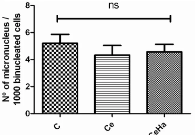
0.01) and CeHA (p < 0.001) groups. Specimens of Ce group also showed lower peak area than in CeHA (p < 0.05) (Fig. 4). Figure 5 showed that no statistical differences were found among the groups considering the number of micronucleus per 1000 binucleated cells.
Figure 3 - Average FT-Raman bone spectrum, showing
the differences in the intensity of the peak attributed to symmetric stretching vibration of primary phosphate (PO4
3-), among the experimental groups: control (C), uncoated apatite Al2O3 ceramic (Ce), and
apatite-coated Al2O3 ceramic (CeHA).
Figure 4 - Mean and standard deviations of the 2940
cm-1 peak area among the experimental groups: control (C), uncoated apatite Al2O3
ceramic (Ce), and apatite-coated Al2O3
Figure 5 - Frequency of occurrence of micronuclei in 1000 binucleated cells (p > 0.05) between the control group (C), uncoated apatite Al2O3 ceramic (Ce), and apatite-coated
Al2O3 ceramic (CeHA).
DISCUSSION
In the present study, FT-Raman, analysis revealed a close relation between the surface of uncoated
and apatite-coated Al2O3 implants and newly
formed bone matrix. The increased intensity of inorganic and organic Raman peaks (Figs. 1 and 3)
in bone formed on apatite-coated Al2O3 implants
showed that those materials were essential for the osseo integration process. The increase in the area
of the Raman peak at 959 cm-1 (bone mineral
content) (Fig. 2) was due to the increased bone
formation, which was stimulated by the
hydroxyapatite coverage of the Al2O3 implant.
According to Lee et al (2010), this acted as a stimulant for bone in growth. The 30 days period for euthanasia in the present study showed to be enough to bone deposition as indicated by Raman spectroscopy evaluation of bone inorganic matrix. These results were in agreement with the previous studies using histological analysis (Pinheiro et al. 2008) and Raman spectroscopy (Lopes et al. 2007).
With respect to the organic matrix evaluated by
the peak area at 2940 cm-1 (related to the CH
bonds of the organic material) higher organic peak areas were found in the bone near the
apatite-coated Al2O3 implants than in the other treatments
showing the influence in collagen deposition (Fig. 4). The organic bone matrix is composed of fibers and essential substance. The fibers that make up are basically of type I collagen (Huesa et al. 2011). Type I collagen is a matrix protein synthesized by osteoblasts that becomes mineralized with the
hydroxyapatite during the later stages of osteogenesis (Pei et al. 2011). Therefore, the higher the deposition of the organic matrix, more mineralization occurs (Huesa et al. 2011), and in the present study this correlation between inorganic component and organic matrix was also found (Figs. 1-4).
The micronucleus test showed that the uncoated
and apatite-coated Al2O3 implants were
non-cytotoxic and safe to in vivo applications. The
results indicated no significant change compared to the control group (Fig. 5). The micronucleus test is one of the main methods for preclinical evaluation of safety of pharmaceuticals indicating whether there is genotoxicity and mutagenicity (Maffei et al. 2008; Bull et al. 2011; Song et al. 2012). The sensitivity of the mutagenicity assay has been used successfully as an indicator of predisposition to cancer (Mafei et al. 2008). Depending on the level of chromatid breaks induced by the mutagenic treatment, subjects may be classified as sensitive or not sensitive what is believed to reflect its ability to repair DNA damage induced by mutagenic agents (Au, 2003). The micronucleus test is a method that allows detailed analysis of chromosomal damage, chromosomal instability, mitotic dysfunction and cell death induced by genotoxins or other factors (Fenech 2006; Mafei et al. 2008). The DNA damage is characterized by a single base changes and in crosslinked, thus with the potential to initiate and promote carcinogenesis (Valko et al. 2006; Zeiger et al. 2009; Song et al. 2012).
CONCLUSION
The micronucleus test showed that the uncoated
and apatite-coated Al2O3 implants were
non-cytotoxic and safe to in vivo applications.
FT-Raman spectroscopy showed that the higher the
deposition of the organic matrix, more
mineralization occurred. The biomaterial proved to be biocompatible and osteoconductive when used as a bone implant.
ACKNOWLEDGMENTS
Drª. Eliandra de Sousa Trichês (Science and Technology Institute– Federal University of São Paulo, SP, Brazil) for providing the uncoated and
apatite-coated Al2O3 foams for biological tests.
REFERENCES
Abarrategi A, Moreno-Vicente C, Martínez-Vázquez FJ, Civantos A, Ramos V, Sanz-Casado JV, et al. Biological properties of solid free form designed ceramic scaffolds with BMP-2: in vitro and in vivo evaluation. PLoS One. 2012; 7(3): e34117. doi:10.1371/journal.pone.0034117. Arora R, Petrov GI, Noojin GD, Thomas PA, Denton ML,
Rockwell BA, et al. Detecting mineral content in turbid medium using non linear bRaman imaging: feasibility study. J Mod Opt. 2011; 58(21): 1914-1921.
Au WW, Mutagen sensitivity assays in population studies, Mutat Res. 2003; 544(2-3): 273-277.
Bull CF, Beetstra-Hill S, Benassi-Evans BJ, Crott JW, Kimura M, Teo T, et al. Application and adaptation of the in vitro micronucleus assay for the assessment of nutritional requirements of cells for DNA damage prevention. Mutagenesis. 2011; 26(1): 193-197.
Campos DM, Anselme K, Soares GDA. In Vitro Biological Evaluation of 3-D Hydroxyapatite/Collagen (50/50 wt. (%)) Scaffolds. Mat Res. 2012; 15(1): 151-158.
Cortes DA, Nogiwa AA, Almanza JM, Ortega S. Biomimetic apatite coating on Mg-PSZ/Al2O3
composites. Effect of the immersion method. Mater Lett. 2005; 59(11): 1352-1355.
Fenech M. Cytokinesis-block micronucleus assay evolves into a cytome assay of chromosomal instability, mitotic dysfunction and cell death. Mutat. Res. 2006; 600(1-2): 58-66.
Huesa C, Yadav MC, Finnilä MAJ, Goodyear SR, Robins SP, Tanner E, et al. PHOSPHO1 is essential for mechanically competent mineralization and the avoidance of spontaneous fractures. Bone. 2011 48(5): 1066-1074.
Iarmarcovai G, Ceppi M, Botta A, Orsière T, Bonassi S. Micronuclei frequency in peripheral blood lymphocytes of cancer patients: A meta-analysis. Mutat Res. 2008; 659(3): 274-283.
Kozielski M, Buchwald T, Szybowicz M, Błaszczak Z, Piotrowski A, Ciesielczyk B. Determination of composition and structure of spongy bone tissue in human head of femur by Raman spectral mapping. J Mater Sci: Mater Med. 2011; (22): 1653-1661.
Lee MJ, Sohn SK, Kim KT, Kim CH, Ahn HB, Rho MS, et al. Effect of hydroxyapatite on bone integration in a rabbit tibial defect model. Clin Orthop Surg. 2010; 2(2): 90-97.
Lopes CB, Pacheco MT, Silveira L Jr, Duarte J, Cangussú MC, Pinheiro AL. The effect of the association of NIR laser therapy BMPs, and guided bone regeneration on tibial fractures treated with wire osteosynthesis: Raman spectroscopy study. J Photochem Photobiol B. 2007; 89(2-3):125-130.
Maffei F, Carbone F, Angelini S, Forti C F, Norppa H, Hrelia P. Micronuclei frequency induced by bleomycin in human peripheral lymphocytes: Correlating BLHX polymorphism with mutagen sensitivity. Mutat Res. 2008; 639(1-2): 20-26.
Meijer GJ, de Bruijn JD, Koole R, van Blitterswijk CA. Cell-Based Bone Tissue Engineering. PLoS Med. 2007; 4(2): 260-264.
Nandi SK, Roy S, Mukherjee P, Kundu B, De DK, Basu D. Orthopaedic applications of bone graft & graft substitutes: a review. Indian J Med Res. 2010; 132: 15-30.
Pei H, Wei-Ping JL, Chang-li Z, Xiao-nong Z, Yao J. Improved osteoblast proliferation, differentiation and mineralization on nanophase Ti6Al4V. Chin Med J. 2011; 124(2): 273-279.
Pinheiro AL, Gerbi ME, Ponzi EA, Ramalho LM, Marques AM, Carvalho CM, et al. Infrared laser light further improves bone healing when associated with bone morphogenetic proteins and guided bone regeneration: an in vivo study in a rodent model. Photomed Laser Surg. 2008; 26(2): 167-174.
Sato M, Aslani A, Sambito MA, Kalkhoran NM, Slamovich EB, Webster TJ. Nanocrystalline hydroxyapatite/titania coatings on titanium improves osteoblast adhesion. J Biomed Mater Res. 2008; 84-A: 265-272.
Song MF, Li YS, Kasai H, Kawai K. Metalilnanoparticle-induced micronuclei and oxidative DNA damage in mice. J Clin Biochem Nutr. 2012; 50(3): 211-216. Sousa E, Ortega FS, Pandolfelli VC. Production and
characterization of alumina foams by the gelcasting process without atmospheric control. Cerâmica. 2009; 55(334): 151-156.
Souza RA, Xavier M, da Silva FF, de Souza MT, Túlio M, Tosato MG, Martins et al. Influence of creatine supplementation on bone quality in the ovariectomized rat model: an FT-Raman spectroscopy study. Lasers Med Sci. 2012; 27(2): 487-495.
Valko M, Rhodes CJ, Moncol J, Izakovic M, Mazur M. Free radicals, metals and antioxidants in oxidative stress-induced cancer. Chem Biol Interact. 2006; 160(1): 1-40.
Wopenka B, Kent A, Pasteris J, Yoon Y, Thomopoulos S. The tendon-to-bone transition of the rotator cuff: a preliminary raman spectroscopic study documenting the gradual mineralization across the insertion in rat tissue samples. Appl Spectrosc. 2008; 62(12): 1285-1294. Zhang L, Tang P, Xu M, Zhang W, Chai W, Wang Y.
Effects of crystalline phase on the biological properties of collagen-hydroxyapatite composites. Acta Biomater. 2010; 6(6): 2189-2199.
Zeiger E, Recio L, Fennell TR, Haseman JK, Snyder RW, Friedman M. Investigation of the low-dose response in the in vivo induction of micronuclei and adducts by acrylamide. Toxicol Sci. 2009; 107(1): 247-257.