EFFECTS OF AMMONIA ON THE
DISEASE SUSCEPTIBILITY OF FISH
Ana Filipa dos Santos Gonçalves
Tese de doutoramento em Ciências Biomédicas
EFFECTS OF AMMONIA ON THE DISEASE
SUSCEPTIBILITY OF FISH
Tese de Candidatura ao grau de Doutor em
Ciências Biomédicas submetida ao Instituto
de Ciências Biomédicas Abel Salazar da
Universidade do Porto.
Orientador – Professor Doutor João Coimbra
Categoria – Professor Catedrático
Afiliação – Instituto de Ciências Biomédicas
Abel Salazar da Universidade do Porto.
Co-orientador – Doutor Jonathan Wilson
Categoria – Investigador Auxiliar
Afiliação
–
Centro
Interdisciplinar
de
Investigação Marinha e Ambiental da
Universidade do Porto
Co-orientador – Professor Mathilakath M.
Vijayan
Categoria – Professor
que a seguir se descriminam:
Gonçalves AF, Páscoa I, Neves JV, Coimbra J, Vijayan MM, Rodrigues P, Wilson JM. The inhibitory effect of environmental ammonia on Danio rerio LPS induced acute phase response. Developmental and Comparative Immunology, 2012; 36 (2): 279-288.
ii
_______________________________________________________
Esta tese de doutoramento não teria sido possível sem a ajuda de muitas pessoas às quais agradeço toda a amizade, apoio e compreensão durante todos estes anos.
Agradeço ao meu orientador, Professor João Coimbra por me ter aberto as portas do seu laboratório no CIIMAR e, com isso, ter permitido o meu desenvolvimento científico mas também pessoal.
I wish to express my deepest gratitude to my co-supervisor, Dr. Jonathan Wilson. Without his guidance, knowledge and assistance; this work would not have been possible.
I would also like to thank my co-supervisor, Prof. Matt Vijayan, who gave invaluable assistance, support and guidance when I was working in his lab.
Ao Professor Pedro Rodrigues por me abrir as portas do IBMC e por todo o apoio, dedicação e conhecimentos transmitidos. Agradeço-lhe a orientação, e as suas preciosas sugestões e críticas que muito valorizaram este trabalho.
Ao Doutor João Neves por toda a ajuda, disponibilidade e dedicação a este trabalho.
À Odete Gonçalves por todo o carinho, amizade e apoio emocional.
À Daniela Lima e Inês Coelho pela amizade e pela ajuda valiosa quando eu já estava perto da recta final.
A todos os amigos e colegas do CIIMAR pela sua amizade, companheirismo e disponibilidade para me ajudarem: Joana Moreira da Silva, Claudia Escórcio, Inês Páscoa, Marisa Passos, Filipe Castro, Emília Afonso, Hélder Nunes, Hugo Santos e restante equipa BOGA.
iii família pelo carinho e apoio incondicional.
Não consegui nem nunca conseguirei descrever em palavras a importância que todos tiveram para a minha vida e para esta tese. Obrigada!
iv
and Environmental Research (CIMAR-UP), Institute of Biomedical Sciences of Abel Salazar (ICBAS) from University of Porto and the Biology Department of University of Waterloo are acknowledged for providing facilities and logistical support.
vi
Dissertation organization
The present PhD thesis is organized in chapters, starting with General Introduction and finishing with General Discussion. Chapters 2, 3 and 4 correspond to the experimental work and are structured as papers. Each chapter will give origin to a separate paper and, within this dissertation, they are free standing, which may lead to a certain degree of repetition throughout the thesis. Tables and figures have sequential numbering from one chapter to the next. The bibliography is consolidated at the end of the thesis.
viii
_______________________________________________________
Ammonia is an unusual toxicant in that it is not only a common environmental pollutant but is also an endogenous by-product of amino acid catabolism within the organism. In aquaculture, ammonia toxicity can be problematic when fish are held at high densities with insufficient water flow, or when using recirculating water systems, which may lead to elevated ammonia levels. Fish can also be exposed to elevated ammonia levels in the environment as a result of an input of biological wastes, runoff from fertilizer application for agriculture activities and atmospheric deposition. High environmental ammonia (HEA) is toxic to fish causing problems at multiple levels of organization, and has been associated with increased vulnerability to different parasitic, viral and bacterial diseases in fishes, although the mechanism(s) is not well understood.
The aim of this Ph.D. thesis was to provide new insights into the complex relationship between HEA levels and increased disease susceptibility using the zebrafish, Danio rerio, as the animal model. The hypotheses addressed were that: i) HEA acts by impairing the acute phase response (APR), and ii) cortisol, the principal corticosteriod in teleosts, mediates that immunosuppression.
The results revealed, for the first time, that HEA exposure impaired lipopolysaccharide (LPS) and bacterial (Edwarsiella tarda) -induction of various acute phase proteins (APPs) in zebrafish. And both acute and chronic exposure to HEA concentrations also significantly elevated whole-body cortisol levels compared with control fish. Significantly, HEA was found to decrease survival during bacterial challenge with E. tarda. In addition, it was demonstrated that exogenous exposure to the stress hormone cortisol, and the elevation of cortisol levels in response to HEA may be playing a key role in the downregulation of important innate immune genes and upregulation of suppressors of cytokine signaling (SOCS) genes. Mifepristone, a glucocorticoid receptor (GR) antagonist, blocked the HEA-cortisol effect through a GR mediated pathway. Given the similar direct effects of HEA and cortisol, it is possible to infer that the ammonia exposure effect on immunosuppression was mediated by cortisol.
ix
associated metabolic costs by restricting activation of cytokine signalling, and diverting energy resources away from the growth promoting action of growth hormone during stress in fish. This would reallocate energy substrates away from anabolic processes, thereby allowing ATP usage for other processes, including production of glucose, a key fuel for coping with the increase energy demand associated with stress.
There are various studies showing that LPS induces socs expression. However, socs induction is not always observed with LPS challenge and there are data showing no changes or even a socs3 down regulation. In this study, LPS challenge supressed socs transcript levels. To this point, the precise role of Socs proteins in LPS responses clearly remains enigmatic.
To summarize, the work presented in this thesis demonstrates that ammonia suppresses the immune response, at least in part, through an increase in cortisol levels in zebrafish, and ultimately increases the disease susceptibility associated with HEA levels in zebrafish. This is of particular relevance for maintenance of healthy fish in aquaculture, and for the monitoring of environmental health in wild fish populations faced with pollution.
x
_______________________________________________________
A amónia é um agente tóxico invulgar na medida em que é um poluente ambiental comum e é também um subproduto do catabolismo dos aminoácidos nos organismos. Em aquacultura é problemática quando os peixes são mantidos em altas densidades, com fluxo mínimo de água, ou quando se utilizam sistemas de recirculação de água, o que pode levar a níveis elevados de amónia. Os peixes também estão expostos a amónia no meio ambiente como resultado de resíduos biológicos, escoamento da aplicação de fertilizantes na agricultura e deposição atmosférica. Amónia ambiental elevada é tóxica para peixes e causa problemas a diferentes níveis: organismo, órgão e célula; e tem sido associada ao aumento da vulnerabilidade a diferentes doenças causadas por parasitas, vírus e bactérias em peixes, embora o mecanismo envolvido não seja bem compreendido.
O objetivo desta tese de doutoramento foi compreender melhor a relação complexa entre níveis elevados de amónia ambiental e a susceptibilidade aumentada a doença, usando o peixe-zebra, Danio rerio, como animal modelo. As hipóteses estudadas foram que a amónia age através da inibição da resposta de fase aguda; e também, que o cortisol, o corticosteroide principal em peixes teleósteos, é o mediador dessa imunossupressão.
Os resultados revelaram, pela primeira vez, que a amónia inibe a indução de várias proteínas de fase aguda, por lipopolissacarídeo bacteriano e por bactéria (Edwardsiella tarda) em peixes-zebra. Exposição aguda ou crónica a concentrações elevadas de amónia elevaram significativamente os níveis de cortisol em comparação com os peixes controle. Além disso, a amónia ambiental diminuiu a sobrevivência durante infeção bacteriana com Edwarsiella
tarda. Também foi demonstrado que a hormona do stress, cortisol, e os
elevados níveis de cortisol em resposta à exposição a amónia podem ser a chave para um aumento da indução de socs, e, uma diminuição da indução de genes importantes da resposta de fase aguda. Mifepristona, um potente antagonista do recetor de glucocorticoides, atenuou a ação do cortisol e os seus efeitos através de uma via mediada pelo recetor de glucocorticoides, nos grupos expostos a amónia. Devido aos efeitos semelhantes da amónia/cortisol, é possível deduzir que o efeito da amónia na imunossupressão foi mediado
xi
pode ser uma resposta adaptativa que limita a resposta inflamatória imunitária e os custos metabólicos associados, ao limitar a ativação da sinalização de citocinas, e também desviando recursos energéticos do crescimento, durante situações de stress em peixes. Isto permite realocar substratos energéticos dos processos anabólicos, permitindo assim o uso de ATP para outros processos, tais como a produção de glucose, para lidar com o aumento da demanda de energia associada ao stress. Existem vários estudos que demonstram que LPS leva à indução de expressão de socs. Contudo, a indução de socs não é sempre observada com LPS, há dados que mostram não existirem diferenças na expressão ou até diminuição da expressão de socs3. Neste estudo, LPS suprimiu a expressão de socs. Neste momento, o papel preciso das proteínas SOCS nas respostas ao LPS claramente permanece enigmático.
Resumindo e concluindo, o trabalho apresentado nesta tese mostra que elevados níveis de amónia ambiental atuam, pelo menos em parte, através do aumento nos níveis de cortisol em peixe-zebra, o que leva uma imunossupressão, e, finalmente, aumenta a susceptibilidade à doença associada com amónia em peixes. Isto é de particular relevância para a manutenção de peixes em aquacultura e à monitorização ambiental nas populações de peixes selvagens sujeitas a poluição.
xii
_______________________________________________________
3β-HSD - 3β-hydroxysteroid dehydrogenase
ACTH - Adrenocorticotropic hormone
APP - Acute Phase Protein
APR - Acute phase response
ATP - Adenosine-5'-triphosphate
C3b / c3b - Complement Component
CT - Cycle threshold
EF1α / ef1α - Elongation factor-1 alpha
CFU - Colony-forming unit
CRH - corticosteroid releasing hormone
CRP / crp - C-reactive protein
GH – Growth hormone
GR / gr – Glucocorticoid receptor
GC - Glucocorticoids
HAMP / hamp – Hepcidin
xiii HPI - hypothalamus-pituitary-interrenal
Iap / iap - Intestinal alkaline phosphatase
IL1β / il1β - Interleukin 1 beta
IL10 / il10 - Interleukin 10
LC50 - Lethal concentration, 50%
LECT2 / lect2 - Leukocyte cell-derived chemotaxin-2
LITAF / litaf - Lipopolysaccharide-induced TNF factor
LPS – Lipopolysaccharide
MS-222 - Tricaine methanesulfonate
NADH – Reduced nicotinanide adenine dinucleotide
NH3 – Unionized ammonia
NH4+ - Ammonium ion
NMDA
-
N-methyl-D-aspartatePBS - Phosphate-buffered saline
PCA - Perchloric acid
RT-PCR – Reverse transcriptase-polymerase chain reaction
xiv SOCS /socs - Suppressor of cytokine signaling
TAN - Total ammonia nitrogen
TBE - Tris-borate-EDTA
TLR / tlr - Toll-like receptor
TNFα / tnfα - Tumor necrosis factor alpha
TRIS - tris(hydroxymethyl)aminomethane
xvi
_______________________________________________________
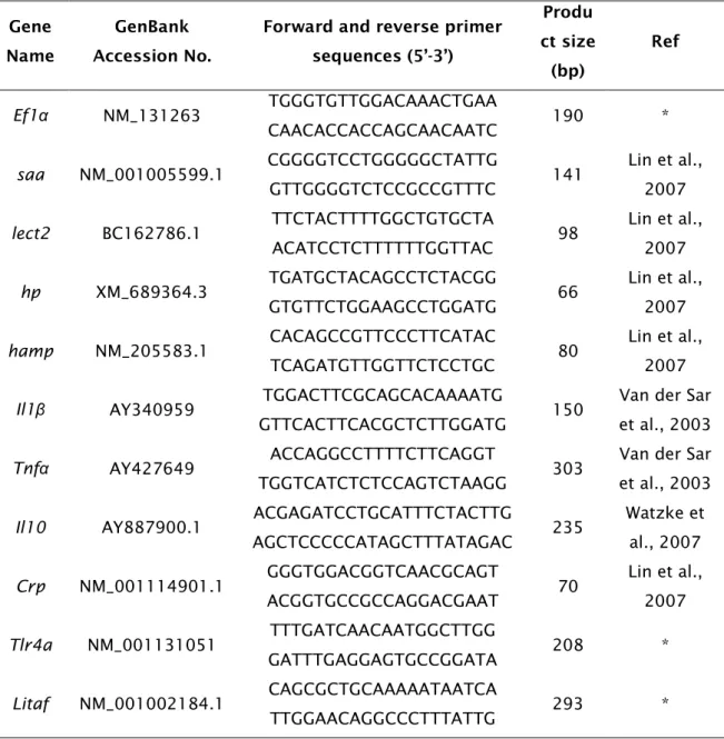
Table 2.1: Primer pairs (sense and anti-sense, respectively) for qPCR with
predicted product size and original gene accession number. Primers with an asterisk (*) were designed with Primer3 and reference sources are given for the remainder.
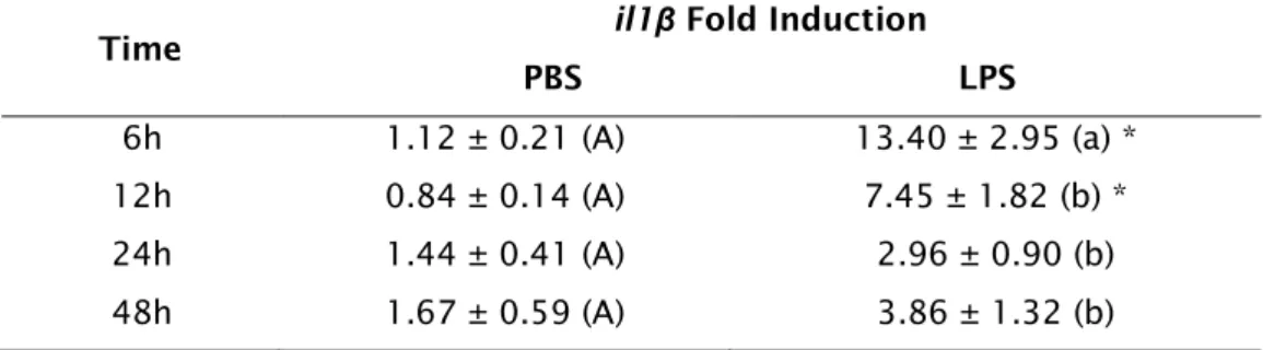
Table 2.2: Data represent il1β time course response after injection of PBS or
10μg/g of LPS, by qPCR analysis. Mean ± standard error of the mean is show in the table (n=7). Values with like characters are not significantly different (PBS upper case and LPS lower case characters). The asterisk (*) indicates a significant difference between the sham and LPS groups at a given time point. Data analysed by 2-way ANOVA and post hoc Student-Newman-Keuls test.
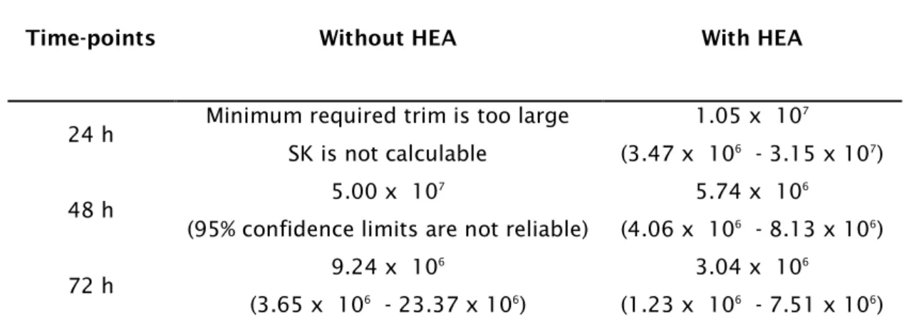
Table 3.1: Edwardsiella tarda LC50 values in the presence or absence of 1 mM
ammonia (pH 8.0) and their 95% confidence limit for different time points (h: hours). Dose of bacteria is in CFU.
Table 3.2: Primer pairs (sense and anti-sense, respectively) for qPCR with
original GenBank accession number. Primers with an asterisk (*) were designed with Primer3 and reference sources are given for the remainder.
Table 4.1: Summary of experiment 2 treatment groups.
Table 4.2: Primer pairs (sense and anti-sense, respectively) for real time qPCR
with original GenBank accession number. Primers with an asterisk (*) were designed with Primer3 and reference sources are given for the remainder.
xviii
_______________________________________________________
Fig. 1.1: Biosynthesis of cortisol in teleost fishes. The shaded area represents
the mitochondrial compartment, the non-shaded area represents the cytosolic compartment. Abbreviations: 3β-HSD, 3β-hydroxysteroid dehydrogenase; P450s, various forms of cytochrome P450 (Mommsen et al., 1999).
Fig. 1.2: The zebrafish, Danio rerio.
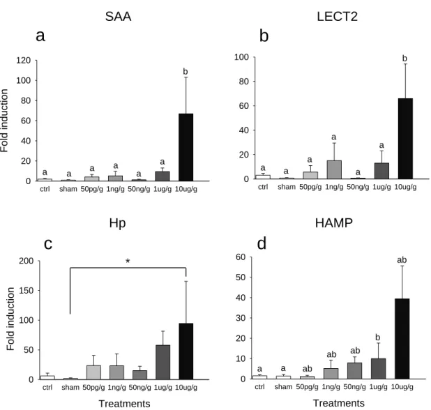
Figure 2.1. Fold induction in response to LPS dose of a) saa; b) lect2; c) hp;
and, d) hamp relative to ef1α, determined by qPCR, in zebrafish viscera (liver, intestine, pancreas and spleen). Fish were i.p. injected with different doses of LPS, PBS (sham injected) or not injected (ctrl). Total RNA was extracted from fish that were sampled 24h post injection. Error bars represent SEM (n=6). Data analysed by 1-way ANOVA and post hoc Student-Newman-Keuls test. Bars with like characters are not significantly different.
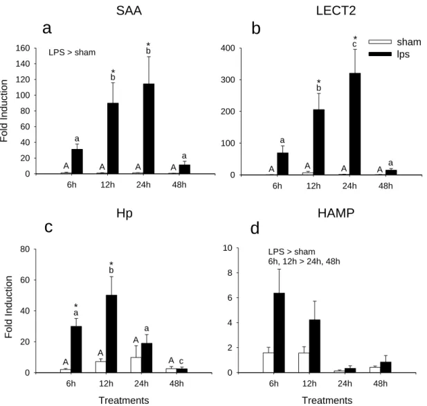
Figure 2.2. Time course fold induction in response to i.p. injection with 10μg/g
LPS (black bars) compared to control fish (sham injected; white bars) as determined by qPCR for a) saa; b) lect2; c) hp; and, d) hamp relative to ef1α. Total RNA was extracted from fish that were sampled at 6, 12, 24, 48h post injection. Error bars represent SEM (n=7). Data analysed by 2-way ANOVA and
post hoc Student-Newman-Keuls test. Bars with like characters are not
significantly different (sham upper case and LPS lower case characters). The asterisk (*) indicates a significant difference between the sham and LPS groups at a given time point.
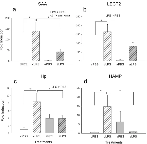
Figure 2.3. Effects of acute ammonia exposure on induction of innate immunity
genes. Following an i.p. injection of 10μg/g LPS (hatched bars) or PBS fish were exposed to 1mM of NH4Cl (pH 8) (grey bars) or control conditions and fold induction of a) saa, b) lect2, c) hp and d) hamp relative to ef1α measured after 24h. Error bars represent SEM (n=7). Data analysed by 2-way ANOVA and post
hoc Student-Newman-Keuls test. Asterisks (*) indicate groups that are
xix
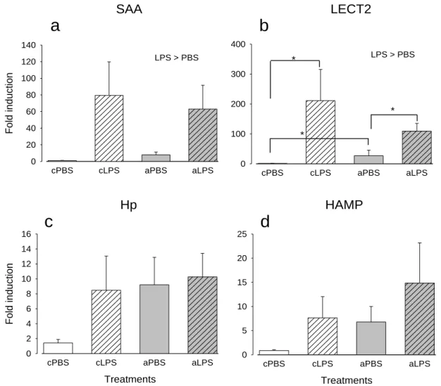
by 10μg/g LPS (hatched bars). Fold induction of a) saa; b) lect2; c) hp; and, d)
hamp relative to ef1α are shown. Error bars represent SEM (n=7). Data analysed
by 2-way ANOVA and post hoc Student-Newman-Keuls test. Asterisks (*) indicate groups that are significantly different.
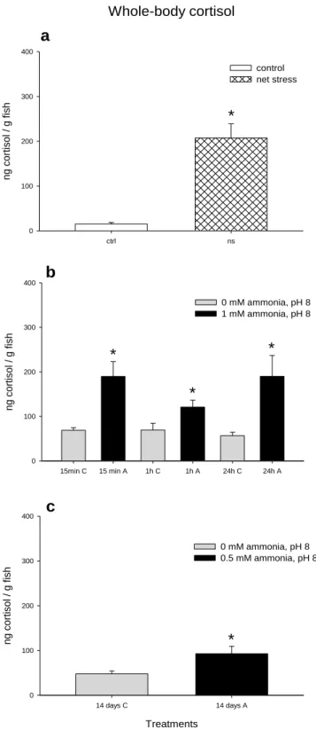
Figure 2.5. Mean whole-body cortisol (ng/g fish) levels of zebrafish of a)
Control or net handling stress groups. b) Control or acute 1mM ammonia exposure after 15min, 1h or 24h, at pH 8. c) Control or chronic 0.5mM ammonia exposure (14 days), at pH 8. Error bars represent SEM (n=6). Data analysed by t-tests (a, c) or two-way ANOVA (b). Asterisks (*) indicate significant differences between treatment groups.
Figure 3.1. Percentage survival in zebrafish injected i.p. with different doses of
E. tarda (104, 5 x 10⁴, 105, 5 x 10⁵ CFU E. tarda in 10ul TSB, control fish were injected with TSB alone). Fish were monitored during 72 h, in 6 h intervals (a) in the absence and (b) in the presence of elevated water ammonia levels (1 mM, pH 8).
Figure 3.2. Effect of acute ammonia exposure on induction of hamp mRNA
expression by E. tarda challenge via either i.p. injection or bath immersion. (a,
c) Bacterial challenge by immersion with 107 CFU/mL E. tarda (hatched bars) or
TSB. (b, d) Bacterial challenge by i.p. injection with 104 CFU E. tarda (hatched bars) or TSB. Following the bacterial challenge fish were exposed to 1 mM of NH4Cl at pH 8.0 (grey bars) or control conditions (white bars) for 24 h. hamp was measured by q-PCR relative to ef1α in (a, b) viscera and (c, d) gill. Data analyzed by 2-way ANOVA and post hoc Student–Newman–Keuls test. Asterisks (*) indicate groups that are significantly different (n = 7).
Figure 3.3. Effect of acute ammonia exposure on induction of lect2 mRNA
expression by E. tarda challenge via either i.p. injection or bath immersion. (a,
c) Bacterial challenge by immersion with 107 CFU/mL E. tarda (hatched bars) or
TSB. (b, d) Bacterial challenge by i.p. injection with 104 CFU E. tarda (hatched bars) or TSB. Following the bacterial challenge fish were exposed to 1 mM of NH4Cl at pH 8.0 (grey bars) or control conditions (white bars) for 24 h. L lect2
xx
(*) indicate groups that are significantly different (n = 7).
Figure 3.4. Effect of acute ammonia exposure on induction of saa mRNA
expression by E. tarda challenge via either i.p. injection or bath immersion. (a,
c) Bacterial challenge by immersion with 107 CFU/mL E. tarda (hatched bars) or
TSB. (b, d) Bacterial challenge by i.p. injection with 104 CFU E. tarda (hatched bars) or TSB. Following the bacterial challenge fish were exposed to 1 mM of NH4Cl at pH 8.0 (grey bars) or control conditions (white bars) for 24 h. saa was measured by q-PCR relative to ef1α in (a, b) viscera and (c, d) gill. Data analyzed by 2-way ANOVA and post hoc Student–Newman–Keuls test. Asterisks (*) indicate groups that are significantly different (n = 7).
Figure 3.5. Effect of acute ammonia exposure on induction of hp mRNA
expression by E. tarda challenge via either i.p. injection or bath immersion. (a,
c) Bacterial challenge by immersion with 107 CFU/mL E. tarda (hatched bars) or
TSB. (b, d) Bacterial challenge by i.p. injection with 104 CFU E. tarda (hatched bars) or TSB. Following the bacterial challenge fish were exposed to 1 mM of NH4Cl at pH 8.0 (grey bars) or control conditions (white bars) for 24 h. hp was measured by q-PCR relative to ef1α in (a, b) viscera and (c, d) gill. Data analyzed by 2-way ANOVA and post hoc Student–Newman–Keuls test. Asterisks (*) indicate groups that are significantly different (n = 7).
Figure 3.6. Effect of acute ammonia exposure on induction of c3b mRNA
expression by E. tarda challenge via either i.p. injection or bath immersion. (a,
c) Bacterial challenge by immersion with 107 CFU/mL E. tarda (hatched bars) or
TSB. (b, d) Bacterial challenge by i.p. injection with 104 CFU E. tarda (hatched bars) or TSB. Following the bacterial challenge fish were exposed to 1 mM of NH4Cl at pH 8.0 (grey bars) or control conditions (white bars) for 24 h. c3b was measured by q-PCR relative to ef1α in (a, b) viscera and (c, d) gill. Data analyzed by 2-way ANOVA and post hoc Student–Newman–Keuls test. Asterisks (*) indicate groups that are significantly different (n = 7).
Figure 3.7. A comparison of the effects of 14 day pre-exposure to sub-lethal
xxi
bars) or TSB. Error bars represent SEM (n = 7). Data analyzed by 2-way ANOVA and post hoc Student–Newman–Keuls test. Asterisks (*) indicate groups that are significantly different.
Figure 4.1. Effect of cortisol and LPS treatment on mRNA expression of socs
genes. Effect of cortisol on (a) socs 1a, (b) socs 2 and (c) socs 3 gene expression in viscera of zebrafish in the presence and absence of LPS. Zebrafish were intraperitoneally injected with cortisol (10 mg/ml) diluted in coconut oil (grey bars), an injection volume of 5μl/g was used. Another group of fish was injected with coconut oil (vehicle alone) and served as the control. After 48h, fish were intraperitoneally injected with 10µg/g LPS (hatched bars) or PBS, an injection of 10μl/g was used. Gene expression was measured by qPCR relative to ef1α. Data analyzed by 2-way ANOVA and post hoc Student–Newman–Keuls test. All values represent means ± SE (n = 7), bars with different letters are significantly different.
Figure 4.2. Effect of cortisol and LPS treatment on mRNA expression of acute
phase proteins. Effect of cortisol on (a) saa and (b) lect2 gene expression in viscera of zebrafish in the presence and absence of LPS. Zebrafish were intraperitoneally injected with cortisol (10 mg/ml) diluted in coconut oil (grey bars), an injection volume of 5μl/g was used. Another group of fish was injected with coconut oil (vehicle alone) and served as the control. After 48h, fish were intraperitoneally injected with 10µg/g LPS (hatched bars) or PBS, an injection of 10μl/g was used. Gene expression was measured by q-PCR relative to ef1α. Data analyzed by 2-way ANOVA and post hoc Student–Newman–Keuls test. All values represent means ± SE (n = 7), bars with different letters are significantly different.
Figure 4.3. Whole-body cortisol (ng/g fish) levels of zebrafish. Effect of cortisol
in the presence and absence of LPS. Zebrafish were intraperitoneally injected with cortisol (10 mg/ml) diluted in coconut oil (grey bars), an injection volume of 5μl/g was used. Another group of fish was injected with coconut oil (vehicle alone) and served as the control. After 48h, fish were intraperitoneally injected with 10µg/g LPS (hatched bars) or PBS, an injection of 10μl/g was used. Data
xxii groups and groups injected with vehicle alone.
Figure 4.4. Effect of ammonia and LPS on socs1 mRNA expression, in the
presence and absence of Mifepristone. Effect of ammonia and LPS on socs1 mRNA transcript level in viscera of zebrafish, in the presence and absence of Mifepristone. Zebrafish were intraperitoneally injected with 100 µg/g Mifepristone, diluted in ethanol, an injection volume of 5μl/g was used. Other fish were injected with saline (0.9% w/v) and served as the control. After 48h, fish were intraperitoneally injected with 10µg/g LPS (grey bars) or PBS, an injection of 10μl/g was used. They were allowed to recover in tanks in the absence or presence of 1mM ammonia (hatched bars) at pH 8.0. Data analyzed by 1-way ANOVA and post hoc Student–Newman–Keuls test. All values represent means ± SE (n = 7). Bars with different letters are significantly different.
Figure 4.5. Effect of ammonia and LPS on mRNA expression of immune
response genes, in the presence and absence of Mifepristone. Effect of ammonia and LPS on (a) saa, (b) lect2, (c) haptoglobin, (d) hepcidin, (e) Il1β and (f) c3b gene expression, in viscera of zebrafish, in the presence and absence of Mifepristone. Zebrafish were intraperitoneally injected with 100 µg/g Mifepristone, diluted in ethanol, an injection volume of 5μl/g was used. Other fish were injected with saline (0.9% w/v) and served as the control. After 48h, fish were intraperitoneally injected with 10µg/g LPS (grey bars) or PBS, an injection of 10μl/g was used. They were allowed to recover in tanks in the absence or presence of 1mM ammonia (hatched bars) at pH 8.0. Data analyzed by 1-way ANOVA and post hoc Student–Newman–Keuls test. All values represent means ± SE (n = 7). Bars with different letters are significantly different.
Figure 4.6. Whole-body cortisol (ng/g fish) levels of zebrafish. Effect of
ammonia and LPS, in the presence and absence of Mifepristone. Zebrafish were intraperitoneally injected with 100 µg/g Mifepristone diluted in ethanol, an injection volume of 5μl/g was used. Other fish were injected with saline (0.9% w/v) and served as the control. After 48h, fish were intraperitoneally injected with 10µg/g LPS (grey bars) or PBS, an injection of 10μl/g was used. They were allowed to recover in tanks in the absence or presence of 1mM ammonia
xxiii letters are significantly different.
xxiv
_______________________________________________________
CHAPTER 1 ... 2 PART I – AMMONIA ... 4 PART II – INNATE IMMUNE SYSTEM ... 7 PART III – CORTISOL ... 10 PART IV - THE EXPERIMENTAL ANIMAL MODEL ... 13 PART V - THESIS AIMS ... 15 CHAPTER 2 ... 18 Abstract ... 20 Introduction ... 21 Material and Methods ... 25
Animals ... 25 Experiment 1 – LPS dose response ... 25 Experiment 2 – LPS time course response ... 25 Experiment 3 – acute ammonia and LPS ... 26 Experiment 4 – chronic ammonia and LPS ... 26 Whole-body cortisol levels after ammonia exposure ... 26 Sampling ... 27 Total RNA extraction and cDNA synthesis ... 27 Primers ... 27 RT-PCR and Quantitative real-time PCR ... 28 Ammonia measurements ... 29 Whole-body cortisol extraction and measurement ... 30 Statistics ... 30
Results ... 31
Experiment 1 – LPS dose response ... 31 Experiment 2 – LPS time course response ... 33 Experiment 3 – acute ammonia and LPS ... 34 Experiment 4 – chronic ammonia and LPS ... 36
Discussion ... 39
LPS induction experiments ... 39 LPS-HEA effects and cortisol levels ... 40
xxv
Abstract ... 46 Introduction ... 47 Material and Methods ... 50
Bacteria and media ... 50 Animals ... 50 Determination of the E. tarda LC50 value in the presence or absence of HEA ... 50 Acute ammonia and E. tarda infection ... 51 Chronic ammonia exposure and E. tarda infection ... 52 Reisolation of bacteria ... 52 Sampling ... 53 Total RNA Extraction and cDNA synthesis ... 53 Primers ... 53 Semi-quantitative and Real-time RT-PCR ... 54 Ammonia measurements ... 55 Statistics ... 55
Results ... 56
Determination of the E. tarda LC50 value in the presence or absence of HEA ... 56 Acute ammonia exposure and Edwarsiella tarda infection ... 57 Effects of chronic ammonia exposure on E. tarda infection ... 64 Whole-body ammonia levels in chronic HEA experiments ... 65
Discussion ... 67 Conclusions ... 71 CHAPTER 4 ... 72 Abstract ... 74 Introduction ... 75 Material and Methods ... 77
Animals ... 77 Experiment 1 – Cortisol and LPS ... 77 Experiment 2 – Mifepristone, LPS and acute HEA ... 77 Sampling ... 78 Total RNA Extraction and cDNA synthesis ... 78 Primers ... 78
xxvi
Ammonia measurements ... 80 Statistics ... 81
Results ... 82
Cortisol and LPS ... 82 Mifepristone, LPS and acute HEA ... 87
Discussion ... 91 Conclusions ... 95 CHAPTER 5 ... 96 Introductory Remarks ... 98 Does ammonia act through immunosuppression of the APR? ... 99 Is cortisol involved in the HEA-mediated suppression of the APR? ... 102 Concluding Remarks and Future Perspectives ... 105 References ... 106
CHAPTER 1
________________________________________________
4
There are various studies showing that exposure to high environmental ammonia levels increase susceptibility disease in fish; however, the mechanism of action of ammonia remains largely unknown. This PhD thesis will try to provide new insights into the complex relationship between ammonia, the innate immune system and the increase disease vulnerability in zebrafish, and its link with the stress hormone cortisol. These factors will be discussed in more detail in the following sections.
PART I – AMMONIA
_______________________________________________________
Ammonia is an unusual toxicant in that it is environmentally relevant and it is also produced naturally as a metabolic waste of protein catabolism within the organism. Proteins are hydrolyzed into amino acids and their deamination results in ammonia. The liver is the major organ for ammonia formation (Pequin and Serfaty, 1963), although also occurring in other organs like kidney and intestine (Walton and Cowey, 1977). Ammonia is also released into the environment through the application of industrial fertilizers and input of biological wastes, industrial emissions, decomposition of vegetation and animals, excretion by aquatic animals and volcanic activity (Ip et al., 2004; USEPA, 1999, Randall and Tsui, 2002). A major concern regarding ammonia toxicity is in aquatic systems, in regions of high human habitation and/or large numbers of farm animals, because urban and agricultural runoff and most biological waste are released into rivers and oceans (Randall and Tsui, 2002). In aquaculture, ammonia toxicity is problematic when using recirculating water systems, or during rearing stages in which fishes are held at high densities with minimal water flow, which may lead to high levels of ammonia.
Ammonia has been shown to have significant adverse neurological and physiological effects in vertebrates. At toxic internal levels, ammonia is detrimental to central nervous system processes, oxidative metabolism, and it may also impair oxygen delivery (Randall and Tsui, 2002; Wilkie, 1997). In aqueous solution ammonia exists as either unionized ammonia (NH3) or ammonium ion (NH4+) in an equilibrium that is largely pH dependent (NH
3 + H +
5
<-> NH4+, pK 9.2). Ammonia causes convulsions, coma and death, probably because elevated NH4+ displaces K+ and depolarizes neurons, causing activation of NMDA
(
N-methyl-D-aspartate) type glutamate receptor, which leads to aninflux of excessive Ca2+ and subsequent cell death in the central nervous system (Randall and Tsui, 2002). Exposure to high environmental ammonia (HEA) also causes gill hyperplasia, anemia, hypercortisolemia and ionoregulatory problems (USEPA, 1999; Ip et al., 2004; Wilkie, 2002).
Fish are generally ammonotelic and rely mainly on passive diffusion down the concentration gradient between the body and water for elimination. If the concentration in surrounding water exceeds that of blood (reverse diffusion gradient), ammonia becomes difficult to remove, and reducing production and/or conversion to nontoxic forms becomes a priority to avoid accumulation to toxic levels. Higher vertebrates make use of uric acid and urea (Wright et al., 1995). However, fish can tolerate relatively higher internal levels of ammonia than mammals, but ammonia accumulation still leads to cell and animal death through its impairment of energy metabolism, by interfering in the Krebs cycle and, more importantly, through its interference with cellular ion and acid-base homeostasis (Ip et al., 2004).
Ammonia is also generally accepted to increase susceptibility to different parasitic, bacterial and viral fish diseases (Carballo et al., 1995; Carballo and Munõz, 1991; Evans et al., 2005; Hanson and Grizzle, 1985; Liu, 2004). For example, juvenile Chinook salmon (Oncorhynchus tshawytscha) previously exposed to 0.12–0.49mM total ammonia nitrogen (TAN)1 levels were more susceptible to Vibrio anguillarum challenge (Ackerman et al., 2006). Also, infection caused by Tetrahymena sp. was significantly higher when guppies (Poecilia reticulata) were exposed to 0.11– 0.32mM TAN (Pimenta-Leibowitz et al., 2005). In addition, Channel catfish, Ictalurus punctatus, injected with
Aeromonas hydrophila and exposed to 0.11mM TAN1 had significantly higher total bacterial counts than controls (Walters and Plumb, 1980). However, the mechanism of action of ammonia remains largely unknown. Detrimental effects are not always observed with ammonia exposure. Lease and collaborators (2003) showed that none of the more traditional toxicity endpoints (growth, whole-body ion content and swimming performance) were significantly affected by a range of TAN up to 1.71mM1 in Lost River suckers. Also, TAN concentrations of 0.24mM1 did not affect the antibody response to Aeromonas
6
(2006) even demonstrated that survival of Deltistes luxatus exposed to
Flavobacterium columnare increased significantly as unionized ammonia
concentrations increased from 0 to 0.72mM TAN1. It is known that elevated ammonia concentrations cause gill damage in various fish species (Smart, 1976; Lang et al., 1987) but it remains unclear if this damage causes an increased susceptibility to infection.
7
PART II – INNATE IMMUNE SYSTEM
_______________________________________________________
The immune system protects organisms from infection with layered defenses of increasing specificity. In simple terms, physical barriers prevent pathogens such as bacteria and viruses from entering the organism. If a pathogen breaches these barriers, the innate immune system provides an immediate, but non-specific response. Innate immune systems are found in all plants and animals (Litman et al., 2005). If pathogens successfully evade the innate response, vertebrates possess a second layer of protection, the adaptive immune system, which is activated by the innate response. Here, the immune system adapts its response during an infection to improve its recognition of the pathogen. This improved response is then retained after the pathogen has been eliminated, in the form of an immunological memory, and allows the adaptive immune system to mount faster and stronger attacks each time this pathogen is encountered (Mayer, 2006).
The innate immune system is a universal and ancient form of host defense against infection (Janeway and Medzhitov, 2002). The elements of the innate immune system include anatomical barriers, secretory molecules and cellular components. The anatomical barriers are very effective in preventing colonization of tissues by microorganisms. However, when there is damage to tissues the anatomical barriers are breached and infection may occur. Once infectious agents have penetrated tissues, another innate defense mechanism comes into play, namely acute inflammation Mayer, 2006).
The acute phase response (APR) is the immediate set of host inflammatory reactions that counteract challenges such as tissue injury, infection and trauma; and it involves metabolic changes in several organ systems (Bayne et al., 2001; Nicolas et al., 1987; Uhlar and Whitehead, 1999). APR has been extensively studied in human and mouse.
One clear indication of the APR is the remarkable increase in the concentrations of many plasma proteins that are synthesized in hepatocytes, known as acute phase proteins (APPs; Gabay and Kushner, 1999; Uhlar and Whitehead, 1999). An APP has been defined as one whose plasma concentration increases (positive acute-phase proteins) or decreases (negative acute-phase proteins) by at least 25 percent during inflammatory disorders. Some APPs were
8
isolated from fish (Bayne and Gerwick, 2001). The APPs function in a variety of defense-related activities, such as limiting the dispersion and killing pathogens, activating the complement system, neutralizing enzymes, scavenging free hemoglobin and radicals, repair of tissue damage, inactivation of proteases and restoration of the healthy (homeostatic) state that prevailed before the stimulus (Bayne and Gerwick, 2001; Gabay and Kushner, 1999; Gruys et al., 2005). In mammals, induction of acute phase proteins (APPs) is mediated by Toll-like receptors (TLRs), pro-inflammatory cytokines and CCAAT/enhancer-binding proteins (or C/EBPs). Upon infection, TLRs can recognize the microbial components thus sensing the invasion of pathogens (Akira and Takeda, 2004). Stimulation of these receptors finally results in the production of pro-inflammatory cytokines, such as interleukin 1, interleukin 6, and TNFα, which are important inducers of APPs (Cohen, 2002). These cytokines in turn induce the production of some important transcription factors of many APP genes (Poli, 1998).
Cytokines are an integral component of the adaptive and innate immune responses. Signal transduction via cytokine receptors is regulated by several mechanisms that control initiation, magnitude and duration of signaling pathways. Cytokine-induced suppressor of cytokine signaling (SOCS) family acts as feedback inhibitors of cytokine receptor signaling by inhibiting a variety of signal transduction pathways (Alexander and Hilton, 2004; Jin et al., 2007).
In this PhD thesis, I stimulated the innate immunity response in zebrafish by injection with Escherichia coli lipopolysaccharides (LPS). LPS are also termed endotoxins and are considered to be a major virulence factor, being responsible for lethal effects and clinical manifestations of diseases in humans and animals (Swain et al., 2008). It has been shown in many studies, that LPS can induce the innate immune response in fish (Huttenhuis et al., 2006; Watzke et al., 2007). LPS is a cell wall component found in most Gram-negative bacteria. However, some Gram-positive bacteria, such as Listeria monocytogenes, also possess endotoxin-like activity (Wexler and Oppenheim, 1979). Higher vertebrates are extremely sensitive to endotoxins, even at low doses, but lower vertebrates such as amphibians and fish are found to be resistant to endotoxic shock. Fish like Oncorhynchus kisutch and Oncorhynchus mykiss are reported to be resistant to Escherichia coli and Aeromonas salmonicida endotoxins in doses up to 80 mg/kg body weight (Wedemeyer et al., 1968).
9
member of the Enterobacteriacae family, which was chosen because it grows at temperatures suitable for zebrafish maintenance and it has been established as a significant pathogen in a variety of fish species. The species most commonly associated with infection with this bacterium are Ictalurus punctatus (channel catfish), Anguilla japonica (Japanese eel) and Paralichthys olivaceus (Japanese flounder; Pressley et al., 2005). Edwarsiella tarda causes Edwardsiellosis, a generalized septicemia in a broad range of hosts, including humans, birds, reptiles, amphibians and aquatic mammals, in addition to fishes (Ling et al., 2001). Natural infection of fish is through waterborne contact with E. tarda. Ling and colleagues demonstrated that the gastrointestinal tract, gills and body surface of fish were the sites of entry by histological and infection kinetics studies (Ling et al., 2001). In addition, E. tarda was chosen for this study because Pressley and colleagues already showed that adult zebrafish were susceptible to challenge by intraperitoneal injection and immersion and determined that E. tarda infection induced a typical acute inflammatory response (Pressley et al., 2005).
10
PART III – CORTISOL
_______________________________________________________
Cortisol is a key modulator of physiological processes; including the stress response, metabolism, growth and the immune response. It is the principal corticosteriod in teleost fishes and its plasma concentrations rise dramatically during stress (Mommsen et al., 1999). In teleosts, cortisol is released from the interrenal tissue (analogous to the adrenal cortex), distributed in the head kidney region; and it is elevated during stress following activation of the hypothalamus-pituitary-interrenal (HPI) axis (Aluru and Vijayan, 2009).
Neuroendocrine response to stress after perception by the sensors of the nervous system involves the immediate secretion of corticosteroid releasing hormone (CRH) by the preoptic nucleus of the hypothalamus. The stimulated CRH receptors in the corticotropic cells of the pituitary gland induce release of adrenocorticotropic hormone (ACTH) into the circulation that subsequently stimulates release of cortisol by the head kidney interrenal cells. ACTH as well as melanocyte-stimulating hormone are derived from cleavage of the pro-opiomelanocortin gene product (Tort, 2011).
Glucocorticoids produce their effect on responsive cells by acting through the glucocorticoid receptor (GR), which regulates the transcription of target genes. In the absence of a ligand, the GR is a transcriptionally inactive cytoplasmic protein that is part of a chaperone-containing multiprotein complex, which maintains a high affinity for the ligand. Upon hormone binding, the GR translocates to the nucleus, where it acts as a transcription factor. The GR binds DNA at glucocorticoid response elements in the promoter regions of corticosteroid-responsive genes, inducing transcription (Kassel and Herrlich, 2007; Schoneveld, 2004).
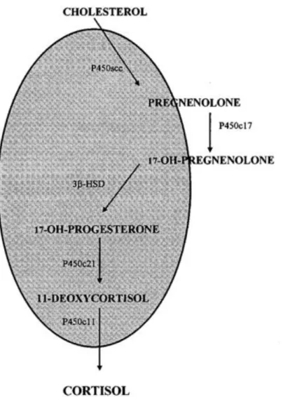
The biosynthesis of cortisol in fish is similar to that in mammals and involves the microsomal enzymatic pathways, including 21-hydroxylation (P450c21), 17α- hydroxylation (P450c17), and 3β-hydroxy steroid dehydrogenation (3β-HSD, Figure 1). In addition, fish possess the mitochondrial inner membrane monooxygenase enzymes, such as the cholesterol side-chain cleavage enzyme (cytochrome P450scc, desmolase) and the 11β-hydroxylase that catalyses the 11β- hydroxylation of deoxycortisol/deoxycorticosterone
11
(cytochrome P450c11) (Figure 1.1; Mommsen et al., 1999).
Fig. 1.1: Biosynthesis of cortisol in teleost fishes. The shaded area represents the mitochondrial compartment, the non-shaded area represents the cytosolic compartment. Abbreviations: 3β-HSD, 3β-hydroxysteroid dehydrogenase; P450s, various forms of cytochrome P450 (Mommsen et al., 1999).
Glucocorticoids (GC) appear to be potent immunosuppressive agents in vertebrates. And in fish, immune suppression has been observed when high levels of cortisol are secreted: cortisol and immune indicators change concurrently after repeated sampling stress in trout causing a reduction of immune competence (Sunyer and Tort, 1995). Severe immune suppression in cold-shock affected sea bream is correlated with high levels of cortisol (Tort et al., 1998). High density stress induces reduced levels of complement together with high cortisol in sea bream (Montero et al., 1999; MacKenzie et al., 2006). Cortisol inhibits inflammatory cytokine expression in vitro (Saeij et al., 2003).
12
Cortisol and lipopolysaccharide (LPS) synergistically stimulate expression of interleukin 1 (IL-1) mRNA in head kidney phagocytes (Engelsma et al., 2003). At the transcript level when macrophages are treated with LPS and cortisol, expression of genes involved in the inflammatory process are mostly down regulated and some of the basic cellular activities, like energetic metabolism, and protein biosynthesis are recovered (MacKenzie et al., 2006). When cortisol and LPS are added to macrophages, cytokine induction is down regulated by cortisol (Castillo et al., 2008). Similarly, LPS-induced expression of TNF decreases by adding cortisol and this effect is reverted by addition of mifepristone (Tort, 2011). Mifepristone (RU 486) is an anti-glucocorticosteroid that exerts its effects at the receptor and possibly the post-receptor level of steroid action. This antihormone has been used in a number of fish studies to block the effects of exogenously added cortisol and to confirm conclusively that cortisol was indeed the active principle behind the noted effects (Mommsen et al., 1999).
13
PART IV - THE EXPERIMENTAL ANIMAL MODEL
_______________________________________________________
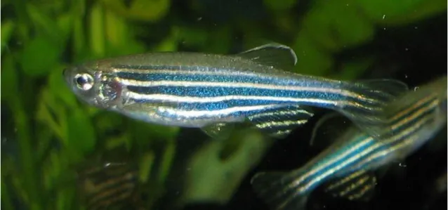
The zebrafish, Danio rerio (Figure 1.2), was the species used in the thesis. It is a tropical, freshwater fish, whose natural habitat is found in India, Pakistan and Bhutan. The zebrafish is a teleost fish and belongs, together with the carp and the goldfish, to the family of Cyprinidae (van der Sar et al., 2004). It is a popular aquarium fish and is one of the most widely used vertebrate models (Phelps and Neely, 2005; Van der Sar et al., 2004).
Fig. 1.2: The zebrafish, Danio rerio.
Zebrafish have a fully developed immune system with both innate and adaptive immune responses, allowing studies that involve both systems (Trede et al., 2004) and is an emerging model species for the study of fish as well as human diseases (Kari et al., 2007). In this regard, several research groups have identified pathogens that can infect and cause disease in zebrafish (Pressley et al., 2005; Neely et al., 2002; Davis et al., 2002; Prouty et al., 2003).
The zebrafish has many advantages when compared to other animal models. Among animals with a fully developed adaptive and innate immune system, the zebrafish is one of the smallest (≤5 cm), allowing large numbers of fish to be housed in a relatively small space. They are prolific, with a given pair able to produce 200–300 new progeny each week (Meeker and Trede, 2008).
14
Under natural conditions, pathogens are thought to infect zebrafish through the gastrointestinal tract, the gills or through the (damaged) fish surface (O’Toole et al.,2004). A gnotobiotic zebrafish model has been established to dissect the molecular interactions between host and microbes in the digestive tract (Rawls et al., 2004). Experimental infection of zebrafish can also be achieved by incubation in media containing the bacteria. This method has been used both for embryos (Davis et al., 2002) and for adult fish (Neely et al., 2002). However, experimental infections are usually introduced by intraperitoneal injection in anaesthetized fish. This enables accurate adjustment of the infection dose.
15
PART V - THESIS AIMS
_______________________________________________________
The proposed goals of this PhD thesis were: to provide new insights into the complexity of the relationship between high environmental ammonia levels and disease susceptibility in fish; to test the hypothesis that ammonia acts through immunosuppression of the innate immune system; and also, to understand if cortisol mediates that suppression, using the zebrafish, Danio
rerio, as the animal model.
In Chapter 2 the hypothesis that ammonia acts through immunosuppression of the innate immune system was tested, trying to provide new insights into the complexity of the relationship between HEA levels and disease susceptibility. To this end, the induction of a number of genes of the APR was analyzed by qPCR. Initially, LPS dose and time-course series were conducted to define experimental parameters to assess the effects of acute (24 h) and chronic (14 d) ammonia exposure on APR. Whole body cortisol levels in response to acute and chronic ammonia exposures were measured to establish if the treatment regime was eliciting organismal stress response.
In Chapter 3 the effects of acute and chronic HEA on response to
Edwarsiella tarda infection were studied in zebrafish. To this end, they were
challenged with intraperitoneal injection or immersion in a bacterial bath in the absence/presence of ammonia. Then, a number of acute phase response (APR) genes were analyzed on viscera (liver, intestine, pancreas and spleen) or gill by qPCR. In addition, the E. tarda LC50 (lethal concentration, 50%) value was assessed for fish exposed to ammonia versus control fish to see if zebrafish mortality was higher in the presence of HEA.
In Chapter 4 it was determined if cortisol mediated the environmental ammonia associated suppression of the acute phase response to LPS. To this end, two experimental studies were performed. In the first experiment, the effect of exogenous cortisol administration on the LPS-induced innate immunity related-genes was assessed in zebrafish. In the second experiment, the effects of glucocorticoid receptor blockade with mifepristone were evaluated in zebrafish exposed to LPS either in the presence or absence of high environmental ammonia.
CHAPTER 2
________________________________________________
THE INHIBITORY EFFECT OF ENVIRONMENTAL
AMMONIA ON DANIO RERIO LPS INDUCED ACUTE
PHASE RESPONSE
Gonçalves, A. F., Páscoa, I., Neves, J. V., Coimbra, J., Vijayan, M. M.,
Rodrigues, P., Wilson, J. M., 2012. The inhibitory effect of environmental
ammonia on Danio rerio LPS induced acute phase response.
Dev. Comp. Immunol. 36 (2), 279–288.
20
Abstract
_______________________________________________________
Ammonia is a toxic by-product of amino acid catabolism and a common environmental pollutant that has been associated with increased disease susceptibility in fish although the mechanism is not well understood. The hypothesis that elevated environmental ammonia acts by impairing the acute phase response was addressed (APR). Specifically, it was determined the impact of sub-lethal acute (24h) and chronic (14d) ammonia exposure on acute phase protein gene expression in zebrafish using a challenge with bacterial LPS (i.p 10μg/g after 24h). A panel of LPS responsive genes (saa, hamp, lect2, hp and
il1β) was identified and evaluated by qPCR. Ammonia was found to impair
induction of saa, hamp and lect2 by 50-90%, providing evidence that ammonia may act potentially through immunosuppression. It was also determined that whole-body cortisol levels of fish subjected to acute (15min, 1h and 24h) and chronic (14d) HEA were higher compared with control fish. These results suggest that the elevated cortisol levels after HEA exposure may have been instrumental in this immunosuppression. Thus providing a mechanistic explanation for the association of increased disease susceptibility that is associated with environmental ammonia exposure.
21
Introduction
_______________________________________________________
Ammonia is an unusual toxicant in that it is environmentally relevant and it is also produced naturally as a metabolic waste of amino acid catabolism within the organism. Ammonia is also released into the environment through the application of industrial fertilizers and input of biological wastes (Ip et al., 2004; USEPA, 1999). In aquaculture, ammonia toxicity may also be problematic during rearing stages in which fish are held at high densities with minimal water flow, or when using recirculating water systems, all of which may lead to elevated levels. In solution ammonia exists either as NH3 or NH4+ in an equilibrium that is largely pH dependent (NH3 + H+ ↔ NH
4
+, pK 9.2). It is toxic to animals if accumulated in tissues and acute toxicity of ammonia is mainly due to its effect on the central nervous system in vertebrates (Randall and Tsui, 2002). Fish are generally ammonotelic and rely mainly on passive diffusion down the concentration gradient between the body and water for elimination. If the concentration in surrounding water exceeds that of blood (reverse diffusion gradient), it becomes difficult to remove, and conversion to nontoxic forms becomes a priority to avoid accumulation to toxic levels. Higher vertebrates make use of uric acid and urea (Wright et al., 1995). However, fish can tolerate higher internal levels of ammonia than mammals, but ammonia accumulation still leads to cell and animal death through its impairment of energy metabolism, by interfering in the Krebs cycle and, more importantly, through its interference with cellular ion and acid-base homeostasis (Ip et al., 2004). Exposure to high environmental ammonia (HEA) also causes gill hyperplasia, anemia, hypercortisolemia and ionoregulatory problems (USEPA, 1999; Ip et al., 2004; Wilkie, 2002). Ammonia causes convulsions, coma and death, probably because elevated NH4+ displaces K+ and depolarizes neurons, causing activation of NMDA type glutamate receptor, which leads to an influx of excessive Ca2+ and subsequent cell death in the central nervous system (Randall and Tsui, 2002).
Ammonia is also generally accepted to increase susceptibility to different parasitic, bacterial and viral fish diseases (Carballo et al., 1995; Carballo and Munõz, 1991; Hanson and Grizzle, 1985). For example, juvenile Chinook salmon (O. tshawytscha) previously exposed to 0.12-0.49mM of total ammonia
22
nitrogen (TAN)1 levels were more susceptible to V. anguillarum challenge (Ackerman et al., 2006). Also, infection caused by Tetrahymena sp. was significantly higher when guppies, Poecilia reticulata were exposed to 0.11-0.32mM TAN1 (Pimenta Leibowitz et al., 2005). In addition, Walters and Plumb (1980) showed that Channel catfish, Ictalurus punctatus, injected with
Aeromonas hydrophila and exposed to 0.11mM TAN1 had significantly higher total bacterial counts than controls. However, the mechanism of action of ammonia remains largely unknown. Detrimental effects are not always observed with ammonia exposure. Lease and collaborators (2003) showed that none of the more traditional toxicity endpoints (growth, whole-body ion content and swimming performance) were significantly affected by range of TAN up to 1.71mM1 in Lost River suckers. Also, TAN concentrations of 0.24mM1 did not affect the antibody response to Aeromonas salmonicida in sunshine bass (Hrubec et al., 1996). Morris and collaborators (2006) even demonstrated that survival of Deltistes luxatus exposed to Flavobacterium columnare increased significantly as unionized ammonia concentrations increased from 0 to 0.72mM TAN1.
The inflammatory response is a complex process that is an important component of the innate immune response. Induction of the acute phase response (APR) in fish is most frequently associated with the activation of the inflammatory response, typically in response to a pathogen challenge (Bayne et al., 2001). In mammals, induction of acute phase proteins (APPs) is mediated by Toll-like receptors (TLRs), pro-inflammatory cytokines and CCAAT/enhancer-binding proteins (C/EBPs). Upon infection, TLRs can recognize the microbial components thus sensing the invasion of pathogens (Akira and Takeda, 2004). Stimulation of these receptors finally results in the production of pro-inflammatory cytokines such as interleukin 1, interleukin 6, and TNFα, which are important inducers of APPs (Cohen, 2002). These cytokines in turn induce the production of some C/EBPs such as C/EBPB and C/EBPD, which are important transcription factors of many acute phase genes (Poli, 1998). The APPs are regarded as having general functions in opsonisation and trapping of micro-organisms and their products, activating the complement system, binding cellular remnants, neutralising enzymes, scavenging free haemoglobin and radicals and modulating the immune response (Gruys et al., 2005).
1 Ammonia concentrations standardized to mM TAN at pH 8 using correction formulas provided
in the ammonia water criteria manual (USEPA, 1999) to facilitate the comparison between different studies.
23
In fish, an innate immunity response is stimulated by injection with bacteria (Lin et al., 2007; Wiens and Vallejo, 2010). Also, immersion of fish in a bacterial suspension has been shown to in- duce the innate immune response (Harriff et al., 2007). However, the injection of bacteria is time-consuming and, in the case of immersion, the use of fish-pathogenic bacteria bears the risk of contaminations of fish culture. In contrast, the stimulation of the innate immune response by bacterial lipopolysaccharides (LPS) is an alternative approach. LPS are also termed endotoxins and are considered to be a major virulence factor, being responsible for lethal effects and clinical manifestations of diseases in humans and animals (Swain et al., 2008). LPS is a cell wall component found in most negative bacteria. However, some Gram-positive bacteria, such as Listeria monocytogenes, also possess endotoxin-like activity (Wexler and Oppenheim, 1979). Higher vertebrates are extremely sensitive to endotoxins, even at low doses, but lower vertebrates such as amphibians and fish are found to be resistant to endotoxic shock. Fish like
Oncorhynchus kisutch and Oncorhynchus mykiss are reported to be resistant to Escherichia coli and Aeromonas salmonicida endotoxins in doses up to 80
mg/kg body weight (Wedemeyer et al., 1968).
The zebrafish, Danio rerio, is the species used in the present study and is a tropical, freshwater fish belonging to the Cyprinidae family. It is a popular aquarium fish and is one of the most widely used vertebrate models (Phelps and Neely, 2005; Van der Sar et al., 2004). Zebrafish have a fully developed immune system with both innate and adaptive immune responses, allowing studies that involve both systems (Trede et al., 2004) and is an emerging model species for the study of fish as well as human diseases (Kari et al., 2007).
In the present study the hypothesis that ammonia acts through immunosuppression of the innate immune system was tested, and also provides new insights into the complexity of the relationship between HEA levels and disease susceptibility. To this end, the induction of a number of genes of the APR by qPCR was examined. Initially, LPS dose and time-course series were conducted to define experimental parameters to assess the effects of acute (24 h) and chronic (14 d) ammonia exposure on APR. The genes saa,
lect2, hp, hamp, il1β, tnfα, il10, crp, tlr4a and litaf were studied in zebrafish.
Whole body cortisol levels in response to acute and chronic ammonia exposures were measured to establish if the treatment regime was eliciting organismal stress response in this species (Alsop and Vijayan, 2008; Fuzzen et al., 2010;
24 Ramsay et al., 2009).
25
Material and Methods
_______________________________________________________
Animals
Adult Danio rerio (0.2–0.9 g body mass) were obtained from a local aquarium fish supplier and held at 26ºC in a 100 L aquarium with aerated Porto city tap water (Na+ 0.5 mM, alkalinity 50 mg/l CaCO3, pH 8). The tank contained an external filtration system supplemented with sterilization by ultraviolet irradiation and 10% of the water was changed daily. The fish were fed with commercial fish food (TetraMin, Tetra, Germany) four times per day on automatic feeders (Eheim 3538, Germany) and were reared on a 12 h light/dark cycle.
Experiment 1 – LPS dose response
In order to determine the optimal dose of LPS for the induction of innate immune system genes, zebrafish were injected with a range of concentrations of LPS. Forty-two zebrafish were anaesthetized (1:10,000 tricaine methanesulfonate, Alpharma, UK, pH 7.5, adjusted with NaHCO3) and intraperitoneally injected (n = 6) with LPS from E. coli (Serotype O55:B5, Sigma) at 50 pg/g, 1 ng/g, 50 ng/ g, 1 µg/g or 10 µg/g wet mass, or phosphate-buffered saline (PBS; 16.2mM Na2HPO4, 3.8mM NaH2PO4, 150mM NaCl, pH 7.3) vehicle alone (sham group). An injection volume of 10 µg/g was used. Another group with no injection served as the control. After 24 h, fish were sacrificed and sampled (see Section x) for gene expression analysis.
Experiment 2 – LPS time course response
In order to determine the time-dependent effects of LPS on the induction of genes of the innate immune system, two groups of twenty-eight zebrafish were anaesthetized, intraperitoneally injected with 10 µg/g LPS (infected group) or PBS (sham group) and fish from each group were sampled at 6, 12, 24 or 48 h after injection (n = 7). This dose was based on the results of Experiment 1.
26
Experiment 3 – acute ammonia and LPS
To elucidate the effects of ammonia exposure on the induction of innate immune response genes by LPS, fish were LPS or sham injected in the presence or absence of elevated water ammonia levels. Twenty-eight zebrafish were anaesthetized and received intraperitoneal injections of either PBS or 10 µg/g LPS. After that they were placed in 5 L plastic aquaria with ammonia concentrations of 0 (control) or 1mM NH4Cl pH 8.0 buffered with 10mM Tris, for 24 h. This concentration of ammonia is known to be sublethal (Páscoa et al., 2008). The ambient ammonia concentration was increased by the addition of a 1M of NH4Cl stock solution. After 24 h the fish were sampled (n = 7; see Section x).
Experiment 4 – chronic ammonia and LPS
Twenty-eight zebrafish were placed in 20 L glass aquaria with 0 (control) or 0.5mM NH4Cl, pH 8.0 buffered with 10mM of Tris, for 14 days. After that time, fish were anaesthetized and received intraperitoneal injections of either PBS or 10 µg/g LPS and were sampled 24 h later (n = 7; see section bellow). Zebrafish were fed twice a day with commercial flake food until 24 h before the injection and were then fasted until the end of the experiment. The ambient ammonia concentration was increased by addition of a stock solution of 1M of NH4Cl. The same stock solution was used throughout the study. Temperature, O2 concentration, pH and water ammonia concentration were measured daily and 25% of water was changed daily. The total ammonia nitrogen (TAN) in the control tank was 0.241 ± 0.039 mM and in the ammonia tank was 0.623 ± 0.036 mM. The fish appetite and behavior were normal throughout the experiment and the ammonia levels did not result in any mortality/morbidity.
Whole-body cortisol levels after ammonia exposure
The effects of both acute and chronic ammonia exposure on whole fish cortisol levels were measured in a separate set of fish. High environmental ammonia exposed or control fish were sampled at 15 min, 1 h, and 24 h acute and 14 d chronic exposure to HEA (1 and 0.5mM TAN, respectively). Conditions were identical as described above. In addition, it was included an acute handling disturbance group as a positive control group to measure cortisol stress response. Briefly, all fish were netted simultaneously, suspended in the air for 3 min, returned to the original tank for 3 min, and resuspended in the air
27
for an additional 3 min exactly as described previously (Ramsay et al., 2009). The stressed fish were returned to the original tank and allowed to recover for 15 min until they were sampled.
Sampling
The zebrafish from each aquarium were heavily anaesthetized (1:5000 tricaine methanesulfonate, pH 7.5, adjusted with NaHCO3), weighed and standard length measured. Fish were then killed by decapitation on ice, with a transverse cut anterior to the pectoral fins. Viscera (that include liver, intestine, pancreas and spleen) were excised and kept in RNA later and remaining carcasses were snap frozen in liquid nitrogen and stored at -80 ºC for later analysis.
Zebrafish for whole body cortisol measurements were anaesthetized with buffered MS-222 (1:5000), blotted on paper towels to remove excess water, weighed and immediately frozen in liquid nitrogen. The samples were stored at -80 ºC.
Total RNA extraction and cDNA synthesis
Total RNA was isolated using a commercial silica based columns (Illustra RNAspin Mini RNA Isolation Kit, GE Healthcare, UK). All steps were performed according to the manufacturer’s instructions, including the on-column treatment of isolated RNA with RNase-free DNase. RNA concentration and purity were assessed by UV spectrophotometry (Genova, Jenway, Dunmow, England) and integrity confirmed by electrophoresis (1.2% formaldehyde agarose gel; Mini-Sub Cell GT Cell, Bio-Rad, Hercules, CA, USA) and stored at -80 ºC until further use. The cDNA for all experiments was synthesized from 1 µg of total RNA with the iScript cDNA Synthesis Kit (Bio-Rad) according to the manufacturer’s protocol.
Primers
Primers for a suite of innate immunity associated genes were designed from gene sequences available in Ensembl (www.ensembl.org) and GenBank (http://www.ncbi.nlm.nih.gov/), or taken directly from published studies (Table 2.1). Primers were designed using Primer3 (Rozen and Skaletsky, 2000) and were initially tested for specificity by RT-PCR.