2012
Tatiana Lima de
Vilhena Magalhães
Costa
Respostas celulares aos erros de tradução
do genoma
Cellular responses to genome
mistranslation
Tatiana Lima de
Vilhena Magalhães
Costa
Respostas celulares aos erros de tradução
do genoma
Cellular responses to genome
mistranslation
Dissertação apresentada à Universidade de Aveiro para cumprimento dos requisitos necessários à obtenção do grau de Doutor em Bioquímica, realizada sob a orientação científica do Doutor Manuel Santos, Professor Associado do Departamento de Biologia da Universidade de Aveiro
Apoio Financeiro: Programa Operacional Potencial Humano (POPH) - QREN, comparticipado pelo Fundo Social Europeu (FSE) e por fundos próprios do MCES.
“One time /one meeting (ichigo-ichie) This gathering never happened in the past and it will never happen in the future, as its exact nature will never be the same again”
Sen no Rikyú (1522-1591) “If we knew what it was we were doing it would not be called research, would it?” “No amount of experimentation can ever prove me right; a single experiment can prove me wrong.”
o júri
Presidente Doutor Vitor José Babau Torres
Professor Catedrático da Universidade de Aveiro
Doutor Vítor Manuel Vieira da Costa
Professor Associado, ICBAS- Instituto de Ciências Biomédicas Abel Salazar da Universidade do Porto
Doutora Maria Alexandra Marques Moreira Mourão do Carmo
Investigadora Principal, IBMC- Instituto de Biologia Molecular e Celular da Universidade do Porto
Doutora Maria Paula Polónia Gonçalves
Professora Associada da Universidade de Aveiro
Doutor Manuel António da Silva Santos
Professor Associado da Universidade de Aveiro
Doutora Paula Cristina Costa Alves Monteiro Ludovico
Professora Auxiliar da Escola de Ciências da Saúde da Universidade do Minho
Doutora Laura Cristina da Silva Carreto
Investigadora Auxiliar do CESAM- Centro de Estudos do Ambiente e do Mar da Universidade de Aveiro
Acknowledgements thinking.
I would like to express my gratitude to the members of the Desjardins laboratory, especially to Christiane, Magali and Joe for the warm welcome in their laboratory, and for making my Canadian days less lonely and more fun. And a big thanks to Xana and Paula for helping with my laboratory experiments, for all the discussions and fruitful inputs that meant so much in this last period of my thesis.
Also, I would like to thank to the all the people in the hallway- Isabel, Sofia, Claúdia, Cristina, Andreia, Marta, and Virgilia - and also to the girls upstairs namely Gabi, Sandra V., Sandra and Margarida, for saving my work when something was missing. A special thanks to Tânia, which was there since the first moment in Aveiro and in the Biology world. Thanks also to Rita Jordão, Márcia Henriques, Rui Adão and also my sweet brother Diogo for being my “study model” in what teaching is concerned. I am sorry if things did not go always as smoothly as they should.
I am most grateful to Dr. Roy Parker and Dr.Daniela Teixeira; Dr.Mathias Petter and Dr. Claudine Kraft; Dr. Oshumi, Dr. Kabeya and Dr. Buchan for sending plasmids and to Dr Klionsky for strains, for the most helpful discussions and interest in my work. And also to Yu-yi Lin for the emails with technical advices and interesting scientific discussions.
During my years in the RNA Biology Laboratory, I have worked with many interesting and fantastic people, several of them became my dear friends. Many thanks to Catarina and Céu for their friendship, encouragement, support, help, advices, critical readings and the most valuable comments. Cristina, João S. (júnior), Rita B., Marisa, Patricia and Laura: thanks for the silly (and not so silly!) conversations, for the company late at night, at weekends and holidays, for the lunches and dinners (Snack, Ramona and Evaristo sempre!), drinks and laughs that we have shared. Thanks for all the help with the experiments too and for making me feel better whenever things seemed upside down. I have the feeling that I have received more than what I gave. Without you it would have been so much difficult! Thanks, João P. (senior) for answering to all my questions. Also, Violeta, thanks for all the administrative work and friendship; Ana S., Jörg, Tobias, and Gabi thank you for the enthusiasm and for your examples.
Thanks, Isabelinha, for all that is behind- the dancing nights, the vacations and wild trips, the horseback riding passion, the talks and the late night pancakes. And today, after all the twists and turns in life and the distance apart, thanks for your continuous support and invaluable friendship.
Thanks to my family too, close relatives and friends in Porto de Mós and surroundings that saw me grow, for all the interest shown in my work and for being such a colorful bunch of people. And, also, to Família Macedo, which has been here for me daily for the last years, making my days much more cheerful.
Mum, Dad and Diogo, thanks for raising me up and making me believe. Thanks for your support, encouragement and trust towards my choices. And for much more that I cannot put into words.
And finally, thank you Bé for being you: someone who urges me on, has faith in me, and makes me smile and laugh. Thanks for being my balance, for your patience and for teaching me to look to the brighter side of life. Thanks for making me incredible happy!
Resumo Erros no processo da síntese proteica podem ter profundos efeitos na fisiologia celular e no desenvolvimento de doenças, nomeadamente doenças neurodegenerativas, cancro e envelhecimento. A introdução de erros durante a síntese de proteínas e, em particular durante o processo da tradução, é designado por “mistranslation” que é um processo pouco estudado e mal compreendido. Neste projecto, construímos leveduras que, sistemática e constitutivamente, treslêem o codão de leucina CUG como serina, o que corresponde a um aumento de erro de 240 vezes relativamente à taxa de erro basal da síntese proteica (0.001%).
Os resultados obtidos demonstram que os erros de tradução induzem a actividade autofágica, acumulação de proteínas insolúveis, produção de espécies reactivas de oxigénio, disrupção funcional e morfológica das mitocôndrias, não ocorrendo, no entanto, destruição selectiva destas. A expressão do gene PNC1, associado ao aumento da longevidade e regulador da actividade da deacetilase Sir2p, é fortemente aumentada em resposta aos erros da tradução. Os genes PNC1 e SIR2 estão envolvidos no controlo da autofagia induzida pelos erros de tradução mas não em situações de stress nutricional. O aumento dos erros de tradução leva à formação de P-bodies, mas não induz a formação de grânulos de stress e reduz a expressão de genes que codificam proteínas ribosomais em vez de se verificar destruição selectiva de ribosomas - ribofagia.
Este estudo mostra que as células de levedura são muito mais resistentes aos erros na tradução do que o esperado. Os resultados mostram um papel fundamental da autofagia na resposta celular aos erros de tradução e indicam que estes têm um forte impacto em alterações morfo-funcionais das mitocondrias, sendo este um dos fenótipos mais marcantes nestas células. Considerando que a maior parte dos mecanismos celulares são conservados entre leveduras e células humanas, este estudo mostra que a levedura é um excelente modelo para estudar a resposta celular aos erros de tradução e sugere que o stress oxidativo, a acumulação de espécies reactivas de oxigénio e a acumulação de proteínas insolúveis podem ser a causa da degeneração celular observada em múltiplas doenças humanas associadas a defeitos na síntese proteica.
autophagy, oxidative stress,proteotoxic stress
abstract Low level protein synthesis errors can have profound effects on normal cell physiology and disease development, namely neurodegeneration, cancer and aging. The biology of errors introduced into proteins during mRNA translation, herein referred as mistranslation, is not yet fully understood. In order to shed new light into this biological phenomenon, we have engineered constitutive codon misreading in S.
cerevisiae, using a mutant tRNA that misreads leucine CUG codons as serine,
representing a 240 fold increase in mRNA translational error relative to typical physiological error (0.0001%).
Our studies show that mistranslation induces autophagic activity, increases accumulation of insoluble proteins, production of reactive oxygen species, and morphological disruption of the mitochondrial network. Mistranslation also up-regulates the expression of the longevity gene PNC1, which is a regulator of Sir2p deacetylase activity. We show here that both PNC1 and SIR2 are involved in the regulation of autophagy induced by mistranslation, but not by starvation-induced autophagy. Mistranslation leads to P-body but not stress-granule assembly, down-regulates the expression of ribosomal protein genes and increases slightly the selective degradation of ribosomes (ribophagy).
The study also indicates that yeast cells are much more resistant to mistranslation than expected and highlights the importance of autophagy in the cellular response to mistranslation. Morpho-functional alterations of the mitochondrial network are the most visible phenotype of mistranslation. Since most of the basic cellular processes are conserved between yeast and humans, this study reinforces the importance of yeast as a model system to study mistranslation and suggests that oxidative stress and accumulation of misfolded proteins arising from aberrant protein synthesis are important causes of the cellular degeneration observed in human diseases associated to mRNA mistranslation.
List of abbreviations…...……….. 11
Background and Thesis Outline……..……….. 12
Chapter 1 - Literature Overview………
18 From genes to proteins: a process (almost) perfect……….
20 Eukaryotic translation…………..………..
22 Translation errors and determinant factors………
28 Protein Quality Control Mechanisms………...
32 Translational Quality Control………
33 Post-translation Quality Control………
35 In the cytosol………
37 In the organelles………...
51 Mistranslation: deleterious and beneficial effects………
57 The mistranslation model……….………
60 References………
63
Chapter 2 - Mistranslation induces protein aggregation and autophagy in yeast
68
Abstract……… 70
Introduction………. 71
Results……….. 74
Mistranslation increases accumulation of insoluble proteins 74
Mistranslation induces ultrastructural alterations in yeast 80
Mistranslation induces autophagy 87
Regulation of autophagy in mistranslating cells 92
The PNC1 and SIR2 genes do not regulated autophagy under starvation 96
Discussion……… 100
Material and Methods………... 105
Yeast Strains and plasmids 105
Protein aggregation determination 107
Immuno-TEM 109
Epifluorescence Microscopy 110
Protein Extraction and Western Blotting 111
DNA microarrays 112
Supplementary Data……….. 117
References……… 128
Chapter 3-mRNA mistranslation causes ROS accumulation and mitochondrial dysfunction……….………. 132
Abstract……… 134
Introduction………... 135
Results……….. 140
mRNA mistranslation up-regulates antioxidant stress response 140 Mistranslation increases ROS production 143 SIR2 role on ROS management in response to mistranslation 150 Mistranslation induces alterations on mitochondrial morphology 162 Mistranslation generates a respiratory deficiency phenotype 166 Mistranslation does not affect yeast chronological aging 171 Discussion……… 173
Material and Methods……… 186
Yeast Strains and manipulation 186 Assessment of intracellular reactive oxygen species (ROS) 189 Plasma membrane Integrity 190 Supplementary Data………... 196
References……… 210
Chapter 4-Mistranslation induces formation of p-bodies in yeast………. 214
Abstract……… 216
Introduction………217
Discussion……….. 237
Material and Methods………... 243
Strains and growth conditions 243
Detection of P-Bodies and Stress Granules 244
Microarray Analysis 245
Quantification of ribosomal proteins 247
Supplementary Data……… 248
References……… 249
Chapter 5-General discussion, conclusions and future perspectives 252 References……….. 274
2D-PAGE two dimensional polyacrylamide gel
electrophoresis
NADPH reduced nicotinamide adenine dinucleotide phosphate
A ampére NADH reduced nicotinamide adenine
dinucleotide
ADH alcohol dehidrogenase NADP+ oxidised nicotinamide adenine dinucleotide phosphate
APS ammonium persulphate nm nanometers
ATG genes
Autophagy Related Genes NP-40 nonylphenyl-polyethylene glycol
ATP adenosine 5’-triphosphate OD600/595 optical density at 600 nm or 595 nm
AZC azetidine-2-carboxylic acid PAS pre-autophagosomal structure
cAMP cyclic adenosine 5’-monohosphate PBS phosphate buffered saline
cDNA complementary deoxyribonucleic acid
PMSF phenylmethylsulfonyl fluoride
Cm centimeter RFU relative fluorescence units
Cy3 cyanine 3 RNA ribonucleic acid
Cy5 cyanine 5 ROS reactive oxygen species
DHE dihydroethidium rpm revolutions per minute
DHR123 dihydrorhodamine 123 rRNA ribosomal ribonucleic acid
DMSO dimethyl sulfoxide RT room temperature
DNA deoxyribonucleic acid SDS sodium dodecyl sulphate
DNP 2, 4-dinitrophenylhydrazine SDS-PAGE
sodium dodecyl sulphate-polyacrylamide gel electrophoresis
DTT dithiotreitol SMD synthetic minimal médium
EDTA ethylenediamine tetracetic acid Suc- LLVY-MCA
succinyl-leucyl-leucyl-valyl-tyrosine 4-methylcoumaryl-7-amide
ER endoplasmatic reticulum TCA trichloroacetic acid
ERAD endoplasmatic reticulum associated degradation
TEM transmission electron microscopy
g (mg,
g, ng) gram (miligram, microgram, nanogram)
TEMED N,N,N,’N,’-tetramethylethylenediamine
GFP green fluorescent protein Tris 2-amino-2-hydroxymethyl-1,3-propanodiol
GTP guanosine 5’-triphosphate tRNA transfer ribonucleic acid
H2O2 hydrogen peroxide U units
HEPES N-[-2-hydroxyethyl]piperazine-N’-[2-ethanesulfonic acid]
Ub ubiquitin
HSP heat shock protein V volt
kDa kilodalton W watt
l (ml, l) liter (mililiter, microliter) YPD Yeast Peptone Dextrose médium
MCA 4-methylcoumaryl-7-amide
mQ milliQ
mRNA messenger ribonucleic acid
mtDNA mitochondrial DNA
NAD oxidised nicotinamide adenine dinucleotide
14 Aims of this study and thesis organization
During the last ten years, our laboratory pioneered a number of studies to elucidate the evolution of genetic code alterations, which are mediated by codon decoding ambiguity, a form of mRNA mistranslation. These studies provide the first insight on how genetic code alterations evolve and raise a number of new biological questions, namely how do organisms tolerate mRNA mistranslation and genetic code alterations? For example, re-construction of a Candida albicans genetic code alteration in the close relative S. cerevisiae showed that codon ambiguity deregulates gene expression, up-regulates the expression of stress genes and induces the accumulation of trehalose and glycogen. These results show that the experimental system used in our laboratory could be an excellent working model for studying the impact of general mistranslation on cells physiology, which is of biomedical relevance. Here we explore this hypothesis to shed new light on the cellular responses to aberrant protein synthesis.
This thesis is written in the form of three manuscripts (chapters 2-4), as follows:
Chapter 1 provides a general introduction and addresses translational mechanisms, translational fidelity and proteome quality control mechanisms. A brief introduction to the working model used in this thesis is provided at the end of the chapter.
Chapter 2 establishes a link between mRNA mistranslation and autophagy in yeast and explores the role of the SIR2 gene on protein aggregation and autophagy.
Chapter 3 describes a link between mistranslation and oxidative stress. The data show that mistranslation increases accumulation of reactive oxygen species (ROS) in both wild-type and in several knockout strains. In addition, we describe the effects of mistranslation on mitochondrial homeostasis.
Chapter 4 describes a link between mistranslation and P-body assembly and explores the formation of stress granules by mistranslation.
15 chapters 2-5. Future perspectives and studies are also presented.
Parts of the work presented in this PhD thesis contributed to the following publications:
Lima-Costa T, Paredes JA, Rondeau C, Desjardins M, Moura GR, Santos MAS , Mistranslation of the genetic code induces autophagy in yeast, International Conference on Yeast Genetics and Molecular Biology. Yeast. 2009 Jul;26(S1): S176-S208
Silva RM, Duarte IC, Paredes JA, Lima-Costa T, Perrot M, Boucherie H, Goodfellow BJ, Gomes AC, Mateus DD, Moura GR, Santos MAS, The yeast PNC1 longevity gene is up-regulated by mRNA mistranslation, PLoS One. 2009; 4(4):e5212.
Silva RM, Paredes JA, Moura GR, Manadas B, Lima-Costa T, Rocha R, Miranda I, Gomes AC, Koerkamp MJG, Perrot M, Holstege FCP, Boucherie H, Santos MAS, Genetic Code Alteration Plays a Critical Role in the Evolution of Candida Genes Quantitative Analysis of Single Amino Acid Changes using a 4000 QTRAP® system(2007),Applied Byosystems technical note.
Silva RM, Paredes JA, Moura GR, Manadas BR, Lima-Costa T, Rocha R, Miranda I, Gomes AC, Koerkamp MJ, Perrot M, Holstege FC, Boucherie H, and Santos MAS. 2007. Critical roles for a genetic code alteration in the evolution of the genus Candida. EMBO J. 26:4555-4565.
The results described in this thesis were presented at the following scientific meetings: VI Simpósio Pós-Graduação do Departamento de Biologia da Universidade de
Aveiro, May, 2007, Aveiro, Portugal (Oral presentation)
EMBO Conference on Protein Synthesis and Translational Control, 12th
-16th Setember, 2007, Heidelberg, Germany (Poster presentation)
RNA 2008-Thirteenth Annual Meeting of the RNA Society, 28thJuly- 3rdAugust,
16 23rd
tRNA Workshop,28th January-1st Feb, 2010, Aveiro, Portugal (Poster presentation) Yeast Genetics and Molecular Biology Meeting, 27thJuly- 1stAugust, 2010, University of British Columbia, Vancouver, Canada (Poster presentation)
18
Chapter 1
20 From genes to proteins: a process (almost) perfect
Gene expression is a highly efficient and accurate molecular process that is tightly
regulated. However, synthesis of functional proteins is not error free. Mistranslation is
a general term used for describing translational errors. Over the last 45 years, many
attempts have been made to quantify the error rate of gene translation, however little
is still known about such errors and their biological relevance remains poorly
understood.
Gene translation errors can occur at replication, transcription, translation and
post-translational level (Figure 1). During replication and transcription, polymerase slippage
or nucleotide misincorporations can occur. Indeed, the bacterium Escherichia coli has a
typical replication error rate of approximately 10-8– 10-9 per base pair (Kunkel and
Bebenek, 2000), and uses specific mechanisms like editing and repair to correct such
errors. In eukaryotes, DNA replication error rates are even lower, being in the order of
10-10– 10-11 (Matsuda et al., 2000). The error rate of transcription in vivo in E. coli has
been estimated at 1.4x10⁻⁴ per nucleotide (Rosenberger and Foskett, 1981; Ninio,
1991) and more recent in vitro studies have shown that the rate of misincorporation of
UTP at G sites during transcription is in the order of 2x10⁻⁶ (Kireeva et al., 2008). On the
other hand, studies in HeLa cells (Fox-Walsh and Hertel, 2009) and
Schizosaccharomyces pombe (Wilhelm et al., 2008) have shown that mRNA splicing can also account for mistranslation through exon skipping and failure in removing introns.
These errors occur at rates of ≈10-2- 10-6(Drummond and Wilke, 2009). Translation, the
21 rate of approximately 1 amino acid per 104 codons (10-4). This means that 15% of all
average-length protein molecules contain at least one misincorporated amino acid
(Reynolds et al., 2010). Even when correctly transcribed and translated, that is, in cases
where proteins have the correct amino acid sequence, post-translational modifications
errors as well as folding errors alter their function, but their relevance and frequency
are still obscure. For instance, Winklhofer and coworkers (Winklhofer et al., 2008) have
suggested that misphosphorylation of the microtubule-binding protein tau is a
pathological signature of Alzheimer’s disease and that it contributes to misfolding and
aggregation of this protein. Additionally, mutations that alter glycosilation have been
described as being extremely deleterious (Freeze, 2006; Freeze, 2006).
As this work is focused on the consequences of erroneous amino acid incorporation
into proteins, a brief overview of the eukaryotic mRNA translation mechanism is
provided below.
22 Figure 1 - Errors in genetic information flow. Errors arise at many stages, from the
transcription of genetic information to the folding and post-translational modification of the finished polypeptide. Estimated error rates are shown. Adapted from Drummond and Wilke, 2009.
Eukaryotic translation
Translation is the last step of gene expression and is divided into three steps: initiation,
elongation and termination. Initiation and termination have mechanistic differences
between eukaryotes, bacteria and archea, while elongation is semi-conserved across
the three kingdoms of life (Kapp and Lorsch, 2004). During initiation, all the events
that are needed for the positioning of the ribosome at the start codon take place.
Polypeptide synthesis occurs during the elongation step and the completed peptide is
released during termination.
During initiation the assembly of a ribosome with an initiator-methionyl-transfer-RNA
(met-tRNAiMet) in its peptidyl (P-) site located on the initiation codon of the mRNA is
accomplished. In order to do so cells use the 5´m7(5´)ppp(5´)N cap structure and the
3’end of the poly(A) tail of the mRNA together with at least 12 eukaryotic initiation
factors (eIFs). In contrast, prokaryotes only need three initiation factors (Kapp and
Lorsch, 2004b). The initiation step can be divided into several steps (Figure 2), namely
(i) assembly of eukaryotic initiation factor 2 (eIF2), GTP and met-tRNAiMet (ternary
complex); (ii) formation of a 43 Svedberg (S) preinitiation complex, comprising the
small (40S) ribosomal subunit, initiation factors ( eIF1, eIF1A, eIF3) and the ternary
complex and probably eIF5; (iii) activation of the mRNA, an ATP-dependent reaction,
23 recruitment of the 43S complex to the (capped) 5’end of the mRNA; (v) scanning of the
5’untranslated region (UTR) of the mRNA by the 43S complexes; (vi) recognition of the
start codon (AUG) and formation of 48S initiation complex, leading to an alteration in
the conformation and with a consequent displacement of eIF1, allowing eIF5-mediated
hydrolysis of eIF2-bound GTP and Pi release; (vii) joining of the large (60S) subunit to
assemble a complete (80S) ribosome, and associated displacement of eIF2–GDP and
other factors (eIF1, eIF3, eIF4B, eIF4F and eIF5) mediated by eIF5B and (viii) hydrolysis
of GTP by eIF5B and release of eIF1A and GDP-bound eIF5B from assembled
elongation-competent 80S ribosomes. At this point, the mRNA is positioned so that
the next codon can be translated during the elongation step of protein synthesis. As
translation is a cyclic process, termination follows elongation and leads to recycling
(Figure 2, step 1), which generates separated ribosomal subunits. It is important to
note that protein synthesis is mainly regulated during initiation, rather than during
24 Figure 2- Details of eukaryote translation initiation. Translation initiation is a complex
process that can be divided into several steps. It comprises ternary complex formation (2), 43S complex formation (3), mRNA activation (4), attachment of pre-initiation complex to mRNA (5), scanning of the 5’UTR (6), recognition of initiation codon (7), joining of 60S subunit and displacement (8), hydrolysis of eIF5B-GTP bound and eIF5B and eIF1A factor release (9) and ribosome recycling (1). Adapted from Jackson et al., 2010.
25 During elongation sequential binding of aminoacyl-tRNAs (aa-tRNAs) to the ribosome
and the formation of peptide bonds between the amino acids occur (Figure 3, A). This
phase is dependent on the activity of several factors, known as elongation factors (EF),
and requires substantial amounts of energy. It starts by the recognition of the
aa-tRNAs by elongation factor eEF-1 (EF-Tu in bacteria), in the presence of GTP. After the
recognition step, the aatRNA-eEF1-GTP complex enters the empty A-site of the
ribosome and the anticodon of the incoming aa-tRNA recognizes the mRNA codon
positioned in the A–site. Universally conserved bases in the small subunit of the
ribosome (16S rRNA, bacteria; 18S eukaryotes) interact with the 2 first bases of the
codon/anticodon complex. This interaction stabilizes a specific ribosomal
conformation, which allows for verification of whether the correct tRNA is bound. A
ribosomal conformational change coupled with the formation of correct
codon-anticodon complexes leads to alterations in the position of active site residues bound
to eEF1A which, in turn, activates its GTPase activity. The eEF1A•GDP complex releases
the aa-tRNA into the A site (Kapp and Lorsch, 2004b). The peptide bond, which is
catalyzed in the ribosomal peptidyl transferase center, is then formed between the
growing polypeptide, which is located in the P-site of the ribosome, and the new
amino acid located in the A-site. At this point, the P-site is free and a new
peptidyl-tRNA occupies the A-site. The ribosome moves one codon forward (mRNA
translocation) thereby exposing new codons for tRNA binding. Simultaneously with the
ribosome movement, the empty tRNA is displaced from the P-site to the E-site as the
peptidyl tRNA is translocated from the A-site to the P-site. This process is facilitated by
elongation factor eEF-2 (EF2-G in prokaryotes) and GTP, which is hydrolyzed by the
26 the next codon on the mRNA is made available for interaction with a new aa-tRNA in
the A-site (Kapp and Lorsch, 2004a). Additionally, ribosome translocation allows
another ribosome to initiate on the 5′ end of the mRNA and begin its round of
translation. Thus, mRNAs usually have several ribosomes attached to them forming a
polysome. These reaction steps are repeated until the ribosome encounters an
in-frame stop-codon (UAA, UAG, UGA). After GTP hydrolysis and the release of the
aa-tRNA onto the ribosome, the eEF1A•GDP complex is released. In order to participate in
successive rounds of polypeptide elongation, it is recycled to its GTP-bound form by
eEF1B multifactor complex. Additionally, fungi possess an additional factor, eEF3, that
possesses both GTPase and ATPase activity (Dasmahapatra and Chakraburtty, 1981;
Skogerson and Engelhardt, 1977) and whose encoding gene is essential for yeast
viability (Qin et al., 1990). This factor is found primarily associated with translating
cytosolic ribosomes and is required for each round of elongation. It is known to
interact with eEF1A (Kovalchuke et al., 1998) and to facilitate the release of the
E-site-deacylated tRNA and the binding of the eEF1A-GTP•aa-tRNA ternary complex to the A
site of the ribosome (Triana-Alonso et al., 1995). The elongation step can be inhibited
by drugs like cycloheximide and lactinidomycin, which specifically block the
translocation step (Schneider-Poetsch et al., 2010).
Whenever a stop codon is found in the ribosomal A site, translation termination occurs
(Figure 3, B). This results in the release of the completed polypeptide chain, following
the hydrolysis of the ester bond that links the polypeptide chain to the P-site tRNA.
The hydrolysis reaction is thought to be carried out by the ribosomal peptidyl
27 release factors are used to recognize the stop signals and terminate protein synthesis.
Eukaryotic cells have 2 RFs (eRF-1 and eRF-3). eRF-1 recognizes the stop codons and
eRF-3 acts together with the first enhancing its activity. The RF bind to the termination
codon in the A site and stimulate hydrolysis of the bond between the tRNA and the
polypeptide chain in the P site. The completed polypeptide and the tRNAs are then
released from the ribosome while the ribosomal subunits and the mRNA template
28 Figure 3- Simplified view of eukaryotic translation elongation and termination. A) Elongation- the elongation factor eEF1A delivers aa-tRNAsto the ribosomal A site. When a
codon/anticodon match is detected, eEF1A deposits the aa-tRNA and is itself released from the ribosome. A peptide bond is then formed. eEF2 promotes the GTP-dependent translocation of the nascent protein chain from the A-site to the P-site of the ribosome, positioning the next codon in the A site and allowing the process to repeat B) Termination - A termination codon at the A site is recognized by a release factor rather than by a tRNA. The result is the release of the completed polypeptide chain, followed by the dissociation of tRNA and mRNA from the ribosome. Note that not all steps are shown, see text for details. Adapted from Kapp and Lorsch,2004b.
Translation errors and determinant factors
Translation is accurate and occurs at biologically relevant rates. As in many other
biological processes, accuracy has its price and therefore, speed and accuracy are a
compromise solution, indicating, therefore, that accuracy is not perfect. Indeed, of all
the steps of the flow of genetic information from genes to proteins, translation is the
most error prone step. Errors occur during tRNA aminoacylation by the aaRS or at the
29 misincorporation, tRNA misacylation, premature termination, stop codon
readthrought, ribosome drop-off and frameshifting are frequent (Drummond and
Wilke, 2009). Aminoacylation errors are mostly due to abnormal recognition of the
tRNAs and amino acids by aaRSs. At the ribosome level missense errors occur when a
wrong amino acid is incorporated into the polypeotide chain, while nonsense errors
are due to stop codon readthough which ultimately results in proteins with extended
C-termini (Figure 4). Processivity errors and frameshifting originate truncated proteins.
Frameshifting errors, i.e., errors that occur due to shifting of reading frames during the
elongation step, normally result in synthesis of truncated proteins due to premature
translation termination (Farabaugh and Bjork, 1999). The mechanisms by which
frameshifting occurs is not yet fully understood, but some models have been
proposed, namely the pause and slip model (Farabaugh and Bjork, 1999), which
postulates that frameshifting is a two step process (Figure 4, B). This type of
translational error occurs at a frequency of 10-5 in E. coli (Curran and Yarus, 1986).
Analysis of premature termination in E. coli and in S. cerevisiae has also shown that this
type of error has a frequency in the order of 10-4 to 10-3 per codon (Arava et al., 2005).
Ribosomal accuracy is therefore energetically costly and affects translational speed
30 Figure 4- Translation errors at the ribosome level- A) During the translation process the
ribosome stops at a codon (in this case ACA), while waiting for a cognate tRNA. At this point 3 things can happen:1) the cognate tRNA arrives and elongation occurs leading accurate translation; 2) a near-cognate tRNA is used and elongation proceeds leading to a missense error (incorporation of a wrong amino acid into the growing peptide chain) or 3) premature termination of translation due to recognition by release factors, spontaneous ribosome drop-off or frameshifting leading to a nonsense error (adapted from Shah and Gilchrist, 2010) B and C)
Frameshifting (Pause-and-Slip model) +1 frameshifting is shown. In the first step (B),
insufficient amount of cognate tRNA results in an empty ribosomal A-site, as the near cognate tRNA forms a suboptimal bond with the mRNA, dropping off easily, leading to a translational pause. In the second step (C), when a weak bond is formed between the near-cognate tRNA and the codon, translocation to the P-site occurs. As the interaction tRNA-mRNA is weak, the near-cognate tRNA can “slip” to the right (+1 frameshiting) or to the left (-1 frameshifting).
31 Amino acid starvation conditions, mRNA structure, codon usage, tRNA abundance and
tRNA modifications are known to increase translational errors (Kramer and Farabaugh,
2007; Stahl et al., 2004; Parker and Precup, 1986; Precup and Parker, 1987) .
Premature termination and ribosome drop-off, for instance, can happen due to
alterations in the concentration of aminoacylated tRNA, as it can transiently stall the
ribosomes (Zhang et al., 2010). Codons corresponding to low abundance tRNAs will be
more error prone than other codons (Drummond and Wilke, 2009; Kramer and
Farabaugh, 2007). Likewise, tRNAs with the right amino acid (cognate) and tRNAs with
the wrong amino-acid (near cognate, single codon-anticodon nucleotide mismatch)
compete for the ribosomal A-site and the latter may introduce missense errors. An
increase in abundance of cognate tRNA decreases missense error and, conversely, an
increase in abundance of near-cognate tRNA has the opposite effect (Shah and
Gilchrist, 2010; Kramer and Farabaugh, 2007; Zaher and Green, 2009). In higher
eukaryotes, tRNA concentration varies among different tissues and stages of
differentiation (Dittmar et al., 2006) and in exponentially growing bacteria tRNA
concentration can change very quickly (Rocha, 2004; Rocha and Danchin, 2004). Base
modifications in tRNAs are known to affect mRNA translation accuracy as they affect
their coding capacity and influence codon-anticodon interactions (Bjork, 1995). Despite
their importance modified nucleosides are apparently not essential in E.coli, which
contrasts to the situation of yeast where the lack of certain modifying enzymes is
lethal (Persson et al., 1992). In S.cerevisiae and bacteria, tRNA modifications have been
suggested to act as biological sensors, changing in quantity and quality accordingly to
32 range of effects, from decreased virulence in bacteria, neuronal system disease in
human, and gene expression and stress response changes in plants.
Protein Quality Control Mechanisms
Errors in DNA replication are repaired by DNA repair systems and errors in
transcription and mRNA processing are fixed by mRNA quality control systems, notably
by the nonsense mediated decay (NMD) (Amrani et al., 2006; Garneau et al., 2007).
Likewise, errors in translation are dealt by protein quality control mechanisms
(Reynolds et al., 2010; Koga et al., 2011; Buchberger et al., 2010). Organisms evolved
strategies to reduce the frequency of errors. For example, genes tend to select
high-fidelity codons which correspond to abundant tRNAs. Furthermore, errors normally
occur between chemically similar amino acid, which minimizes their impact on protein
structure (Drummond and Wilke, 2009). Therefore, organisms normally tolerate errors
because these do not lead to significant changes in fitness. This is known as
translational-robustness selection, i.e, a selection pressure that causes proteins to be
tolerant to missense errors. In other words, translationally robust proteins fold and
function even when mistranslated.
In addition, both aaRS and ribosomes possess mechanisms to avoid and correct errors.
If these mechanisms fail and abnormal proteins are synthetized, post-translational
quality control mechanisms are activated. The following paragraphs elucidate briefly
33 Translational Quality Control
Aminoacyl-tRNA-synthetases discriminate between cognate and near-cognate amino
acids (Figure 5, A). However, this can be a tricky task, because some amino acids are
very similar and only differ by as much as one functional group. For instance valine
(val) and isoleucine (ile) differ by a single methyl group. Quality control starts at the
active site of the enzyme, where amino acid selection occurs. Studies in S. cerevisiae
and E.coli have shown that the activation and attachment of the wrong amino acid to a
tRNA can be corrected by pre-transfer editing or post transfer editing (Ling et al.,
2009). The first implies the hydrolysis of mischarged aminoacyl adenylates with
consequent release of the non-cognate amino acid, AMP and PPi and can be tRNA
dependent or tRNA independent (Reynolds et al., 2010). The second case is tRNA
dependent, as it requires the tRNA CCA 3’ end to move from the synthetic site to the
editing site, allowing the hydrolysis of the RNA-amino acid bond. The tRNA is released
from the active site and the aa-tRNA rebinds to the aaRS active site. Additionally, some
aaRS have the ability to compete with the EF-1A for aa-tRNAs, meaning that a
resampling of the mysacilated tRNAs that passed the first editing mechanism exists
(Reynolds et al., 2010). EF-1A also contributes to translation accuracy as it binds
cognate and non-cognate aa-tRNAs with different affinities (Dale and Uhlenbeck,
2005b; Dale and Uhlenbeck, 2005a).
After aa-tRNA synthesis and EF-1A binding, the complex is transferred to the ribosome.
Although not able to discriminate between mischarged tRNAs, this organelle can
34 interaction is monitored in two 2 steps (Figure 5, B). When the ternary complex binds,
the ribosomal 30S subunit monitors the geometry of the interaction. A correct
interaction allows for multiple conformational changes that accelerate GTP hydrolysis
on eEF-1A. On the other hand, a mismatch will block the conformational change,
delaying GTP hydrolysis and increasing the chances of complex dissociation. After GTP
hydrolysis, the aa-tRNA is released and either enters the ribosomal 80S subunit or is
rejected. An additional quality control step is performed when the new peptidyl-tRNA
is translocated from the A site to the P site (Reynolds et al., 2010). The ribosome is still
checking for codon-anticodon mismatches and, if they occur, there is a general
specificity loss at the A site. This results in either amplification of errors (Zaher and
Green, 2009) or premature termination and accelerated peptide release. Besides
guaranteeing codon-anticodon interaction accuracy, the ribosome must also ensure
the correct reading frame in order to avoid frameshifting errors. Studies indicate that
35 Figure 5- Quality control mechanisms at the translation level. A) At the aminoacyl-tRNA step formation. When non-cognate amino acids are activated, the aminoacyladenilates can be
hydrolyzed by the aaRS editing site. Alternatively, misacylated tRNA can be released from the aaRS and be edited by trans-acting factors or aaRS resampling; B) At the ribosome level – when incorrect codon-anti-codon interactions occur the most frequent outcome is ternary complex dissociation. On other cases, these incorrect interactions can result in GTP hydrolysis and translocation. If the wrong interactions persist, resulting in wrong amino acid incorporation, ribosomal A site specificity is compromised, release factors bind and the peptide is released. Adapted from Reynolds et al 2010.
Post-translation Quality Control
Despite the defence mechanisms mentioned above, errors still occur ultimately
resulting in protein misfolding or malfunction. Therefore, cells possess protein quality
control (PQC) mechanisms that act downstream of translation, namely chaperones,
the ubiquitin-proteasome pathway, ERAD and autophagy. Their function is to destroy
36 (Figure 6). These PQC’s are highly dynamic and react rapidly to proteome quality needs
by increasing or decreasing the transcription and translation of their various
components. In addition, they may be tissue specific which may explain why certain
mutations in proteins that are expressed in all tissues only give rise to cellular
dysfunction in one or other tissue. Some components are constitutively expressed, as
for instance Hsp70p or the proteasome, while others only appear upon stress. A brief
overview of these systems is provided in the following paragraphs.
Figure 6- A schematic illustration of protein proteostasis alterations and its relationships to PQC in the cell. When no failures occur at the protein synthesis level, correctly folded
proteins are produced. On the other hand, when proofreading activity or any other security system fails, misfolded proteins are released from the ribosome. PQC functions to minimize the
37 production of abnormal proteins in the cell, remove unsalvageable abnormal proteins, and prevent abnormal proteins from damaging the cell. Chaperones facilitate the folding of nascent polypeptides and the unfolding/refolding of misfolded proteins, prevent the misfolded proteins from aggregating, and escort terminally misfolded proteins for degradation by the UPS. The UPS degrades both misfolded/damaged proteins and most unneeded native proteins in the cell. This process involves two steps: first, covalent attachment of ubiquitin to a target protein by a cascade of chemical events and then the degradation of the target protein by the proteasome. The autophagy pathway participates in PQC by helping remove protein aggregates formed by the misfolded proteins that have escaped from the surveillance of chaperones and the UPS. When misfolded proteins accumulate in the lumen of the endoplasmatic reticulum UPR and ERAD are activated. For simplicity, not all the described and reciprocal relations between the different systems are displayed. See text for details. Adapted from Yang et al., 2010.
In the cytosol
Chaperone Systems
Although cells possess multiple quality control networks, preference is given to
chaperone mediated repair (Figure 7) (Buchberger et al., 2010; Stolz and Wolf, 2010).
This may be explained by thermodynamics as both degradation and refolding are ATP
dependent processes and the overall energetic balance of refolding should be
favorable, when compared to degradation followed by de novo synthesis. Additionally,
under severe conditions, cell survival can be endangered as degradation implies the
substitution of missing protein functions by de novo synthesis, being this last process
comparatively slower than protein repair. Chaperones and, specifically, heat-shock
proteins (HSPs), are engaged in folding both newly synthesized and denaturated
proteins. They are known to interact with exposed hydrophobic surfaces. In general,
aggregation is reduced but in some cases proteins are targeted to the
38 buffer phenotypic change during development, regulate the onset of tumorigenesis,
defend the cell against the effects of stress (such as heat shock, exposure to toxins,
heavy metals or viral infections) and assist in translocating newly synthesised proteins
to their functional destination (Tomala and Korona, 2008; Calderwood and Ciocca,
2008; Mosser and Morimoto, 2004).
Chaperones can be divided into several classes, according to their molecular weight.
Some of the major chaperones (Hsp70p, Hsp90p, small Hsps) are present at high
concentrations in non-stressed cells reaching 1–5% of total cellular protein while
others are only produced under stress conditions. One of the most prominent class of
chaperones is the HSP70 family. This family comprises two different forms of proteins
namely the Hsp70p, which are expressed during cellular stress and are homologous
heat shock cognate proteins and the Hsc70p, which are constitutively expressed. These
proteins can be found in the cytosol (Hsc70p and inducible Hsp70p of higher
organism), ER (BiP/kar2) or mitochondria (Hsp75p) (Hartl and Hayer-Hartl, 2002), and
in a variety of situations, e.g., protein folding, prevention of protein aggregation,
membrane translocation and autophagy (Stolz and Wolf, 2010). The activity of HSP70
is ATP dependent and works in cycles of binding to the substrate and ATP hydrolysis,
but Hsp70p hydrolyses ATP inefficiently by itself and needs the help of Hsp40p and
nucleotide exchange factors (NEFs) to facilitate it. Therefore, these proteins can be
usually found together. When bound to ATP, HSP70p chaperones have low affinity for
their substrates but ADP increases the affinity towards the substrates (Stolz et al.,
39 Hsp40s, also termed J proteins due to their founding member (bacterial DnaJ), act
mainly as co-factors and, as stated above, stimulate the ATP hydrolysis step within the
Hsp70p reaction cycle. Some Hsp40 proteins, such as bacterial DnaJ and yeast Ydj1,
can prevent aggregation by themselves, through ATP-independent transient and rapid
association with substrates (Cheetham and Caplan, 1998; Fan et al., 2003). This
chaperone family can be found in different cellular compartments and has different
substrate affinities.
HSP100 family members are known to interact with other chaperones, namely with
Hsp70p/Hsp40p during protein disaggregation and also with co-chaperones, namely
Sti1p, Cpr7p and Cnsp1p (Bbas-Terki et al., 2001). Within the HSP100 family of
proteins, yeasts express a ~104 kDa form which is necessary to protect cells from
various stress conditions such as heat, heavy metals and ethanol. Mutational studies
have shown that Hsp104p is not required for normal growth and is expressed at low
levels under normal growth conditions (Bosl et al., 2006; Lindquist and Kim, 1996).
Also, at normal temperatures, Hsp104p regulates the formation and inheritance of
yeast prions. Hsp104p can exist in either inactive monomeric, dimeric or trimeric forms
or in an active ring-shaped hexamer form (Bosl et al., 2006). These different forms are
considered to exist in a dynamic equilibrium. Several studies have demonstrated that
these conformational alterations are regulated by nucleotide and ionic strength as well
as by one of the two nucleotide binding domains that characterize this protein family
(NBD2). The translocation of the peptide through the protein channel is thought to be
driven by the energy provided by ATP hydrolysis, performed by the two NBD, which
40 interact with the Hsp104p system (Cashikar et al., 2005; Haslbeck et al., 2005; Mogk et
al., 2003a; Mogk et al., 2003b). Small heat shock proteins (sHSPs) bind substrates
rather unspecifically and prevent irreversible aggregation of proteins. However, it
remains to be clarified whether the Hsp100 chaperones interact physically with either
Hsp40p/70p or sHSPs, or if their relation is only at the functional level.
The members of the Hsp90p family are mainly committed to ATP binding and
hydrolysis and, therefore, are characterized by an ATP binding domain in their
N-terminal region. These proteins have a helical domain at the C-N-terminal domain, that
allows for dimerization, which is necessary for ATP binding. Additionally, these
proteins possess a large hydrophobic patch which is attributed to substrate binding
(Stolz and Wolf, 2010). Their function is tightly regulated by numerous co-chaperones
(Hessling et al., 2009; Taipale et al., 2010) and they bind to a wide range of substrates,
including signal transduction kinases and transcription factors.
Other chaperones, like the Hsp60 (chaperonin) family member TRiC/CCT (TCP1-ring
complex or chaperonin containing TCP1), recognize a smaller range of substrates.
TRiC/CTT is composed of a double-ring structure of eight different subunits in each ring
forming a large cavity in which the polypeptide is folded to a native or near-native
form, in a protected environment (Spiess et al., 2004; Buchberger et al., 2010). Its
substrate specificity is not well defined and it is not present in the ER.
Although not completely clarified, the emerging picture shows that all chaperone
41 protein homeostasis. In yeast, the expression of this protein family is regulated by
heat-shock transcription factor 1 (HSF1).
Figure 7- Hsps in the eukaryotic cytoplasm. Unfolded proteins are recognized by Hsp70 that
acts together with Hsp40 co-chaperones. More mature folding intermediates are recognized by Hsp90 or TRiC/CTT. Small Hsps promote solubilization and contribute to ameliorate protein aggregation driven by Hsp100 family. Adapted from Buchberger et al., 2010
42 The ubiquitin-proteasome system
A second line of cytosolic PQC is the ubiquitin-proteasome system (UPS) which
represents the main proteolytic pathway in the cell. The UPS is also implicated in
intracellular signaling, transcriptional control and regulation of cell death and is highly
conserved among eukaryotes. Proteasome substrates are initially tagged with ubiquitin
(Ub), preferentially at a lysine side chain. The process is initiated by activation of
ubiquitin molecules by the ubiquitin-activating enzyme E1, which forms a high energy
Ub-thiol ester bond in the presence of ATP. The activated Ub is then transferred to an
Ub-conjugating enzyme, E2, which forms an E2-thiol ester bond. Finally, ubiquitin is
transferred to the target substrate protein through an isopeptide linkage between the
conserved C-terminal glycine residue of ubiquitin and the ε-amino group of lysine
substrate residues. In many cases, the transfer of ubiquitin from E2 to target proteins
requires the involvement of an ubiquitin ligase, E3. The Ub signal is recognized by
intrinsic proteasome receptors and the target proteins are degraded with consequent
release of free and reusable ubiquitin. Some proteins may require the attachment of
an adaptor molecule before recognition by of the Ub signal by the proteasome and
while other proteins do not need to be ubiquitinated for UPS degradation, namely
mutant α factor precursor (pαF) and ornithine decarboxylase (ODC) (Coffino, 2001;
Wolf and Hilt, 2004).
The 26S proteasome is a large multicatalytic protease complex of approximately 700
kDa (Elofsson et al., 1999) which is present both in the cytoplasm and the nucleus
(Navon and Ciechanover, 2009). It consists of the 20S proteolytic core particle (20S, CP)
43 proteasome and is involved in recognition, binding and unfolding of ubiquitinated
proteins and in the regulation of the opening of the 20S core particle (Heinemeyer et
al., 1991; Voges et al., 1999; Wolf and Hilt, 2004). It is a highly conserved organelle and
has three major catalytic activities, namely a chymotrypsin-like activity, a trypsin-like
activity and a post-glutamyl peptide hydrolyzing (PGPH) activity, being capable of
catalyzing cleavage of peptide bonds on the carboxyl side of basic, acidic, and
hydrophobic amino acid residues in both natural peptides and synthetic substrates
(Zhu Y and Gao Q, 2009).
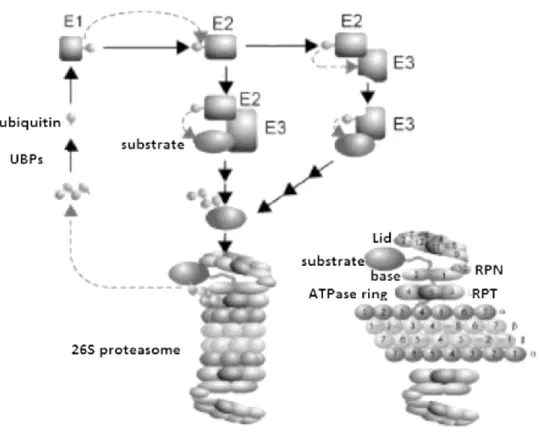
Figure 8- The yeast ubiquitin proteasome system Attachment of ubiquitin to target proteins
requires three enzymatic steps. Ubiquitin activating enzymes (E1) activate ubiquitin in an ATP dependent reaction. An activated ubiquitin moiety is then formed and is transferred and bound to ubiquitin-conjugating enzymes (E2) that serve as carrier proteins. Ubiquitin protein ligases (E3) catalyze the covalent attachment of ubiquitin to target protein. Multiple cycles of ubiquitination result in synthesis and attachment of polyubiquitin chains that serve as a
44 recognition signal for the degradation of the target protein by the 26S proteasome. The 26S proteasome is formed by the 20S catalytic core complex and two 19S regulatory complexes capping the 20S complex at both ends. The 20S complex is composed of four axially stacked rings. Each outer ring consists of seven nonproteolytic subunits. Each of the two inner rings is formed by seven proteolytic subunits. The 19S complex consists of the base and lid subcomplex. The base subcomplex contains six nonredundant ATPases of the AAA superfamily. The lid subcomplex contains at least eight subunits including deubiquitylating enzymes and receptors for ubiquitylated proteins. Polyubiquitinated target proteins enter the 19S regulatory complex and are recognized, deubiquitynated, unfolded, and translocated into the central cavity of the 20S catalytic core complex, where they are degraded by different hydrolytic activities. Degradation peptides are released from the 26S proteasome by diffusion and further degraded to single amino acids by cytosolic peptidases or, in higher eukaryotes, are used for major histocompatibility class I antigen presentation. Adapted from Wolf and Hilt, 2004.
Autophagy
Another defense system that cells possess to overcome protein dysfunction problems
is autophagy. The term autophagy refers to the cellular processes of self-digestion
which involve the uptake of cellular components for degradation in the
vacuole/lysosome (yeasts/mammals) (Levine and Kroemer, 2008). Under vegetative
growth conditions, autophagy occurs at a basal level in both yeast and mammalian
cells, but the lack of nutrients or other types of stress trigger the process. It can be
divided in four pathways, namely macroautophagy, microautophagy,
chaperone-mediated autophagy (CMA) and cytosol-to-vacuole targeting pathway (CVT). The first
two types are conserved between yeast and mammals, while the other two have only
been described in mammals or yeast, respectively. Each of these pathways differ in the
way the cytoplasmatic substrates are delivered to the vacuole (Huang and Klionsky,
2007). During macroautophagy (referred as autophagy in this thesis), a portion of the
cytoplasm is sequestered by a double-membrane structure named autophagosome
45 vacuole (Figure 9). In contrast, microautophagy allows direct uptake of cytoplasmic
material by invaginations of the vacuolar membrane, while chaperone-mediated
autophagy involves the translocation of specific cytosolic proteins that contain a
specific signal motif – KFERQ- across the lysosomal membrane (Figure 10).
Cytosol-to-vacuole targeting is used for the delivery of the Cytosol-to-vacuole resident hydrolases through
vesicules smaller than autophagosomes.
More than 30 genes (ATG genes, AuTophaGy related), which are mainly conserved in
higher eukaryotes, are involved in autophagy regulation. Among the ATG genes, a
subset is required for autophagosome formation in all subtypes of autophagy, which is
usually referred as the core autophagy machinery. This machinery is composed of
three major functional groups: (1) Atg9p and its cycling system, which includes Atg9p,
the Atg1p kinase complex (Atg1p and Atg13p), Atg2p and Atg18p; (2) the
phosphatidylinositol 3-OH kinase (PI(3)K) complex (vacuolar protein sorting (Vps)34,
Vps15p, Atg6p(Vps30) and Atg14p) and (3) the ubiquitin-like protein (Ubl) system,
which includes two Ubl proteins (Atg8p and 3Atg12p), an activating enzyme (Atg7p),
two analogues of ubiquitin-conjugating enzymes (Atg10p and Atg3p), an Atg8p
modifying protease (Atg4p), the protein target of Atg12p attachment (Atg5p) and
Atg16p (Kundu and Thompson, 2008).
The autophagic process can be divided into several sequential steps, namely induction,
cargo selection and packaging, nucleation and vesicle formation, targeting, docking
and fusion, breakdown and export, which share similarities between yeasts and
46 Figure 9- Schematic overview of the autophagic pathway. Autophagy can be broken down in several discrete steps. Induction: requires the dephosphorylation of Atg13p which
interacts with Atg1p, up-regulating the latter kinase activity. Cargo selection and packaging: are specific of selective autophagy pathways and involve several proteins. Nucleation of
vesicle formation: lipids and proteins that constitute the autophagosome and Cvt vesicles are
brought together. Several organelles (ER, mitochondria) have been suggested to be involved in this step, as a lipid source. Vesicle Expansion and Completion: Two ubiquitin-like conjugation systems involving the ubiquitin-like proteins Atg8p and Atg12p are needed for vesicle expansion. Targeting, docking and fusion: The machinery involved in this step includes the SNARE proteins Vam3p, Vam7p, Vti1p and Ykt6p; the Rab protein Ypt7p; members of the class C Vps/HOPS complex; the NSF, SNAP and GDI homologues Sec17p, Sec18p and Sec19p and Ccz1p and Mon1p. In mammalian cells, the initial fusion may involve an endosome.
Breakdown and export; autophagosomes are degraded in the interior of the vacuole. Adapted
from Huang and Klionsky, 2007.
Until very recently, starvation induced autophagy was considered a non-selective
pathway, but numerous observations have shown that autophagy can be a selective
process which can eliminate specific proteins, complexes and organelles. These use the
conserved core autophagy machinery.The mechanisms that ensure the accurateness in
47 some specific post-translational modifications and specific key factors may play an
important role in this process (Kraft et al., 2009). The selective types of autophagy can
be divided into two groups: organellar (ribophagy, mitophagy, pexophagy,
reticulophagy and piecemeal autophagy of the nucleus) and non-organellar types of
autophagy (Cvt pathway, aggrephagy and xenophagy). A brief explanation of each
process is provided below.
The selective degradation of ribosomes is known as ribophagy (Kraft et al., 2008). In S.
cerevisiae, the degradation of ribosomes occurs faster than cytosolic proteins, which suggests a selective autophagosomal degradation pathway. Indeed, it has been shown
that – under nutrient-limiting conditions – mature ribosomes are both non-selectively
degraded by the bulk autophagic pathway and rapidly and selectively degraded by a
specific macroautophagic process (Figure 10). During extended periods of starvation,
ribophagy seems to be essential for cell survival implying that the selective removal of
excessive, non-functional or wrongly-assembled ribosomes may be required to
attenuate protein synthesis and to provide an important source of new building blocks
to maintain cellular homeostasis. Human homologues of the ribophagy process have
been identified but do not have yet known functions. However, a critical regulator of
autophagy, the Atg1p mammalian homologue Ulk1p, has been implicated in the
autophagic clearance of ribosomes during reticulocyte maturation.
When the degradation target is mitochondria, the term mitophagy is used (Figure 10).
These organelles are essential as they supply energy to the cell by carrying out
48 However, mitochondria are also a source of potential harmful reactive oxygen species
that damage lipids, proteins and DNA and have been implicated in aging, cancer and
neurodegeneration. Therefore, controlling the number and quality of mitochondria is
crucial for cellular homeostasis. Several different mechanisms have been proposed to
mediate mitophagy: non-specific macroautophagy, selective macroautophagy (Priault
et al., 2005), selective microautophagy and non selective microautophagy.
Interestingly, mitophagy is preferentially mediated by microautophagy in yeasts and
macroautophagy in mammals. In either case, the regulatory mechanisms are largely
unknown, but osmotic swelling and organelle fragmentation caused by depletion of
the mitochondrial inner membrane protein Mdm38, induce mitophagy (Tatsuta and
Langer, 2008). It has been suggested that fission of the swollen organelle triggers the
autophagic machinery, which is corroborated by the finding that inhibition of the
fission protein Dnm1p blocks mitophagy. In mammals, loss of mitochondrial
membrane potential and opening of the mitochondrial permeability transition pore
seem to be common prerequisites for mitophagy.
Pexophagy is the selective degradation of peroxisomes (Figure 10). These organelles
are involved in many aspects of lipid metabolism and the elimination of peroxides and
their number also needs to be tightly controlled. In P.pastori, at least two basic modes
of selective peroxisome degradation have been described, namely macro- and
micropexophagy, analogous to macro- and microautophagy. In mammals, pexophagy
has also been described but it remains unclear if it is a selective process. Peroxisome
unbalance and pexophagy have been described to play important roles in several
49 The ER also appears to be a selective target for autophagic degradation (Bernales et
al., 2006), named reticulophagy. This organelle is the entry site of secretory proteins
and most of the integral membrane polypeptides, where they are properly folded and
modified. Reticulophagy is responsible for removal of surplus ER upon UPR
inactivation. It contributes to cells physiology in two ways, namely by reducing ER size
after folding stress induced enlargement and by sequestering damaged parts of the ER
containing potentially toxic aberrant proteins. This pathway can also occur as a result
of starvation, but apparently starvation and UPR induced reticulophagy differ at the
morphological level. Also, the two processes depend on different Atg proteins
(Mijaljica et al., 2006; Ogata et al., 2006; Bernales et al., 2006).
Piecemeal autophagy of the nucleus (PMN) occurs under starvation conditions and the
term refers to the process where nonessential parts of the S.cerevisiae yeast nucleus
are targeted for degradation in the vacuole. It resembles microautophagy as the cargo
is sequestered into an invagination of the vacuolar membrane. So far, no analogous
mechanism and no homologous genes have been identified in mammalian cells.
The Cvt pathway is the best characterized type of selective autophagy. It refers to a
biosynthetic process that transports certain resident hydrolases such as
aminopeptidase I and α-mannosidase to the vacuole where they are enzimatically
processed into their mature form. This pathway has only been identified in S.
50 The term aggrephagy refers to the breakdown of toxic protein aggregates in
pathological conditions (Figure 10) and is important in the prevention of inclusion
bodies formation in healthy individuals (Kraft et al., 2009). It has only been described
in higher eukaryotes.
Xenophagy is related to MHC class II crosspresentation and is only known in higher
eukaryotes. Interestingly, several pathogens manipulate the autophagic process in
their mammalian host cells in order to survive and to establish a persistent infection.
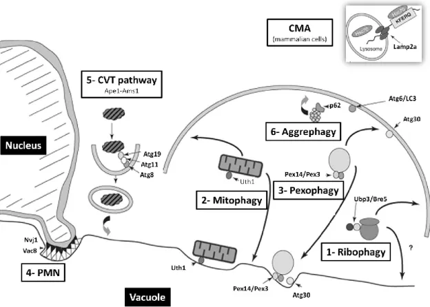
Figure 10- Selective types of autophagy. 1- Ribophagy: refers to ribosome degradation and
depends on Atg1p and Atg7p, which are core components of the autophagy machinery, Ccz1p which is necessary for autophagosome fusion to the vacuole and Ubp3p and its cofactor Bre5p which is involved in 60s ribosomal subunit degradation. Rps5p, Cdc48p and Ufd3p may also be involved in this pathway. Ubiquitination of ribosomal subunits or associated factors by Rsp5p may provide a specific engulfment signal and a subsequent Ubp3/Bre5-dependent