Francisco José Ribeiro Mourão
Endoscopic Submucosal Dissection
for Gastric Superficial Lesions
Mestrado Integrado em Medicina
Área: Gastrenterologia
Trabalho efetuado sob a Orientação de:
Professor Doutor Mário Dinis Ribeiro
E sob a Coorientação de:
Professor Doutor Pedro Pimentel Nunes
Trabalho organizado de acordo com as normas da revista:
European Journal of Gastroenterology & Hepatology
for Gastric Superficial Lesions
Aos meus pais,
Aos meus mestres,
Endoscopic Submucosal Dissection for Gastric Superficial Lesions
Francisco Ribeiro-Mourão
1, Nuno Veloso
1,2, Mário Dinis-Ribeiro
1,2, Pedro
Pimentel-Nunes
1,2, 31
CINTESIS/ CIDES, Porto Faculty of Medicine, PORTUGAL;
2 Department of Gastroenterology, Portuguese Oncology Institute – Porto, PORTUGAL; 3
Department of Physiology, Porto Faculty of Medicine, PORTUGAL;
Running head: ESD for gastric superficial lesions
Conflicts of interest and source of funding:
None to declare.
Wordcount: 2472
Correspondence author and reprinting requests:
Francisco Ribeiro Mourão, Bsc
CINTESIS/ CIDES
Faculdade de Medicina do Porto (CIM-FMUP)
Rua Dr.Plácido da Costa, s/n
4200- 450 Porto – PORTUGAL
Tel: + 351 91 4808259
ABSTRACT
Background and aims: Endoscopic Submucosal Dissection (ESD), an endoscopic technique used
for treatment of gastric superficial lesions, has been gaining importance on western countries. Procedural times have an impact on various outcomes. Our aim is to define which factors from patients, lesions and procedure can predict longer procedural times.
Methods: In a cohort of 127 lesions resected by ESD with IT-knife by experienced
gastroenterologists, characteristics from the patient (age, gender, presence of co-morbidities, usage and suspension of anti-platelet drugs and general physical condition), lesion (size, histopathological diagnosis at biopsy, location, macroscopic type and submucosal invasion) and procedure (complications) were retrospectively analyzed for its impact on time of procedure. Univariate and multivariate analysis were performed.
Results: Lesions larger than 20mm (p<0.001), on the upper third of the stomach (p=0.035) and
with an ASA score of 3 (p=0.031) were considered influential factors for a longer procedure time and specifically for a time of procedure longer than 90 minutes. Existence of intra-procedure complications was also a predictor for a procedure time >90 minutes. Lesion’s size >20mm and location in the upper third were independently associated with a procedure time longer than 90 minutes (OR 4.91[95%CI 2.29-10.50] and OR 18.26 [95%CI 2.02-164.78], respectively)
Conclusion: The time of procedure of ESD for gastric superficial lesions is influenced by size of
lesion (>20 mm) and location (upper third of stomach), which predict a time longer than 90 minutes. This can be useful for better management of workflow, operation, training of teams and anesthesic procedures.
KEYWORDS: stomach neoplasms, gastric cancer, endoscopic gastrointestinal surgical
INTRODUCTION
Endoscopic Submucosal Dissection (ESD) is an endoscopic technique used for treatment of gastric superficial lesions [1]. It has been widely used in countries such as Korea and Japan, but its use only widespread in the Western countries in the last decade [2]. Although having successful results [3-5], ESD requires a high level of expertise in order to reach the desired outcomes [6, 7] .
Specifically, longer procedural times are related to a higher level of complications [8] such as delayed bleeding [9], perforation [10, 11], post-operative pneumonia [11-13] and other clinical complications related to premedication and a heavy workload for patients [7]. Moreover, previous retrospective studies have shown that time of procedure can be influenced by different factors such as existence of fibrosis [14, 15], presence of ulceration [7, 15-17], area of the resected specimen [7, 16-19], location on the upper portion of the stomach [7, 16-19], adhesion [19] and presence of a scar [7]. Therefore, it is essential to take these factors into account in the pre-operatory period, since they can influence the workflow for ESD such as allocation of type of rooms and anesthesic procedures, and level of training of teams [11].
The present work aims at addressing the procedure time of ESD for removal of superficial gastric lesions and to define which patients’ characteristics, lesions’ features and procedure variables may be predictive of longer procedural times.
MATERIALS AND METHODS
A. Type of study and selection of patients
Our study reports a retrospective cohort of 162 consecutively patients (with 195 gastric neoplastic lesions) that were referred to the Portuguese Institute of Oncology – Porto (IPO) from March 2003 to April 2013 for assessment and treatment of gastric superficial neoplasias. This study was conducted in accordance with the ethical principles of the Declaration of Helsinki, in compliance with good clinical practice.
All patients were referred for endoscopic treatment after a multidisciplinary oncology group decision and full medical and anesthesiology evaluation. Both oral and written informed consent was given by patients. All the endoscopic procedures performed on IPO during this period were screened by their report on the institute database, followed by analysis of the clinical record of the patient.
For the purpose of this study, only cases treated by ESD, without ulcerative findings on the lesion, and technically performed with IT-knife were selected. Fifty three procedures were excluded because they were treated by Endoscopic Mucosal Resection (EMRc). Of the ESD procedures, one was excluded because Flex-knife was used along with IT knife, six were excluded because Diathermic loop was used along with IT-knife and two were excluded because cap was used along with IT-Knife. Six procedures were excluded due to incomplete information regarding time of procedure.
Two operators effectuated the endoscopic procedures (MDR and PPN). MDR received training in Japan and in live animal courses before introducing the technique in the Hospital. PPN had animal training and then gradually begun the endoscopic procedures under MDR supervision in 2010 [20, 21]. Lateral margins of each lesion were always determined by chromoendoscopy with indigo carmine 1% [22-24] or with virtual chromoendoscopy using HR-NBI (applying Pimentel-Nunes et al classification for delimitation of lesions [25]) and small marks were made 2-5 mm from the edges of the lesion using needle-knife coagulation. The technique of Endoscopic Submucosal Dissection (ESD) was initiated after the submucosal injection of the lesion with an epinephrine and saline solution (1:100000) and a few drops of methylene blue. After that to obtain access to the submucosal layer 3 to 4 small mucosal incisions using needle-knife were made. Then, an IT-knife (Olympus) in the Endocut mode was used to do the circumference of the lesion outside the coagulation markers. Complete dissection of the lesion was performed using Endocut mode (Olympus electrosurgical unit) with further submucosal injections as needed being made throughout the procedure.
All procedures were performed under deep sedation or general anesthesia (with propofol and fentanyl) supervised by an anesthesiology team.
C. Definitions: procedural time and potential predictive factors
Procedural time was defined as the time of anesthesia, in minutes, reported in patients’ clinical files by the anesteshiology team. Thereafter, two groups were created according to a time shorter or longer than 90 minutes of procedure.
In order to determine the influential factors on procedure time the following variables were analyzed:
- Regarding the lesion: Gross cross-sectional dimension (measured endoscopically) in
millimeters(mm), followed by sub-grouping in lesions ≤20 mm (absolute indication for endoscopic resection on differentiated lesions without ulcerative findings [26]) and >20 mm, histopathological findings at diagnostic biopsy (low-grade dysplasia, high grade dysplasia or
T1a), Location (upper, middle and lower third of the stomach), macroscopic type (organized by Paris classification [27], followed by sub-grouping in depressed and non-depressed lesions) and Histopathological definitive classification defined by the presence or absence of submucosal invasion.
- Regarding the patient: age, gender, presence of co-morbidities, previous suspension of
anti-platelet drugs and general condition of the patient, evaluated by the American Society of Anaesthesiology score (ASA 1,2,3,4 or 5) [28].
- Regarding the procedure: existence and type of complications.
D. Statistical Analysis
Statistical Package for Social Sciences (SPSS 21.0 Package Facility, SPSS Inc, IL, USA) was used for data support and analysis. Analysis was performed using descriptive statistics methods, as well as Kruksal-Wallis test for analysis with time as a continuous variable, and Chi-Square and Fisher’s Exact test for analysis of dichotomic variables. Logistic regression was used to estimate OR for individual variables in multivariate analysis. A value of p<0.05 was considered to be statistically significant.
RESULTS
Characteristics of lesions
Of the 127 lesions, 55% were performed on male patients with an average age of 69 (±10.9) years old and a median of 71 (IQR 61-77). Eight (6%) lesions were located on the upper third, 27 (21%) on the middle third and 92 (72%) were located on the lower third. The median time of procedure was of 85 minutes (IQR 55-130). The median tumor size was of 20 mm (IQR 15-30).
Factors predictive of ESD procedure time
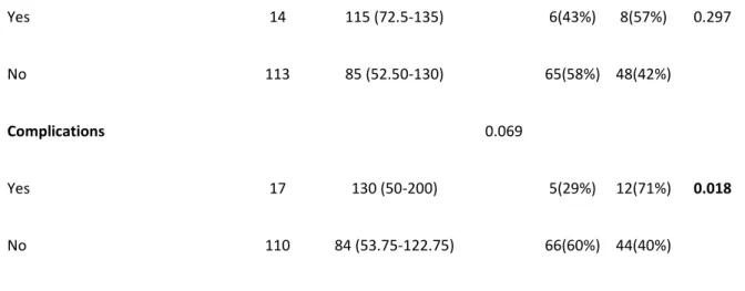
Tumor size, location and the ASA score were significantly associated with procedure time (see Table 1). Specifically, a procedure for a lesion >20 mm, located at the upper third of the stomach and in a patient with an ASA of 3 were associated with longer procedure time, with results significantly different from the other characteristics on the same group. Other patients’, lesions’ and characteristics of procedure are shown on Table 1.
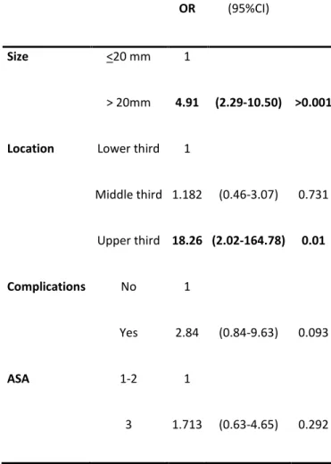
Procedures were furthermore analysed in two different groups - those taking less than and those taking more than 90 minutes. These results are consistent with the findings on the previous analysis, as the majority of the cases (72%) with a lesion ≤20 mm lasted 90 minutes or less, while the majority of the cases with a lesion >20 mm (66%) lasted for more than 90 minutes (p<0,001). Also, lesions located on the upper third took more than 90 minutes (88%), compared to lesions at other locations (p<0.035) and gross majority of lesions in patients with ASA1 took less than 90 minutes to remove (74%) while lesions on patients with ASA 3 took more than 90 minutes on 60% of the cases (p=0,031). Moreover, this analysis also shows that intra-procedure complications pushed the procedure time to more than 90 minutes as 71% of the procedures with complications took more than 90 minutes (p=0.018). In multivariate analysis (Table 2), those patients harbouring lesions larger than 20 mm and located to the upper third showed an increased risk of 4.91 times [95% CI 2.29-10.50] and 18.26 times [95% CI 2.02-164.78], respectively and independently. The occurrence of complications during the
procedure and the ASA score do not seem to be independently predicting procedural time longer than 90 minutes.
DISCUSSION
To our knowledge this is the first study relating ESD time of procedure with factors from the lesion, patient and the procedure itself in Western countries. We showed that lesions with more than 20 mm, located on the upper third of the stomach and in patients with other co-morbidities have a time of procedure significantly higher than smaller lesions, on middle and lower stomach and in patients with less co-morbidities. Larger lesions and lesions on the upper third are independent predictors for longer procedure time. Our results may permit to establish these factors relevant for planning and management of these patients.
Predictive factors of prolonged time of procedure
Comparing the different characteristics of the lesions, a cross sectional dimension >20 mm had longer procedure times when compared to lesions ≤20 mm [120 (IQR 80-147.50) minutes vs 65 (IQR 45-110) minutes, p<0.001). Moreover, when the lesion was located on the upper third the median time was of 145 (IQR 115-253.75) minutes, significantly different from times recorded for lesions on the lower third with a median time of 80 (IQR 46.25-120) minutes, p=0.022. In what relates to the general condition of the patient, measured by the ASA Score, ASA 3 patients had a median procedure time of 120 (IQR 62.5-165) minutes, clearly longer than patients with ASA 1 with a median time of 65 (IQR 40-95) minutes, p=0.011. We also showed that these same characteristics tend to be associated with a procedure longer than 90 minutes. In fact, the size and location were independently associated with a time longer than 90 minutes whereas the risk profile of patients and/or the evidence of complications (bleeding) during the procedures were not independent.
The reasons why the first two factors can act as predictors for a longer time of procedure can be easily explained - a larger lesion will obviously require a higher area to be dissected and therefore more time; the location at the upper third, due to the position required for the scope and the wall characteristics, require more technical skills [7, 29, 30]. Nevertheless, for the other two factors we may
have different reasons not to observe them as independent predictive factors – bleeding is expected more often in lesions in the upper third [31] and ASA 3 patients prevalence is very low and they tend to be older [median age of 75 (IQR 69.5-80) vs 70 (IQR 59-76) on ASA 1 and 2, p=0.005] what may lead to larger [median dimension of 30 mm (IQR 18-30) on ASA 3 vs 20 mm (IQR 15-25) on ASA 1 and 2 , p=0.037] and more advanced lesions.
Predictive factors compared to western series
Moreover, the majority of our findings are in accordance with previous findings in eastern series, specifically in what regards to size and location of lesion [7, 15-19].
Goto et al. [16] have even developed a formula to predict the time of procedure based on size of lesion, location on the upper third and presence of ulceration. Comparing to our results, and considering the non-existence of ulcerated lesions on this series, for a lesion of 20 mm or more and located on the upper third, its predicted time of procedure is never less than 86 minutes which is in accordance with our findings that those two factors are associated with a procedure time longer than 90 minutes.
Ahn et al. [18] also presented results that are consistent with our findings as the predicted times of procedure for lesions on the upper third with more than 30 mm are always superior to 90 minutes. However, it doesn’t have the same conclusion to lesions between 21 and 30 mm.
Regarding the intra-procedure complications predicting a longer time of procedure (>90 minutes) it is in agreement with previous findings by Yamamoto et al. [32] stating that uncontrolled hemorrhage makes the procedure lengthier.
We have also linked a higher ASA score to a prolonged procedure time. However, this finding contradicts Kim et al. [33] if we assume that a ASA 3 is similar to their’s high risk group defined as having one or more co-morbidity states. However, it is not clear if this contradiction is real or if it is due to different classification systems and it was not confirmed as an independent factor.
Limitations
One of the limitations of our study has to do with the standard used to calculate time of procedure, based on the time of anesthesia. This means that our times of procedure can be slightly superior to the ones found on other studies that, for instance, count the time only on the beginning of lesion’s marking. Nevertheless, the mean time of procedure on this series is similar to times reported on different eastern series. However, it can have an impact on the finding of the relation with the ASA score, as this one could relate directly to time of anesthesia and not with time of procedure. Also, it has also been reported on literature that many times the ASA Score is subjective to inter-observer variations [34]. Therefore, this finding should be looked with special attention.
However, future studies should focus on the analysis of time of anesthesia and time of procedure itself alone, evaluated at the same time, to give us a perspective on the impact of complications of the procedure itself or anesthesic complications on the global time.
Another limitation has to do with the evaluation of size being done with a cut-off point in the 20 mm, a methodological option that has to do with the size of our series not allowing comparisons in smaller groups.
A different limitation has to do with the fact that we only considered IT-knife for analysis for the scope of this study as previous studies refer that different knives have different times of procedure associated [35]. However, the option here was purely methodological as we had cases on our series with other knives, but the choice of other knives or concomitant knives with IT-knife was based on the fact that lesions were identified as more complicated and lengthy, that made us to opt to focus on only one knife, so this bias was not present on this study. Anyway, further studies comparing times of procedure with different knives can be an interesting are for research.
Finally, we do not have consistently recorded data for fibrosis and existence of scar throughout the observation period, bringing to the surface the limitation of this study being a retrospective study and subsequently the comparisons with other works. The definition of long-term prospective studies on this area with the focus on studying factors influencing time of procedure are the key for obtaining consistent and comparable data worldwide.
Conclusion
In summary, we found that lesions on the upper stomach, greater than 20 mm and in patients with significant co-morbidities can increase the time of procedure and it is expected that it will last more than 90 minutes, with the first two being independent predictors. It is important to keep in mind if these 3 factors are present on a certain lesion before the procedure, so an adequate planning of operation, human resources and anesthesic method can be performed, therefore allowing a increased efficacy and efficiency.
ACKNOWLEDGEMENTS
We hereby acknowledge all of those involved in the treatment of these patients, namely the Anesthesiology Department of IPO, all the nurses in Gastroenterology Department and the multidisciplinary teams that with us treated these patients.
REFERENCES
1. Tanabe, S., K. Ishido, K. Higuchi, T. Sasaki, C. Katada, M. Azuma, et al., Long-term outcomes of endoscopic submucosal dissection for early gastric cancer: a retrospective comparison with conventional endoscopic resection in a single center. Gastric Cancer, 2014. 17(1): p. 130-6. 2. Ribeiro-Mourao, F., P. Pimentel-Nunes, and M. Dinis-Ribeiro, Endoscopic submucosal dissection
for gastric lesions: results of an European inquiry. Endoscopy, 2010. 42(10): p. 814-9.
3. Min, B.H., J.H. Lee, J.J. Kim, S.G. Shim, D.K. Chang, Y.H. Kim, et al., Clinical outcomes of endoscopic submucosal dissection (ESD) for treating early gastric cancer: comparison with endoscopic mucosal resection after circumferential precutting (EMR-P). Dig Liver Dis, 2009.
41(3): p. 201-9.
4. Ahn, J.Y., H.Y. Jung, K.D. Choi, J.Y. Choi, M.Y. Kim, J.H. Lee, et al., Endoscopic and oncologic outcomes after endoscopic resection for early gastric cancer: 1370 cases of absolute and extended indications. Gastrointest Endosc, 2011. 74(3): p. 485-93.
5. Nonaka, S., I. Oda, T. Nakaya, C. Kusano, H. Suzuki, S. Yoshinaga, et al., Clinical impact of a strategy involving endoscopic submucosal dissection for early gastric cancer: determining the optimal pathway. Gastric Cancer, 2011. 14(1): p. 56-62.
6. Park, Y.M., E. Cho, H.Y. Kang, and J.M. Kim, The effectiveness and safety of endoscopic submucosal dissection compared with endoscopic mucosal resection for early gastric cancer: a systematic review and metaanalysis. Surg Endosc, 2011. 25(8): p. 2666-77.
7. Chung, I.K., J.H. Lee, S.H. Lee, S.J. Kim, J.Y. Cho, W.Y. Cho, et al., Therapeutic outcomes in 1000 cases of endoscopic submucosal dissection for early gastric neoplasms: Korean ESD Study Group multicenter study. Gastrointest Endosc, 2009. 69(7): p. 1228-35.
8. Mannen, K., S. Tsunada, M. Hara, K. Yamaguchi, Y. Sakata, T. Fujise, et al., Risk factors for complications of endoscopic submucosal dissection in gastric tumors: analysis of 478 lesions. J Gastroenterol, 2010. 45(1): p. 30-6.
9. Toyokawa, T., T. Inaba, S. Omote, A. Okamoto, R. Miyasaka, K. Watanabe, et al., Risk factors for perforation and delayed bleeding associated with endoscopic submucosal dissection for early gastric neoplasms: analysis of 1123 lesions. J Gastroenterol Hepatol, 2012. 27(5): p. 907-12.
10. Yoo, J.H., S.J. Shin, K.M. Lee, J.M. Choi, J.O. Wi, D.H. Kim, et al., Risk factors for perforations associated with endoscopic submucosal dissection in gastric lesions: emphasis on perforation type. Surg Endosc, 2012. 26(9): p. 2456-64.
11. Akasaka, T., T. Nishida, S. Tsutsui, T. Michida, T. Yamada, H. Ogiyama, et al., Short-term outcomes of endoscopic submucosal dissection (ESD) for early gastric neoplasm: multicenter survey by osaka university ESD study group. Dig Endosc, 2011. 23(1): p. 73-7.
12. Isomoto, H., K. Ohnita, N. Yamaguchi, E. Fukuda, K. Ikeda, H. Nishiyama, et al., Clinical outcomes of endoscopic submucosal dissection in elderly patients with early gastric cancer. Eur J Gastroenterol Hepatol, 2010. 22(3): p. 311-7.
13. Park, C.H., H. Kim, Y.A. Kang, I.R. Cho, B. Kim, S.J. Heo, et al., Risk factors and prognosis of pulmonary complications after endoscopic submucosal dissection for gastric neoplasia. Dig Dis Sci, 2013. 58(2): p. 540-6.
14. Jeong, J.Y., Y.H. Oh, Y.H. Yu, H.S. Park, H.L. Lee, C.S. Eun, et al., Does submucosal fibrosis affect the results of endoscopic submucosal dissection of early gastric tumors? Gastrointest Endosc, 2012. 76(1): p. 59-66.
15. Oda, I., T. Odagaki, H. Suzuki, S. Nonaka, and S. Yoshinaga, Learning curve for endoscopic submucosal dissection of early gastric cancer based on trainee experience. Dig Endosc, 2012. 24
Suppl 1: p. 129-32.
16. Goto, O., M. Fujishiro, S. Kodashima, S. Ono, and M. Omata, Is it possible to predict the procedural time of endoscopic submucosal dissection for early gastric cancer? J Gastroenterol Hepatol, 2009. 24(3): p. 379-83.
17. Nagata, S., Y.F. Jin, M. Tomoeda, M. Kitamura, M. Yuki, H. Yoshizawa, et al., Influential factors in procedure time of endoscopic submucosal dissection for gastric cancer with fibrotic change. Dig Endosc, 2011. 23(4): p. 296-301.
18. Ahn, J.Y., K.D. Choi, J.Y. Choi, M.Y. Kim, J.H. Lee, K.S. Choi, et al., Procedure time of endoscopic submucosal dissection according to the size and location of early gastric cancers: analysis of 916 dissections performed by 4 experts. Gastrointest Endosc, 2011. 73(5): p. 911-6.
19. Lu, Z.S., Y.S. Yang, D. Feng, S.F. Wang, J. Yuan, J. Huang, et al., Predictive factors of endoscopic submucosal dissection procedure time for gastric superficial neoplasia. World J Gastroenterol, 2012. 18(47): p. 7009-14.
20. Dinis-Ribeiro, M., P. Pimentel-Nunes, M. Afonso, N. Costa, C. Lopes, and L. Moreira-Dias, A European case series of endoscopic submucosal dissection for gastric superficial lesions. Gastrointest Endosc, 2009. 69(2): p. 350-5.
21. Deprez, P.H., J.J. Bergman, S. Meisner, T. Ponchon, A. Repici, M. Dinis-Ribeiro, et al., Current practice with endoscopic submucosal dissection in Europe: position statement from a panel of experts. Endoscopy, 2010. 42(10): p. 853-8.
22. Dinis-Ribeiro, M., A. da Costa-Pereira, C. Lopes, and L. Moreira-Dias, Feasibility and cost-effectiveness of using magnification chromoendoscopy and pepsinogen serum levels for the follow-up of patients with atrophic chronic gastritis and intestinal metaplasia. J Gastroenterol Hepatol, 2007. 22(10): p. 1594-604.
23. Dinis-Ribeiro, M., Chromoendoscopy for early diagnosis of gastric cancer. Eur J Gastroenterol Hepatol, 2006. 18(8): p. 831-8.
24. Dinis-Ribeiro, M., A. da Costa-Pereira, C. Lopes, L. Lara-Santos, M. Guilherme, L. Moreira-Dias, et al., Magnification chromoendoscopy for the diagnosis of gastric intestinal metaplasia and dysplasia. Gastrointest Endosc, 2003. 57(4): p. 498-504.
25. Pimentel-Nunes, P., M. Dinis-Ribeiro, J.B. Soares, R. Marcos-Pinto, C. Santos, C. Rolanda, et al., A multicenter validation of an endoscopic classification with narrow band imaging for gastric precancerous and cancerous lesions. Endoscopy, 2012. 44(3): p. 236-46.
26. Japanese Gastric Cancer, A., Japanese gastric cancer treatment guidelines 2010 (ver. 3). Gastric Cancer, 2011. 14(2): p. 113-23.
27. The Paris endoscopic classification of superficial neoplastic lesions: esophagus, stomach, and colon: November 30 to December 1, 2002. Gastrointest Endosc, 2003. 58(6 Suppl): p. S3-43. 28. Owens, W.D., J.A. Felts, and E.L.J. Spitznagel, ASA Physical Status Classifications: A Study of
29. Kaltenbach, T., R. Soetikno, C. Kusano, and T. Gotoda, Development of expertise in endoscopic mucosal resection and endoscopic submucosal dissection. Techniques in Gastrointestinal Endoscopy, 2011. 13(1): p. 100-104.
30. Murata, A., K. Okamoto, K. Muramatsu, and S. Matsuda, Endoscopic submucosal dissection for gastric cancer: the influence of hospital volume on complications and length of stay. Surg Endosc, 2013.
31. Oda, I., H. Suzuki, S. Nonaka, and S. Yoshinaga, Complications of gastric endoscopic submucosal dissection. Dig Endosc, 2013. 25 Suppl 1: p. 71-8.
32. Yamamoto, S., N. Uedo, R. Ishihara, N. Kajimoto, H. Ogiyama, Y. Fukushima, et al., Endoscopic submucosal dissection for early gastric cancer performed by supervised residents: assessment of feasibility and learning curve. Endoscopy, 2009. 41(11): p. 923-8.
33. Kim, B.J., T.H. Chang, J.J. Kim, B.H. Min, J.H. Lee, H.J. Son, et al., Efficacy and safety of endoscopic submucosal dissection for early gastric cancer in patients with comorbid diseases. Gut Liver, 2010. 4(2): p. 186-91.
34. Mak, P.H., R.C. Campbell, M.G. Irwin, and A. American Society of, The ASA Physical Status Classification: inter-observer consistency. American Society of Anesthesiologists. Anaesth Intensive Care, 2002. 30(5): p. 633-40.
35. Lee, J.H., S.J. Hong, J.Y. Jang, S.E. Kim, and S.Y. Seol, Outcome after endoscopic submucosal dissection for early gastric cancer in Korea. World J Gastroenterol, 2011. 17(31): p. 3591-5.
Time
Characteristics n median (IQR) p value < 90 min > 90 min P value
Procedures (total) 127 85 (55-130) Gender 0.443 Male 70 97.5 (50-140) 34(49%) 36(51%) 0.065 Female 57 80 (60-120) 37(65%) 20(35%) Age§ 71 (61-77) 0.705 <= 65 42 85 (43.75 -121.25) 25(60%) 17(40%) 0.564 >65 85 90 (60-140) 46 (54%) 39(46%) ASA 0.011* ASA 1 31 65 (40-95) 23(74%) 8 (26%) 0.031* ASA 2 71 90 (55-130) 38(54%) 33 (46%) ASA 3 25 120 (62.50-165) 10(40%) 15(60%) Co-morbidities 0.125 Yes 92 90 (30-112.75) 47(51%) 45(49%) 0.076 No 35 72.5 (55-140) 24(69%) 11(31%) Anti-platelets 0.153 Yes 25 105 (60-187.5) 12(48%) 13(52%) 0.374 No 102 85 (48.75-126.25) 59(58%) 43(42%) Suspension of anti-platelets 0.764
Yes 14 117.5 (60-200) 5(36%) 9(64%) 0.416 No 8 102.5 (52.50-201.25) 4(50%) 4(50%) Size of lesion <0.001 <= 20 mm 74 65 (45-110) 53(72%) 21(28%) <0.001 >20 mm 53 120 (80-147.5) 18(34%) 35(66%) Histopathology at biopsy 0.521 LGD 40 75 (46.25-125) 26(65%) 14(35%) 0.367 HGD 57 90 (52.50-137.50) 29(51%) 28(49%) Adenocarcinoma 30 90 (60-130) 16(53%) 14(47%) Type of lesion 0.891 Naive 121 85 (55-130) 68(56%) 53(44%) 0.542 Recidive 6 95 (29.75-156) 3(50%) 3(50%) Location 0.022** Upper third 8 145 (115-253.75) 1 (12%) 7(88%) 0.035** Middle third 27 90 (60-180) 15(56%) 12(44%) Lower third 92 80 (46.25-120) 55(60%) 37(40%) Macroscopic features 0.833 Depressed lesions 60 87.5 (60-130) 36(60%) 24(40%) 0.379
Non depressed lesions 67 85 (45-140) 35(52%) 32(48%)
Yes 14 115 (72.5-135) 6(43%) 8(57%) 0.297
No 113 85 (52.50-130) 65(58%) 48(42%)
Complications 0.069
Yes 17 130 (50-200) 5(29%) 12(71%) 0.018
No 110 84 (53.75-122.75) 66(60%) 44(40%)
* Statistically significant for comparison between ASA1 and ASA3
** Statistically significant for comparison between Upper third and Lower third § Median Age (IQR)
IQR, interquartile range; min, minutes; LGD, Low-grade dysplasia; HGD, High-Grade dysplasia
TABLE 1 – Characteristics of patients, lesions and procedure with univariate analysis for predictors of
OR (95%CI)
Size <20 mm 1
> 20mm 4.91 (2.29-10.50) >0.001
Location Lower third 1
Middle third 1.182 (0.46-3.07) 0.731 Upper third 18.26 (2.02-164.78) 0.01 Complications No 1 Yes 2.84 (0.84-9.63) 0.093 ASA 1-2 1 3 1.713 (0.63-4.65) 0.292
OR, Odds Ratio; CI, Confidence Interval
AGRADECIMENTOS
Ao Professor Doutor Mário Dinis Ribeiro por ter, desde cedo, alimentado o gosto pela investigação científica e por ter sido, mais do que o orientador deste trabalho, um orientador de todo o meu percurso académico e uma fonte de inspiração e encorajamento, nunca tendo desistido de acreditar na minha capacidade de trabalho.
Ao Professor Doutor Pedro Pimentel Nunes, por todos os desafios que foi colocando e por todo o apoio na concepção e desenvolvimento do trabalho.
Ao Dr. Nuno Veloso, por ter começado a germinação deste trabalho comigo e por ter sido catalisador de momentos de boa disposição durante o trabalho.
Aos médicos e enfermeiras do serviço de Gastrenterologia do Instituto Português de Oncologia do Porto por me terem recebido de braços abertos em todos os momentos deste trabalho e por me incluírem como um membro da sua equipa.
À Daniela Linhares, pelo apoio que me deu para realizar este trabalho, pelos momentos em que me ouviu sem contestar, em desabafo, e por aqueles em que me ouviu e me chamou à atenção para o caminho certo.
À Ana Moço e à Mariana Vieira por todas as palavras de encorajamento e pela amizade cultivada ao longo de vários anos.
Ao Jorge Barbosa, pelo apoio durante o processo de escrita e pelos olhos implacáveis na revisão do trabalho.
À Direção da Associação Nacional de Estudantes de Medicina, durante o ano de 2013, por me ter apoiado em todas as decisões e me ter substituído nas tardes de ausência em que me encerrava para colher dados.
Aos meus pais, fonte de inspiração desde tenra idade, e que sempre foram uma fonte de motivação e pressão para o trabalho, mas também de conforto e amizade.
Ao João Dias por ter vivido esta aventura desde há vários anos e por me ter motivado e encorajado a ser perfeccionista e realizado.
and Submission of
Manuscripts to the
European Journal of
Gastroenterology &
Hepatology
Copyright Transfer and Disclosure Form(PDF) Reprint Ordering
Permissions Requests
Note: These instructions comply with those formulated by the International Committee of Medical Journal Editors. For further details, authors should consult the “Uniform
Requirements for Manuscripts Submitted to Biomedical Journals” at http://www.icmje.org.
Aims and scope
The European Journal of Gastroenterology & Hepatology publishes papers reporting original clinical and scientific research which are of a high standard and which contribute to the advancement of knowledge in the field of gastroenterology and hepatology. The journal publishes four types of manuscripts: in-depth reviews by invitation only, original papers, letters to the Editor and case reports.
Table of contents
Points to consider before submission Redundant or duplicate publication Conflicts of interest
Permissions to reproduce previously published material Patient consent forms
Ethics committee approval Authorship
Compliance with NIH and Other Research Funding Agency Accessibility Requirements Copyright assignment Submissions Presentation of Papers Title Page Abstracts Keywords Text Acknowledgements References Tables Illustrations
Legends for illustrations Units of measurement Abbreviations and symbols Supplemental Digital Content Offprints
Letters to the Editor
Points to consider before submission
Please think carefully about the following points and make the appropriate declarations.
Redundant or duplicate publication
We ask you to confirm that your paper has not been published in its current form or a substantially similar form (in print or electronically, including on a web site), that it has not been accepted for publication elsewhere, and that it is not under consideration by another publication. The International Committee of Medical Journal Editors has provided details of what is and what is not duplicate or redundant publication (www.icmje.org). If you are in doubt (particularly in the case of material that you have posted on a web site), we ask you to proceed with your submission but to include a copy of the relevant previously published work or work under consideration by other journals. In your covering letter to the editors, draw attention to any published work that concerns the same patients or subjects as the present paper.
Conflicts of interest
Authors must state all possible conflicts of interest in the manuscript, including financial, consultant, institutional and other relationships that might lead to bias or a conflict of interest. If there is no conflict of interest, this should also be explicitly stated as none declared. All sources of funding should be acknowledged in the manuscript. All relevant conflicts of interest and sources of funding should be included on the title page of the manuscript with the heading “Conflicts of Interest and Source of Funding:”. For example: Conflicts of Interest and Source of Funding: A has received honoraria from Company Z. B is currently receiving a grant (#12345) from Organization Y, and is on the speaker’s bureau for Organization X – the CME organizers for Company A. For the remaining authors none were declared.
(www.icmje.org/update.html). The form is readily available on the manuscript submission page www.editorialmanager.com/ejgh and can be completed and submitted electronically. Please note that authors may sign the copyright transfer agreement form electronically. For additional information about electronically signing this form, go to
http://links.lww.com/ZUAT/A106.
Permissions to reproduce previously published material
Authors should include with their submission copies of written permission to reproduce material published elsewhere (such as illustrations) from the copyright holder. Authors are responsible for paying any fees to reproduce material.
Patient consent forms
Patients have a right to privacy that should not be infringed without informed consent. Identifying details (written or photographic) should be omitted if they are not essential, but patient data should never be altered or falsified in an attempt to attain anonymity. Complete anonymity is difficult to achieve, and a consent form should be obtained if there is any doubt. For example, masking the eye region in photographs of patients is inadequate protection of anonymity. When informed consent has been obtained it should be indicated in the published article.
A statement to the effect that such consent had been obtained must be included in the ‘Methods’ section of your paper and an example of the consent form you used must be uploaded with your manuscript.
Ethics committee approval
You must state clearly in your submission in the Methods section that you conducted studies on human participants must with the approval of an appropriate named ethics committee. Please also look at the latest version of the Declaration of Helsinki. Clinical studies should be in accordance with the latest publication of ‘Good Clinical Practice’. Similarly, you must confirm that experiments involving animals adhered to ethical standards and must state the care of animal and licensing guidelines under which the study was performed.
Authorship
All authors must sign copyright forms accompanying their submission to confirm that they have read and approved the paper, that they have met the criteria for authorship as established by the International Committee of Medical Journal Editors, that they believe that the paper represents honest work, and that they are able to verify the validity of the results reported.
Compliance with NIH and Other Research Funding Agency Accessibility Requirements
A number of research funding agencies now require or request authors to submit the post-print (the article after peer review and acceptance but not the final published article) to a repository that is accessible online by all without charge. As a service to our authors, LWW will identify to the National Library of Medicine (NLM) articles that require deposit and will transmit the post-print of an article based on research funded in whole or in part by the National Institutes of Health, Wellcome Trust, Howard Hughes Medical Institute, or other funding agencies to PubMed Central. The Copyright Transfer Agreement provides the mechanism.
Copyright assignment
Papers are accepted for publication on the understanding that exclusive copyright in the paper is assigned to the Publisher. Authors are asked to submit signed copyright assignment form with their paper. They may use material from their paper in other works published by them after seeking formal permission.
Submissions
All manuscripts and materials must be submitted through the web-based tracking system at
https://www.editorialmanager.com/ejgh/. The site contains instructions and advice on how to use the system. Authors should NOT in addition then post a hard copy submission to the editorial office, unless you are supplying artwork, letters or files that cannot be submitted electronically, or have been instructed to do so by the editorial office. For those authors who have no option but to submit by mail please contact the Editorial Office
Double spacing should be used throughout the manuscript, which should include the following sections, each starting on a separate page: title page, abstract and keywords, text, acknowledgements, references, individual tables and captions. Margins should be not less than 3 cm. Pages should be numbered consecutively, beginning with the Title Page, and the page number should be placed in the top right hand corner of each page. Abbreviations should be defined on their first appearance in the text; those not accepted by international bodies should be avoided.
Before submitting to the journal make sure you have provided:
description of authors contributions to the study information who performed statistical analysis
Presentation of Papers Title Page
The Title Page should carry the full title of the paper and a short title, of no more than 45 characters and spaces, to be used as a ‘running head’ (and which should be so identified). The first name, middle initial and last name of each author should appear. If the work is to be attributed to a department or institution, its full name should be included. Any
disclaimers should appear on the Title Page, as should the name and address of the author responsible for correspondence concerning the manuscript and the name and address of the author to whom requests for reprints should be made. Finally, the Title Page should include a statement of conflicts of interest and source of funding, and when none state “none declared”.
Abstracts
The second page should carry a structured abstract of no more than 250 words. The abstract should state the Objective(s) of the study or investigation, basic Methods (selection of study subjects or laboratory animals; observational and analytical methods), main Results (giving specific data and their statistical significance, if possible), and the principal Conclusions. It should emphasise new and important aspects of the study or observations.
Keywords
The abstract should be followed by a list of 3–10 keywords or short phrases which will assist the cross-indexing of the article and which may be published. When possible, the terms used should be from the Medical Subject Headings list of the National Library of Medicine (http://www.nlm.nih.gov/mesh/meshhome.html).
Text
Full papers of an experimental or observational nature may be divided into sections headed Introduction, Methods (including ethical and statistical information), Results and Discussion (including a conclusion), although reviews may require a different format.
Word limit for original studies and reviews is 5000 words, case reports 3.500 words and letters 1500 words (tables and figures are not counted).
Acknowledgements
Acknowledgements should be made only to those who have made a substantial contribution to the study. Authors are responsible for obtaining written permission from people acknowledged by name in case readers infer their endorsement of data and conclusions.
References
References should be numbered consecutively in the order in which they first appear in the text. They should be assigned Arabic numerals, which should be given in brackets, e.g. [17]. References should include the names of all authors when six or fewer; when seven or more, list only the first six names and add et al. References should also include full title and source information. Journal names should be abbreviated as in MEDLINE (NLM Catalog,
http://www.ncbi.nlm.nih.gov/nlmcatalog). Articles in journals
Standard journal article:
Simopoulos AP. The traditional diet of Greece and cancer. Eur J of Cancer Prev 2004;13 :219-230.
Tan MP, Newton JL, Chadwick TJ, Gray JC, Nath S, Parry SW. Home orthostatic training in vasovagal syncope. Europace 2010;12:240–246.
More than six authors:
Schaefer M, Schmidt F, Folwaczny C, Lorenz R, Martin G, Schindlbeck N, et al. Adherence and mental side effects during hepatitis C treatment with interferon alfa and ribavirin in psychiatric risk groups. Hepatology 2003;37:443–451.
Supplements:
Matthews G, Kronborg IJ, Dore GJ. Treatment for hepatitis C virus infection among current injection drug users in Australia. Clin Infect Dis 2005;40(Suppl 5):S325–S329
Books
Book:
Whitehead WE, Schuster MM. Gastrointestinal Disorders. Behavioral and Physiological Basis
techniques, findings. 1999 Stuttgart Thieme Verlag:13–175.
Online
Snyder CL, Young DO, Green PHR, Taylor AKPagon RA, Bird TC, Dolan CR, Stephens K. Celiac disease GeneReviews [Online, 03 July 2008]. 1993 Seattle University of Washington. Personal communications and unpublished work should not feature in the reference list but should appear in parentheses in the text. Unpublished work accepted for publication but not yet released should be included in the reference list with the words ‘in press’ in parentheses beside the name of the journal concerned. References must be verified by the author(s) against the original documents.
Tables
Each table should be typed on a separate sheet in double spacing. Tables should not be submitted as photographs. Each table should be assigned an Arabic numeral, e.g. (Table 3) and a brief title. Vertical rules should not be used. Place explanatory matter in footnotes, not in the heading. Explain in footnotes all non-standard abbreviations that are used in each table. Identify statistical measures of variations, such as standard deviation and standard error of the mean.
Be sure that each table is cited in the text. If you use data from another published or unpublished source, obtain permission and acknowledge the source fully.
Illustrations
A) Creating Digital Artwork
1. Learn about the publication requirements for Digital Artwork:
http://links.lww.com/ES/A42
2. Create, Scan and Save your artwork and compare your final figure to the Digital Artwork Guideline Checklist (below).
3. Upload each figure to Editorial Manager in conjunction with your manuscript text and tables.
B) Digital Artwork Guideline Checklist
Here are the basics to have in place before submitting your digital artwork:
Artwork should be saved as TIFF, EPS, or MS Office (DOC, PPT, XLS) files. High resolution PDF files are also acceptable.
Crop out any white or black space surrounding the image.
Diagrams, drawings, graphs, and other line art must be vector or saved at a resolution of at least 1200 dpi. If created in an MS Office program, send the native (DOC, PPT, XLS) file.
Photographs, radiographs and other halftone images must be saved at a resolution of at least 300 dpi.
Photographs and radiographs with text must be saved as postscript or at a resolution of at least 600 dpi.
Each figure must be saved and submitted as a separate file. Figures should not be embedded in the manuscript text file.
Remember:
Cite figures consecutively in your manuscript using Arabic numerals in parentheses, e.g. (Fig. 2).
Number figures in the figure legend in the order in which they are discussed.
Upload figures consecutively to the Editorial Manager web site and enter figure numbers consecutively in the Description field when uploading the files.
If hard copies are submitted they should have a label pasted to the back bearing the figure number, the title of the paper, the author's name and a mark indicating the top of the figure.
Illustrations should be presented to a width of 82 mm or, when the illustration demands it, to a width of 166 mm.
Photomicrographs must have internal scale markers.
If photographs of people are used, their identities must be obscured or the picture must be accompanied by written consent to use the photograph.
If a figure has been published before, the original source must be acknowledged and written permission from the copyright holder for both print and electronic formats should be submitted with the material. Permission is required regardless of authorship or publisher, except for documents in the public domain.
Figures may be reduced, cropped or deleted at the discretion of the editor. Colour illustrations are acceptable but authors will be expected to cover the extra reproduction costs (for current charges, contact the publisher).
Legends for illustrations
Units of measurement
Measurements of length, height, weight, and volume should be reported in metric units (metre, kilogram, or litre) or their decimal multiples. Temperatures should be given in degrees Celsius. Blood pressures should be given in millimetres of mercury.
All haematologic and clinical chemistry measurements should be reported in the metric system in terms of the International System of Units (SI). Editors may request that alternative or non-SI units be added by the authors before publication.
Abbreviations and symbols
Use only standard abbreviations. Avoid abbreviations in the title and abstract. The full term for which an abbreviation stands should precede its first use in the text unless it is a standard unit of measurement.
Supplemental Digital Content
Supplemental Digital Content (SDC): Authors may submit SDC via Editorial Manager to LWW journals that enhance their article's text to be considered for online posting. SDC may include standard media such as text documents, graphs, audio, video, etc. On the Attach Files page of the submission process, please select Supplemental Audio, Video, or Data for your uploaded file as the Submission Item. If an article with SDC is accepted, our production staff will create a URL with the SDC file. The URL will be placed in the call-out within the article. SDC files are not copy-edited by LWW staff, they will be presented digitally as submitted. For a list of all available file types and detailed instructions, please visit
http://links.lww.com/A142.
SDC Call-outs
Supplemental Digital Content must be cited consecutively in the text of the submitted manuscript. Citations should include the type of material submitted (Audio, Figure, Table, etc.), be clearly labeled as "Supplemental Digital Content," include the sequential list number, and provide a description of the supplemental content. All descriptive text should be included in the call-out as it will not appear elsewhere in the article.
Example:
We performed many tests on the degrees of flexibility in the elbow (see Video,
Supplemental Digital Content 1, which demonstrates elbow flexibility) and found our results inconclusive.
List of Supplemental Digital Content
A listing of Supplemental Digital Content must be submitted at the end of the manuscript file. Include the SDC number and file type of the Supplemental Digital Content. This text will be removed by our production staff and not be published.
Example:
Supplemental Digital Content 1.wmv
SDC File Requirements
All acceptable file types are permissible up to 10 MBs. For audio or video files greater than 10 MBs, authors should first query the journal office for approval. For a list of all available file types and detailed instructions, please visit http://links.lww.com/A142.
Offprints
Offprints may be purchased using the appropriate form that will be made available with proofs. Orders should be sent when the proofs are returned; orders received after this time cannot be fulfilled.
Letters to the Editor
Letters commenting on papers in EJGH will be considered for publication. They should be submitted within 4 weeks of the appearance of the original item and be 300 words, or shorter. Such letters will be passed to the authors of the original paper, who will be offered an opportunity to reply.
Letters of general interest, up to 450 words long, will be peer reviewed if they contain original data. They may contain one table, or one figure and have no more than five references and up to five authors. Proofs will be sent out on acceptance.
Please include with either category of letter a declaration of conflict of interest, if any, e.g., conflict of interest: none (or declare conflict).
Open access
LWW’s hybrid open access option is offered to authors whose articles have been accepted for publication. With this choice, articles are made freely available online immediately upon publication. Authors may take advantage of the open access option at the point of acceptance to ensure that this choice has no influence on the peer review and acceptance process. These articles are subject to the journal’s standard peer-review process and will be accepted or rejected based on their own merit.
Councils UK (RCUK) is $2,540. The publication fee is charged on acceptance of the article and should be paid within 30 days by credit card by the author, funding agency or
institution. Payment must be received in full for the article to be published open access. Any additional standard publication charges, such as for color images, will also apply.
Authors retain copyright
Authors retain their copyright for all articles they opt to publish open access. Authors grant LWW a license to publish the article and identify itself as the original publisher.
Creative Commons license
Articles opting for open access will be freely available to read, download and share from the time of publication. Articles are published under the terms of the Creative Commons License Attribution-NonCommerical No Derivative 3.0 which allows readers to disseminate and reuse the article, as well as share and reuse of the scientific material. It does not permit commercial exploitation or the creation of derivative works without specific permission. To view a copy of this license visit: http://creativecommons.org/licenses/by-nc-nd/3.0.
Compliance with NIH, RCUK and other research funding agency accessibility requirements
A number of research funding agencies now require or request authors to submit the post-print (the article after peer review and acceptance but not the final published article) to a repository that is accessible online by all without charge. As a service to our authors, LWW identifies to the National Library of Medicine (NLM) articles that require deposit and transmits the post-print of an article based on research funded in whole or in part by the National Institutes of Health, Howard Hughes Medical Institute, or other funding agencies to PubMed Central. The revised Copyright Transfer Agreement provides the mechanism. LWW ensures that authors can fully comply with the public access requirements of major funding bodies worldwide. Additionally, all authors who choose the open access option will have their final published article deposited into PubMed Central.
RCUK funded authors can choose to publish their paper as open access with the payment of an article process charge, or opt for their accepted manuscript to be deposited (green route) into PMC with an embargo.
With both the gold and green open access options, the author will continue to sign the Copyright Transfer Agreement (CTA) as it provides the mechanism for LWW to ensure that the author is fully compliant with the requirements. After signature of the CTA, the author will then sign a License to Publish where they will then own the copyright.
It is the responsibility of the author to inform the Editorial Office and/or LWW that they have RCUK funding. LWW will not be held responsible for retroactive deposits to PMC if the author has not completed the proper forms.
FAQ for open access
http://links.lww.com/LWW-ES/A48
Home Recommendations Brow se Manuscript Preparation Preparing for Submission
Preparing for
Submission
PAGE CONTENTS 1. General Principles 2. Reporting Guidelines 3. Manuscript Sections a. Title Page b. Abstract c. Introduction d. Methods e. Results f. Discussion g. References h. Tables i. Illustrations (Figures) j. Units of Measurementk. Abbreviations and Symbols
1. General Principles
The text of articles reporting original research is usually divided into Introduction, Methods, Results, and Discussion sections. This so-called “IMRAD” structure is not an
arbitrary publication format but a reflection of the process of scientific discovery. Articles often need subheadings within these sections to further organize their content. Other types of articles, such as meta-analyses, may require different formats, while case reports, narrative reviews, and editorials may have less structured or unstructured
formats. FAQ How do I format a specific citation? Recommendations Browse About the Recommendations Roles & Responsibilities Publishing & Editorial Issues Manuscript Preparation Preparing for Submission Sending the Submission Translations Archives Subscribe to Changes RecommendationsConflicts of Interest Journals
Following the ICMJE Recommendations
About ICMJE
News & Editorials
extracting portions of articles in electronic versions. Supplementary electronic-only material should be
submitted and sent for peer review simultaneously with the primary manuscript.
2. Reporting Guidelines
Reporting guidelines have been developed for different study designs; examples include CONSORT for randomized trials, STROBE for observational studies,
PRISMA for systematic reviews and meta-analyses, and
STARD for studies of diagnostic accuracy. Journals are encouraged to ask authors to follow these guidelines because they help authors describe the study in enough detail for it to be evaluated by editors, reviewers, readers, and other researchers evaluating the medical literature. Authors of review manuscripts are encouraged to describe the methods used for locating, select¬ing, extracting, and synthesizing data; this is mandatory for systematic reviews. Good sources for reporting guidelines are the
EQUATOR Network and the NLM's Research Reporting Guidelines and Initiatives.
3. Manuscript Sections
The following are general requirements for reporting within sections of all study designs and manuscript formats.
a. Title Page
General information about an article and its authors is presented on a manuscript title page and usually includes the article title, author information, any disclaimers, sources of support, word count, and sometimes the number of tables and figures.
Article title. The title provides a distilled description of the
complete article and should include information that, along with the Abstract, will make electronic retrieval of the article sensitive and specific. Reporting guidelines recommend and some journals require that information about the study design be a part of the title (particularly important for randomized trials and systematic reviews and meta-analyses). Some journals require a short title, usually
KEEP UP-TO-DATE Request to receive an E-mail when the Recommendations are updated. Subscribe to Changessubmission system. Electronic submission systems may restrict the number of characters in the title.
Author information: Each author's highest academic
degrees should be listed, although some journals do not publish these. The name of the department(s) and institution(s) or organizations where the work should be attributed should be specified. Most electronic submission systems require that authors provide full contact
information, including land mail and e-mail addresses, but the title page should list the corresponding authors' telephone and fax numbers and e-mail address.
Disclaimers. An example of a disclaimer is an author's
statement that the views expressed in the submitted article are his or her own and not an official position of the
institution or funder.
Source(s) of support. These include grants, equipment,
drugs, and/or other support that facilitated conduct of the work described in the article or the writing of the article itself.
Word count. A word count for the paper's text, excluding
its abstract, acknowledgments, tables, figure legends, and references, allows editors and reviewers to assess whether the information contained in the paper warrants the paper's length, and whether the submitted manuscript fits within the journal's formats and word limits. A separate word count for the Abstract is useful for the same reason.
Number of figures and tables. Some submission systems
require specification of the number of Figures and Tables before uploading the relevant files. These numbers allow editorial staff and reviewers to confirm that all figures and tables were actually included with the manuscript and, because Tables and Figures occupy space, to assess if the information provided by the figures and tables warrants the paper's length and if the manuscript fits within the journal's space limits.
Conflict of Interest declaration. Conflict of interest
information for each author needs to be part of the manuscript; each journal should develop standards with regard to the form the information should take and where it will be posted. The ICMJE has developed a uniform conflict
it. Despite availability of the form, editors may require conflict of interest declarations on the manuscript title page to save the work of collecting forms from each author prior to making an editorial decision or to save reviewers and readers the work of reading each author's form.
b. Abstract
Original research, systematic reviews, and meta-analyses require structured abstracts. The abstract should provide the context or background for the study and should state the study's purpose, basic procedures (selection of study participants, settings, measurements, analytical methods), main findings (giving specific effect sizes and their
statistical and clinical significance, if possible), and principal conclusions. It should emphasize new and important aspects of the study or observations, note important limitations, and not overinterpret findings. Clinical trial abstracts should include items that the CONSORT group has identified as essential. Funding sources should be listed separately after the Abstract to facilitate proper display and indexing for search retrieval by MEDLINE. Because abstracts are the only substantive portion of the article indexed in many electronic databases, and the only portion many readers read, authors need to ensure that they accurately reflect the content of the article.
Unfortunately, information in abstracts often differs from that in the text. Authors and editors should work in the process of revision and review to ensure that information is consistent in both places. The format required for
structured abstracts differs from journal to journal, and some journals use more than one format; authors need to prepare their abstracts in the format specified by the journal they have chosen.
The ICMJE recommends that journals publish the clinical trial registration number at the end of the abstract. The ICMJE also recommends that, when a registration number is available, authors list that number the first time they use a trial acronym to refer to the trial they are reporting or to other trials that they mention in the manuscript.
c. Introduction
tested by, the study or observation. Cite only directly pertinent references, and do not include data or conclusions from the work being reported.
d. Methods
The guiding principle of the Methods section should be clarity about how and why a study was done in a particular way. The section should include only information that was available at the time the plan or protocol for the study was being written; all information obtained during the study belongs in the Results section.
i. Selection and Description of Participants Clearly describe the selection of observational or
experimental participants (healthy individuals or patients, in¬cluding controls), including eligibility and exclusion criteria and a description of the source population. Because the relevance of such variables as age, sex, or ethnicity is not always known at the time of study design, researchers should aim for inclusion of representative populations into all study types and at a minimum provide descriptive data for these and other relevant demographic variables. If the study was done involving an exclusive population, for example in only one sex, authors should justify why, except in obvious cases (e.g., prostate
cancer).” Authors should define how they measured race or ethnicity and justify their relevance.
ii. Technical Inform ation
Specify the study's main and secondary objectives–usually identified as primary and secondary outcomes. Identify methods, equipment (give the manufacturer's name and address in parentheses), and procedures in sufficient detail to allow others to reproduce the results. Give references to established methods, including statistical methods (see below); provide references and brief descrip¬tions for methods that have been published but are not well-known; describe new or substantially modified methods, give the reasons for using them, and evaluate their limitations. Identify precisely all drugs and chemicals used, including generic name(s), dose(s), and route(s) of administration. Identify appropriate scientific names and gene names.
Describe statistical methods with enough detail to enable a knowledgeable reader with access to the original data to judge its appropriateness for the study and to verify the reported results. When possible, quantify findings and present them with appropriate indicators of mea¬surement error or uncertainty (such as confidence intervals). Avoid relying solely on statistical hypothesis testing, such as P values, which fail to convey important information about effect size and precision of estimates. References for the design of the study and statistical methods should be to standard works when possible (with pages stated). Define statistical terms, abbreviations, and most symbols. Specify the statistical software package(s) and versions used. Distinguish prespecified from exploratory analyses, including subgroup analyses.
e. Results
Present your results in logical sequence in the text, tables, and figures, giving the main or most important findings first. Do not repeat all the data in the tables or figures in the text; emphasize or summarize only the most important observations. Provide data on all primary and secondary outcomes identified in the Methods Section. Extra or supplementary materials and technical details can be placed in an appendix where they will be accessible but will not interrupt the flow of the text, or they can be published solely in the electronic version of the journal. Give numeric results not only as derivatives (for example, percentages) but also as the absolute numbers from which the derivatives were calculated, and specify the statistical significance attached to them, if any. Restrict tables and figures to those needed to explain the argument of the paper and to assess supporting data. Use graphs as an alternative to ta¬bles with many entries; do not duplicate data in graphs and tables. Avoid nontechnical uses of technical terms in statistics, such as “random” (which implies a randomizing device), “normal,” “significant,” “correlations,” and “sample.”
Separate reporting of data by demographic variables, such as age and sex, facilitate pooling of data for subgroups across studies and should be routine, unless there are compelling reasons not to stratify reporting, which should be explained.
Emphasize the new and important aspects of the study and the conclusions that follow from them in the context of the totality of the best available evidence. Do not repeat in detail data or other information given in other parts of the manuscript, such as in the Introduction or the Results section. For experimental studies, it is useful to begin the discussion by briefly summarizing the main findings, then explore possible mechanisms or explanations for these findings, compare and contrast the results with other relevant studies, state the limitations of the study, and explore the implications of the findings for future research and for clinical practice.
Link the conclusions with the goals of the study but avoid unqualified statements and conclusions not adequately supported by the data. In particular, distinguish between clinical and statistical significance, and avoid making statements on economic benefits and costs unless the manuscript includes the appropriate economic data and analyses. Avoid claiming priority or alluding to work that has not been completed. State new hypotheses when war¬ranted, but label them clearly.
g. References
i. General Considerations Related to References Authors should provide direct references to original
research sources whenever possible. Although references to review articles can be an efficient way to guide readers to a body of literature, review articles do not always reflect original work accurately. On the other hand, extensive lists of references to original work on a topic can use excessive space. Fewer references to key original papers often serve as well as more exhaustive lists, particularly since
references can now be added to the electronic version of published papers, and since electronic literature searching allows readers to retrieve published literature efficiently. Do not use conference abstracts as references: they can be cited in the text, in parentheses, but not as page footnotes. References to papers accepted but not yet published should be designated as “in press” or
“forthcoming.” Information from manuscripts submitted but not accepted should be cited in the text as “unpublished observations” with written permission from the source.