UNIVERSIDADE DE TRÁS-OS-MONTES E ALTO DOURO
KETAMINE AND MEDETOMIDINE EFFECTS ON BRAIN:
BEHAVIORAL, HISTOPATHOLOGICAL AND
ELECTROPHYSIOLOGICAL STUDIES
TESE DE DOUTORAMENTO
EM CIÊNCIAS VETERINÁRIAS
PATRÍCIA DO CÉU OLIVEIRA RIBEIRO
Orientador: Professor Doutor Luís Miguel Joaquim Marques Antunes Co-orientador: Professora Doutora Paula Cristina Avelar Rodrigues
II
O conteúdo desta tese é da responsabilidade do autor.
Tese de acordo com o regulamento de concessão do grau de doutor em ciências veterinárias pela Universidade de Trás-os-Montes e Alto Douro.
Trabalho financiado pela FCT e co-financiado por COMPETE: -01-0124- FEDER-009497, referente à bolsa de doutoramento (BD/48883/2008) e ao projecto: CVT/099022/2008.
IV
A
GRADECIMENTOS
Primeiramente, eu quero agradecer à Fundação para a Ciência e para a Tecnologia e à FEDER por ter suportado financeiramente este trabalho, com a aprovação de uma bolsa individual de doutoramento (BD/48883/2008), e a aprovação de um projecto (CVT/099022/2008).
Além disso, quero fazer um agradecimento especial ao Professor Luis Antunes, meu orientador, por me ter dado a oportunidade de ter trabalhado com ele todos estes anos, transmitindo-me o seu vasto leque de conhecimento científico. Muito obrigada, pelos bons conselhos e pelas discussões científicas que tivemos, e que em muito contribuíram para o meu crescimento pessoal e profissional.
Também quero agradecer à minha co-orientadora Professora Paula Avelar Rodrigues por me ter dado o privilégio de trabalhar com ela. Uma pessoa extremamente sábia, que com muita paciência me iniciou e acompanhou na área de histologia. Muito obrigada pelo apoio constante e pelos conselhos e críticas sensatas.
A todos os membros do grupo de Ciências de Animais de Laboratório do IBMC, especialmente à doutora Anna Olsson, que me recebeu amavelmente no grupo, permitindo a realização dos testes comportamentais na sua sala de testes.
À Ana Valentim, que me acompanhou desde o início da minha jornada na investigação científica, ensinando-me tudo o que sabia e repetindo com paciência todas as vezes que eu solicitava. Pelo apoio que me deu durante a realização dos testes comportamentais e durante a redacção desta tese. Muito obrigada Ana…
Ao Nuno Franco, pela ajuda que me deu na resolução dos vários problemas informáticos com que me deparei, pelos conselhos e pela amizade.
A todos os membros do grupo de Neuroprotecção do IBMC, especialmente à Doutora Teresa Summavielle.
Às professoras: Maria dos Anjos Pires, Anabela, Maria de Lurdes, Fernanda, Justina e Isabel Pires do Laboratório de Histopatologia a e Anatomia Patológica da UTAD, que me acolheram com toda a paciência e simpatia, disponibilizando-me sempre o equipamento necessário para a execução dos estudos histopatológicos.
Ao Doutor Rodrigo Cunha e Doutor Henrique Silva do CNC. Ao primeiro por me ter acolhido no laboratório, disponibilizando-me todo o material necessário para a realização dos
V
Quero também fazer um agradecimento especial, ao Doutor Ângelo Tomé, do CNC de Coimbra, por toda a sua simpatia e paciência com que me ensinou todas as questões práticas relacionadas com a electrofisiologia cerebral. Transmitindo-me desde cedo a extrema importância do perfeccionismo para o sucesso da técnica. Sem dúvida nenhuma que é um excelente professor e investigador.
À dedicada e muito profissional Lígia Lourenço, do Laboratório de Histopatologia e Anatomia Patológica da UTAD, que muito prestativa me ajudou imenso na realização prática das técnicas histopatológicas elaboradas ao longo desta tese, muitas vezes em horário pós-laboral e sempre com um sorriso no rosto.
À D. Ana e à D. Glória do Laboratório de Histopatologia e Anatomia Patológica da UTAD, por toda a sua simpatia e assistência.
A todas as minhas amigas e amigos, desde os da infância até aos da universidade. Com eles sorri, chorei, brinquei, aprendi e compartilhei aventuras imagináveis. Obrigada a todos…
Ao meu fantástico companheiro de viagem, Bruno Ferreira, por todo o seu amor, compreensão, paciência e incondicional apoio. Pela segurança que me transmite, por tomar a iniciativa sempre que eu estou indecisa, por me ensinar a controlar a minha impulsividade e principalmente por me fazer feliz. Ele encontra sempre uma maneira de me fazer sorrir. Muito obrigada Amor…
A toda a minha família, de onde destaco a minha querida avozinha Emilia, uma lutadora, que foi a minha segunda mãe, cuidando sempre de mim com um sorriso enorme.
E finalmente, eu quero agradecer e dedicar este trabalho aos meus pais: Maria de Fátima Lobo e Mário Ribeiro. Pelos quais eu agradeço a DEUS, visto que, para mim, são os melhores do mundo.
Primeiro ao meu carinhoso paizinho, que infelizmente já não está entre nós, mas que continua a cuidar de mim. Carinhoso, brincalhão e rígido quando necessário, é aquela pessoa da qual sempre terei orgulho. Sempre me incentivou a estudar, acreditando que um dia eu seria “doutora por extenso” … Então cá vai paizinho! Espero que fiques feliz por eu terminar mais esta etapa da minha vida... e muito obrigada por tudo…
Depois, à minha querida mãe; que talvez fosse melhor tratá-la por minha companheira ou minha melhor amiga, e sem a qual eu não teria conseguido terminar esta etapa. Sempre presente, meiga e paciente; incentivando-me a lutar pelos meus objectivos e apoiando-me
VI
incondicionalmente. Ela chora com as minhas tristezas e festeja as minhas alegrias. Não tenho palavras para agradecer tudo o que fizeste e fazes diariamente por mim. Muito obrigada GRANDE MÃE…
VIII
G
ENERAL
A
BSTRACT
Anesthesia is essential to perform surgical procedures, providing analgesia, hypnosis and muscular relaxation. Moreover, it is a requirement for several procedures in neurobiological research.
The N-metil D-Aspartate (NMDA) receptor antagonist, ketamine, and the α2
-adrenoceptor agonist, medetomidine, are examples of drugs frequently used in veterinary clinics and in research to induce anesthesia, analgesia and sedation. Usually, these drugs are used in combination, improving perioperative hemodynamic stability and reducing the anesthetic requirements. However, it was described that anesthesia with NMDA receptor antagonists may induce deficits of memory in neonates, being uncertain the effects of a single administration of ketamine in adult brain. Moreover, there is a lack of knowledge regarding the effect of a single administration of ketamine combined with the medetomidine on brain. Therefore, the main purpose of this thesis is to explore the impact of a single administration of different doses of ketamine alone or combined with medetomidine on brain of adult mice. To achieve this aim were performed four different studies: two using behavioral, histopathological and immunohistochemistry tests (chapter 3 and 4) and two using electrophysiological tests (chapter 5 and 6).
In the first study, described in chapter 3, were evaluated the effects of different doses of ketamine alone and it combined with medetomidine on memory and neurodegeneration. This study included anesthetic doses of ketamine combined with medetomidine (25 mg/kg of ketamine + 1mg/kg of medetomidine, 75 mg/kg of ketamine + 1mg/kg of medetomidine), subanesthetic doses of ketamine alone (25mg/kg and 75 mg/kg) and a sedative dose of medetomidine alone (1 mg/kg). Some mice were tested in a battery of behavioral tests (T-maze, vertical pole and open field test), and others were used for histopathological (hematoxylin and eosin staining) and immunohistochemical analyses (expression of procaspase-3, activated caspase-3 and brain-derived neurotrophic factor).
The second study, described in chapter 4, complemented the first study, since it included an anesthetic dose of ketamine alone (150 mg/kg) and behavioral tests more complexes (radial-maze test), which allowed to assess different types of memory. Moreover, the neurodegenerative evaluation included some regions of the brain that in the first study were not evaluated.
IX
synaptic transmission and synaptic plasticity (paired-pulse facilitation and long-term potentiation), in hippocampal slices of adult mice. These drugs were applied directly in the bath of slices. For basal synaptic excitatory transmission and paired-pulse facilitation were tested concentrations from 1 µM to 600 µM of ketamine, from 1 µM to 200 µM of medetomidine and from 30 µM +1 µM to 600µM +24 µM of ketamine + medetomidine combination. For long-term potentiation were tested concentrations from 3 µM to 100 µM of ketamine, from 0.1 µM to 0.4 µM of medetomidine and the combination of 3 µM of ketamine with 0.1µM of medetomidine.
In the fourth study, described in chapter 6, different doses of ketamine (25 mg/kg and 75 mg/kg) were administered in adult mice to assess the impact of this drug on hippocampal long-term potentiation 24 hours after anesthesia. This study complemented the previous study (chapter 5) because the ketamine was administered directly in mice by an i.p. injection, similarly to first and second study.
The results of chapter 3 and 4 showed that a single administration of ketamine and medetomidine, alone or in combination, did not affect the memory neither neurodegeneration. These findings were supported by the results obtained in chapter 6, that showed that the ketamine did not affected the long-term potentiation, an essential mechanism for memory formation, 24 hours after anesthesia. Moreover, the combination of ketamine with medetomidine provided a good hemodynamic stability and prevented the hyperlocomotion that arose during the administration of ketamine alone.
The results of chapter 5 showed that ketamine and medetomidine alone or in combination mainly affected the long-term plasticity (long-term potentiation) rather than paired-pulse facilitation (short-term plasticity), at the exact moment of drugs applications. This fact suggested the importance of postsynaptic mechanisms, assessed by long-term potentiation, in detriment of presynaptic mechanisms, assessed by paired-pulse facilitation, in the action of these drugs on hippocampus.
In conclusion, this thesis showed that a single administration of ketamine and medetomidine, alone or in combination, did not affect memory in adult mice. Thus, the ketamine/medetomidine combination showed to be a safe anesthetic combination for clinical and research purposes when adult mice are used.
X
Palavras-Chave: ketamine, medetomidine, memory, mice, neurodegeneration, synaptic
plasticity.
XII
R
ESUMO
G
ERAL
A anestesia é essencial para a realização de procedimentos cirúrgicos, promovendo analgesia, hipnose e relaxamento muscular; sendo um requisito na realização de vários procedimentos na investigação neurobiológica.
O antagonista dos receptores N-metil D-Aspartato (NMDA), cetamina, e o agonista dos receptores α2-adrenergicos, medetomidina, são exemplos de fármacos frequentemente
utilizados em clínicas veterinárias e em investigação científica para induzir anestesia, analgesia e sedação. Normalmente, estas drogas são utilizadas em combinação, melhorando a estabilidade hemodinâmica e reduzindo os requisitos anestésicos. No entanto, está descrito que a anestesia com antagonistas dos receptores NMDA pode induzir défices de memória em recém-nascidos, sendo ainda incerto o efeito de uma única administração de cetamina na memória dos adultos. Além disso, existe uma lacuna na literatura relativamente ao efeito da administração de cetamina em combinação com medetomidina no cérebro. Assim, o objetivo principal deste trabalho foi explorar o impacto de uma única administração de diferentes doses de cetamina isolada ou em associação com medetomidina no cérebro de murganhos adultos. Para atingir esse objectivo foram realizados quatro estudos diferentes: dois usando testes comportamentais, histopatológicos e imuno-histoquímicos (capítulo 3 e 4) e dois usando testes electrofisiológicos (capítulo 5 e 6).
No primeiro estudo, descrito no capítulo 3, foram avaliados os efeitos de diferentes doses de cetamina sozinha e combinada com medetomidina na memória e na neurodegeneração. Este estudo incluiu doses anestésicas de cetamina combinada com medetomidina (25 mg/kg de cetamina + 1 mg/kg de medetomidina e 75 mg/kg de cetamina + 1 mg/kg de medetomidina), doses subanestésicas de cetamina isolada (25 mg/kg e 75 mg/kg) e uma dose sedativa de medetomidina isolada (1 mg/kg). Alguns murganhos foram testados numa bateria de testes comportamentais (testes “T-maze”, “vertical pole” e “open field”) e outros utilizados para histopatologia (hematoxilina e eosina) e imuno-histoquímica (expressão de procaspase-3, caspase-3 activada e de factor neurotrófico derivado do cérebro).
O segundo estudo, descrito no capítulo 4, complementou o primeiro, visto incluir uma dose anestésica de cetamina isolada (150 mg/kg) e testes comportamentais mais complexos (teste “radial-maze”) que permitiram avaliar diferentes tipos de memória. A avaliação
XIII
No terceiro estudo, abordado no capítulo 5, foram estudados os efeitos de diferentes concentrações de cetamina, medetomidina e da combinação cetamina/medetomidina na transmissão sináptica basal e na plasticidade sináptica (facilitação por pulso pareado e potenciação a longo prazo). Os fármacos foram aplicados na solução que contém as fatias de hipocampo. Para a transmissão sináptica basal e para a facilitação de pulso pareado foram testadas concentrações entre 1 e 600 µM de cetamina, entre 1 e 200 µM de medetomidina e desde 30 µM + 1 µM até 600 µM + 24 µM da combinação de cetamina+medetomidina. Para a potenciação de longo prazo foram testadas concentrações entre 3 e 100 µM de cetamina, entre 0,1 e 0,4 µM de medetomidina e a combinação de 3 µM de cetamina com 0,1 µM de medetomidina.
No quarto estudo, descrito no capítulo 6, duas doses de cetamina (25 mg/kg e 75 mg/kg) foram administradas em murganhos adultos para avaliar o impacto deste fármaco na potenciação a longo prazo no hipocampo, 24 horas após a anestesia. Este estudo complementou os estudos anteriores visto a cetamina ter sido administrada diretamente nos murganhos por injeção intraperitoneal.
Os resultados dos capítulos 3 e 4 mostraram que uma única administração de cetamina e medetomidina, isoladamente ou em combinação, não afecta a memória nem a neurodegeneração. Estes resultados foram apoiados pelos resultados obtidos no capítulo 6, que mostraram que a cetamina não afeta a potenciação a longo prazo, um mecanismo essencial para a formação da memória, 24 horas após a anestesia. Além disso, a combinação de cetamina com medetomidina forneceu boa estabilidade hemodinâmica durante a anestesia e preveniu a hiperlocomoção observada aquando da administração isolada de cetamina.
Os resultados do capítulo 5 mostraram que a cetamina e a medetomidina, isoladamente ou em combinação, afectam principalmente a plasticidade de longo prazo em detrimento da plasticidade de curto prazo, aquando da aplicação dos fármacos directamente no hipocampo. Este fato sugere a importância dos mecanismos pós-sinápticos, avaliados por potenciação de longo prazo, em detrimento de mecanismos pré-sinápticos, avaliados por facilitação por pulso pareado, na ação desses fármacos no hipocampo.
Em conclusão, este trabalho mostrou que uma única administração de cetamina e medetomidina, isoladamente ou em combinação, não afecta a memória de murganhos adultos. Assim sendo, a combinação de cetamina/medetomidina mostrou-se uma combinação
XIV
anestésica segura para fins clínicos e de pesquisa científica quando são utilizados murganhos adultos.
Palavras-Chave: cetamina, medetomidina, memória, murganhos, neurodegeneração,
plasticidade sináptica.
XVI
T
ABLE OF CONTENTS
AGRADECIMENTOS ... IV
GENERAL ABSTRACT ... VIII
RESUMO GERAL ... XII
TABLE OF CONTENTS ... XVI
LIST OF FIGURES ... XX
LIST OF TABLES ... XXII
LIST OF ABREVIATIONS ... XXIV
CHAPTER 1 - GENERAL INTRODUCTION ... 1
1.1ANESTHESIA ... 1 1.1.1 Ketamine ... 2 1.1.2 Medetomidine ... 5 1.1.3 Ketamine/Medetomidine combination ... 7 1.1.4 Atipamezole ... 8 1.2 MEMORY ... 8
1.2.1 Neuroanatomy and memory ... 10
1.2.2 Memory and synaptic plasticity ... 13
1.2.3 Neurotransmitter systems, synaptic plasticity and memory formation ... 14
1.3ANESTHESIA AND MEMORY ... 18
1.4 IMPACT OF ANESTHESIA ON BRAIN ... 19
1.5AIMS OF THE THESIS ... 21
CHAPTER 2 – GENERAL MATERIAL AND METHODS ... 23
2.1ANIMALS,HUSBANDRY AND FOOD RESTRICTION ... 25
2.2ANESTHESIA ... 25
2.2.1 Anesthesia administered by intraperitoneal injection (protocol used in chapter 3, 4 and 6) ... 26
2.2.2 Anesthesia applied in hippocampal slices (protocol used in chapter 5) ... 28
2.3BEHAVIOR TASKS ... 29
2.3.1 Open field (used in protocol of chapter 3 and 4) ... 29
2.3.2 Vertical pole test (used in protocol of chapter 3 and 4) ... 30
2.3.3 T-maze test (used in protocol of chapter 3) ... 31
2.3.4 Radial-maze test (used in protocol of chapter 4) ... 32
2.4HISTOPATHOLOGICAL (HEMATOXYLIN-EOSIN STAINING) AND IMMUNOHISTOCHEMICAL (PROCASPASE -3, ACTIVATED CASPASE-3 AND BRAIN-DERIVED NEUROTROPHIC FACTOR) STUDIES ... 33
XVII
AFFECTED BY SEDATIVE AND ANESTHETICS DOSES OF KETAMINE/MEDETOMIDINE
COMBINATIONS IN ADULT MICE ... 39
3.1ABSTRACT ... 39
3.2INTRODUCTION ... 40
3.3MATERIAL AND METHODS ... 41
3.4RESULTS ... 42
3.5DISCUSSION ... 47
CHAPTER 4- A SINGLE INTRAPERITONEAL INJECTION OF KETAMINE DOES NOT AFFECT SPATIAL WORKING AND REFERENCE MEMORY OR NEURODEGENERATION IN ADULT MICE ... 51
4.1ABSTRACT ... 51
4.2INTRODUCTION ... 52
4.3MATERIAL AND METHODS ... 53
4.4RESULTS ... 54
4.5DISCUSSION ... 58
CHAPTER 5- STUDY OF THE EFFECTS OF KETAMINE AND MEDETOMIDINE ALONE OR IN COMBINATION ON EXCITATORY SYNAPTIC TRANSMISSION AND ON SYNAPTIC PLASTICITY (PPF AND LTP) IN HIPPOCAMPAL SLICES OF ADULT MICE ... 61
5.1.EFFECTS OF DIFFERENT CONCENTRATIONS OF KETAMINE ON EXCITATORY SYNAPTIC TRANSMISSION AND ON SYNAPTIC PLASTICITY (PPF AND LTP) ... 61
5.1.1 Abstract ... 61
5.1.2. Introduction ... 62
5.1.3 Materials and Methods ... 63
5.1.4 Results ... 64
5.1.5 Discussion ... 68
5.2.EFFECTS OF DIFFERENT CONCENTRATIONS OF MEDETOMIDINE ON EXCITATORY SYNAPTIC TRANSMISSION AND ON SYNAPTIC PLASTICITY (PPF AND LTP) ... 70
5.2.1 Abstract ... 70
5.2.2 Introduction ... 71
5.2.3 Materials and Methods ... 72
5.2.4 Results ... 73
5.2.5 Discussion ... 78
5.3EFFECT OF KETAMINE/MEDETOMIDINE COMBINATION ON BASAL EXCITATORY SYNAPTIC TRANSMISSION AND ON SYNAPTIC PLASTICITY (PPF AND LTP) IN HIPPOCAMPAL SLICES OF ADULT MICE 80 5.3.1Abstract ... 80
XVIII
5.3.3 Materials and Methods ... 81
5.3.4 Results ... 82
5.3.5 Discussion ... 87
CHAPTER 6- STUDY OF THE EFFECTS OF AN INTRAPERITONEAL INJECTION OF KETAMINE ON LONG-TERM POTENTIATION IN HIPPOCAMPAL SLICES OF ADULT MICE – A PILOT STUDY ... 89
6.1ABSTRACT ... 89
6.2.INTRODUCTION ... 89
6.3MATERIALS AND METHODS ... 90
6.4RESULTS ... 91
6.5DISCUSSION ... 93
CHAPTER 7 – GENERAL DISCUSSION ... 95
CHAPTER 8- GENERAL CONCLUSION ... 103
FUTURE STUDIES ... 104
REFERENCES ... 105
APPENDIX A ... 117
List of outputs ... 117
APPENDIX B ... 123
Outputs from chapter 3 ... 123
APPENDIX C ... 135
Outputs from chapter 4 ... 135
APPENDIX D ... 149
Outputs from chapter 5 ... 149
APPENDIX E ... 157
XX
L
IST OF FIGURES
Figure 1.1 Schematic representation of the three main components of general
anesthesia ………... 1
Figure 1.2 Molecular structure of two isomers of ketamine ……….... 3
Figure 1.3 Ketamine’s biotransformation ………...………. 3
Figure 1.4 Molecular structures of two isomers of medetomidine………... 6
Figure 1.5 Taxonomy of mammalian memory systems. ……….. 9
Figure 1.6 Line drawing of the rat brain shows the septotemporal and transverse axes of the hippocampal formation. ……….. 12
Figure 1.7 Connections in the Hippocampal Memory System. ………... 14
Figure 1.8 Glutamate receptors and synaptic plasticity ...……… 16
Figure 1.9 Schematic representations of the action of α2-adrenoceptors that
induce intrinsic plasticity at postsynaptic terminal ...……….. 18
Figure 2.1 Intraperitoneal injection ……….. 26
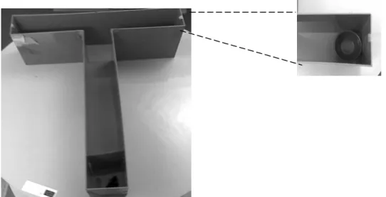
Figure 2.2 Anesthetic setup for in vivo experiments. ………... 27
Figure 2.3 Open field arena with a mouse. ………..……… 30
Figure 2.4 Mice descending the pole in the vertical pole test. .……….... 30
Figure 2.5 T-maze apparatus ……….... 31
Figure 2.6 Radial arm maze apparatus ………. 33
Figure 2.7 Mouse brain and its stereotaxic coordinates .……….. 35
Figure 2.8 Electrophysiological setup ……….. 36
Figure 2.9 A print screen of the LTP software, with an example of a reanalysis of an experiment of the long-term potentiation, and a schematic example of a signal of field excitatory postsynaptic potential……… 37
Figure 3.1 Timeline of the behavior tests. ……….... 41
Figure 3.2 Number of trials necessary to complete the T-maze task at 28 hours, 1 week and 2 weeks after intraperitoneal injection.……… 43
Figure 3.3 Latency time to turn and completely descend the vertical pole in seconds, four hours after anesthesia. ……….. 44
Figure 3.4 Speed in the open field test induced by different treatment groups… 45
XXI
Figure 4.1 Timeline for the behavior tests protocol. ……… 53
Figure 4.2 Time to turn and completely descend the vertical pole in seconds, 4 and 20 hours after injection………. 55
Figure 4.3 Animals’ speed in the open field after control (saline), 25, 75 or 150
mg/kg of ketamine administration measured during 20 minutes at 24, 48 and 72 hours post-anesthesia. ……….. 56
Figure 4.4 Histopathological and immunohistochemical results. ……… 57
Figure 5.1 Effects of different concentration of ketamine on basal synaptic
transmission. ………... 65
Figure 5.2 Effects of different concentration of ketamine on paired-pulse
facilitation. ……….. 66
Figure 5.3 Effects of ketamine on long-term potentiation induced by high
frequency stimulation. ……… 67
Figure 5.4 Effects of different concentration of medetomidine on basal
synaptic transmission. ………. 74
Figure 5.5 Effects of different concentration of medetomidine on paired-pulse
facilitation. ……….. 75
Figure 5.6 Effects of medetomidine on long-term potentiation induced by high
frequency stimulation. ……… 77
Figure 5.7 Effects of different concentration of ketamine combined with medetomidine on basal synaptic transmission. ………... 83
Figure 5.8 Percentage of inhibition of the fEPSP slope caused by different concentrations of ketamine and medetomidine combined or alone. ... 84
Figure 5.9 Time-course of the effects of increasing cumulative concentrations of ketamine combined with medetomidine on paired pulse
facilitation. ……….. 85
Figure 5.10 Effects of ketamine/medetomidine combination on long-term potentiation induced by high frequency stimulation. ………. 86
Figure 6.1 Effects of ketamine on long-term potentiation induced by high frequency stimulation, 24 hours after intraperitoneal administration of saline solution (control) or ketamine. ………. 92
C
XXII
L
IST OF TABLES
Table 1.1 Description of the major brain structures’ functions……….. 11
Table 2.1 Summary of all the experiments performed………... 24
Table 2.2 Scores of clinical signs used to evaluate depth of anesthesia………… 27
Table 3.1 Hemodynamic and oxygen saturation data……… 43
Table 4.1 Number of reference memory (Ref.) and working memory (Work.) errors in radial-maze test for 12 consecutive days after intraperitoneal injection of saline or different doses of ketamine……….. 55
XXIV
L
IST OF
A
BREVIATIONS
AC adenylate cyclase
aCSF artificial cerebrospinal fluid
AMPA α-amino-3-hydroxy-5-methyl-4-isoxazole-propionate ANOVA analysis of variance
ATP adenosine triphosphate
BDNF brain-derived neurotrophic factor
CA cornu ammonis
Ca2+ calcium
CaMKII calcium/calmodulin-dependent kinase II
cAMP cyclic adenosine monophosphate
cm centimeter
CNS central nervous system
CREB cAMP response element binding protein
DG dentate gyrus
EC50 concentration of a ligand that causes 50% of the maximal response
EC entorhinal cortex
EMEA FDA
European Medicinal Evaluation Agency food and drug administration
fepsp field excitatory postsynaptic transmission
GABA γ-amino butyric acid
H&E hematoxylin–eosin
HFS high frequency stimulation
Ih hyperpolarization-activated currents i.p. intraperitoneal K+ potassium LC locus coeruleus LTP long-term potentiation mf mossy fibers min. minute
XXV
NMDA N-methyl-D-aspartate
NA noradrenaline or norepinephrine
Na+ sodium
Nacl sodium chloride
NAPK mitogen-activated protein kinase
PBS phosphate buffered saline
PKA protein kinase A
PKC protein kinase C
PPF paired-pulse facilitation
pp perforant pathway
PWR pedal withdrawal reflex
Rc recurrent collateral
RR righting reflex
Sc schaffer collateral/commissural axons
SD standard deviation
S second
SUB subicular complex
SVR systemic vascular resistance
1
C
HAPTER
1
-
G
ENERAL INTRODUCTION
1.1A
NESTHESIAOn March 30, 1842, Crawford Long administered the first surgical anesthetic, ether, in Jefferson, Georgia. However, unfortunately, Long did not publish his results until 1849 (Long, 1849) and consequently history remembers October 16, 1846 as the date of the birth of anesthesia and Thomas Morton as the “discoverer of anesthesia”. At the Massachusetts general hospital, Morton gave the first successful public demonstration of general anesthesia. He anesthetized Edward Abbot, so that John Warren could remove a vascular tumor from his neck. John Warren then uttered the famous words, “Gentlemen, this is no humbug” (Jeffrey A. Norton, 2008; Robinson and Toledo, 2012). From that moment on, the use of anesthesia spread around the world and human surgery was revolutionized.
Some years later, in 1850s, G.H. Dadd claimed veterinary anesthesia as an important refinement and a scientific principle (Holland, 1973) and finally animals could also benefit from anesthesia routinely. Vertebrate, including humans, and some invertebrate animals share similar pathways and perception of pain (Smith and Lewin, 2009); so also non-human animals should benefit from analgesics and anesthetics when submitted to painful situations.
Several definitions were described for the concept of “anesthesia” but no consensus exists on its definition till date. Anesthesia has been defined as a continuum or progressive depression of the central nervous system (CNS) (TaskForce, 2006) and its effects are commonly described as the combination of three main components: hypnosis, analgesia and muscle relaxation (figure 1.1).
Figure 1.1 Schematic representation of the three main components of general anesthesia. Hypnosis
Analgesia Muscular
2
To achieve all these effects with a reasonable safety margin, a combination of different drugs are generally used. Moreover, when an anesthetic protocol is performed using a combination of drugs, lowering the dose of each drug is necessary to produce the desired effects and to reduce the risk of side-effects such as hemodynamic instability (Arras et al., 2001). This is the concept of balanced anesthesia, the use of a combination of anesthetics that act synergistically in the desired effects and with reduced side-effects (Tonner, 2005).
Each anesthetic drug may induce one or more components of general anesthesia, depending on the molecular targets to which it was specifically developed and presented in a region of the nervous system (Hemmings et al., 2005).
In this work, two different anesthetic drugs were used: ketamine and medetomidine. When these two drugs are administered in combination, the three components of general anesthesia can be achieved, depending of the concentration used.
1.1.1KETAMINE
Ketamine was first synthesized in 1962 by the Belgian chemist Calvin Stevens at Parke Davis Labs. It was approved by the Food and Drug Administration (FDA) in February of 1970 (Mozayani, 2002). Later the ketamine was also approved by the European Medicinal Evaluation Agency (EMEA). Ketamine has been described as an anesthetic agent that produces a dissociative anesthesia, i.e. produces catalepsy, and analgesia, but not necessarily a common loss of consciousness; the subject seems to be disconnected of the environment (Pender, 1972; Kohrs and Durieux, 1998). These symptoms are the reason why ketamine is also used as a drug of abuse (Morgan et al., 2004).
Nevertheless, recent research continues to clarify different aspects of ketamine pharmacology, and new clinical uses have been suggested for this drug such as its use as an anti-depressant (Berman et al., 2000).
Physicochemical characteristics
Ketamine, 2-(2-chlorophenyl)-2-(methylamino)-cyclohexanone, is available as a racemic mixture in a commercial preparation (Imalgene ®, Ketanest®, Ketaject®, Ketalar®) (Mozayani, 2002). There are two optical isomers of the ketamine: S(+) ketamine and R(-) ketamine (figure 1.2). Clinically, the anesthetic potency of the S(+)-isomer is approximately three to four times more than of the R(-)-isomer, which is attributable to the higher affinity of
CHAPTER 1- GENERAL INTRODUCTION
3
the S(+)-isomer to the phencyclidine binding sites on the N-methyl-D-aspartate (NMDA) receptors (Mozayani, 2002). Ketamine is water- and lipid-soluble, allowing its administration in various routes and providing extensive distribution in the body (Sinner and Graf, 2008). It may be administered by the intravenous, intramuscular, oral, and rectal routes without signs of irritation (Haas and Harper, 1992). In animals it may also be administered by the intraperitoneal (i.p.) route (Cruz et al., 1998).
Figure 1.2 Molecular structure of two isomers of ketamine (ChemDoodle® v5.0, iChemLabs, LLC)
Metabolization and Elimination
Biotransformation of ketamine is mediated by hepatic microsomal enzymes resulting in multiple metabolites. The most important pathway involves N-demethylation of ketamine, by cytochrome P450, to norketamine, an active metabolite with one third of the anesthetic potency of ketamine (Mozayani, 2002). Norketamine is then hydroxylated and conjugated to water soluble compounds that are excreted in the urine (90%) (Mozayani, 2002). Five percent is eliminated in the feces and the remaining 5% are excreted unchanged in the urine (Mozayani, 2002).
Dehydronorketamine is an important breakdown product of ketamine, but it is not an important metabolite in vivo (Reich and Silvay, 1989) (figure 1.3).
4
Mechanisms of action
Pharmacologically, ketamine is classified as a noncompetitive NMDA antagonist, but the full range of effects cannot be explained by this inhibition alone. The actions of ketamine are mediated by NMDA, opioid, monoaminergic and muscarinic cholinergic receptors (Hirota and Lambert, 1996). Moreover, ketamine also interacts with voltage-sensitive Ca2+ channels and, at higher doses, it can block the sodium channels (Hirota and Lambert, 1996). All these interactions contribute to the anesthetic action of ketamine.
More specifically, ketamine produces dissociative anesthesia by blockage of the NMDA receptor channel pore and by inhibiting the phencyclidine binding site. The effects of ketamine on the opiate receptor may contribute to the analgesic state (Hirota and Lambert, 1996) and the interaction between ketamine and muscarinic receptors, in a dose-dependent manner, may contribute to the neuromuscular rigidity (Haas and Harper, 1992).
Clinical uses
Ketamine is frequently used as an anesthetic/analgesic agent in clinical practice and in research with laboratory animals. In clinical practice, it is used in painful diagnostic procedures, obstetrics, in asthmatic patients, when traumatic and hypovolemic shock occur, and in burn situations (Kuznetsova et al., 1984; Adams and Hempelmann, 1990; Ikechebelu et al., 2003). Moreover, it is very important for emergency procedures and when the equipment for drug delivery and patients´ motorization are limited, since ketamine maintain the hemodynamic stability (Copeland and Dillon, 2005). Ketamine is also recommended for post-operative analgesia (Angst and Clark, 2010; Carstensen and Moller, 2010; Trupkovic et al., 2011) and in the treatment of chronic pain (Sinner and Graf, 2008).
In laboratory animals, ketamine is frequently used as part of the anesthetic protocols. It is combined with other drugs in a high variety of surgeries and short procedures (Cruz et al., 1998) such as collection of blood, biopsies, implementation of monitoring devices, aseptic urine collection, among other situations.
Effects on body system
Ketamine stimulates the sympathetic nervous system (Sinner and Graf, 2008). It has potent vasodilatation properties, which increases intracranial pressure and compromises intracranial complacence (Haas and Harper, 1992).
CHAPTER 1- GENERAL INTRODUCTION
5
Regarding cardiovascular system, ketamine appears as an stimulant (Sinner and Graf, 2008). Several investigators have reported increases in heart rate, pulmonary arterial pressure, pulmonary vascular resistance, systemic vascular resistance and in systemic arterial pressure (Greeley et al., 1986; Haas and Harper, 1992).
In respiratory system, ketamine is a mild respiratory depressant with unique properties. It has the ability to maintain functional residual capacity upon induction of anesthesia, thus decreasing the chances of intraoperative hypoxemia (Haas and Harper, 1992). Moreover, it increases lung compliance and decrease airway resistance. Ketamine causes bronchodilation (Sinner and Graf, 2008) and increases salivary and tracheobronchial mucous gland secretions (Haas and Harper, 1992).
Concerning the urinary system, no studies were found about the specific action of anesthetic doses of ketamine in this system. However, in humans, it has been reported that when ketamine is used as recreational drug, it could induce ulcerative cystitis or vesicopathy (Middela and Pearce, 2011; Morgan et al., 2012). Bilateral hydronephrosis and renal papillary necrosis have also been reported in some cases (Morgan et al., 2012).
Regarding gastrointestinal system, ketamine can cause nausea and vomiting. This probably happens because ketamine inhibit neuronal uptake and increase serotonergic activity (Aroni et al., 2009).
1.1.2 MEDETOMIDINE
Medetomidine was developed in the 1970s in Finland and it was approved by the FDA, for veterinary use, in March of 1996. Later, the medetomidine was also approved by the EMEA. It produces sedative and analgesic effects (Sinclair, 2003).
Physicochemical characteristics
Medetomidine, 4-1-[(2,3-dimethylphenyl)ethyl]-1H-imidazole, is found as a commercial preparation (Domitor®, Dormilan®) of a racemic mixture of two steroisomers, medetomidine and levo-medetomidine (figure 1.4) (Sinclair, 2003). The dextro-isomer, dexmedetomidine, is the active isomer of medetomidine formulation and when administered at half the dose induces similar effects to medetomidine. The levo-isomer, levomedetomidine only have pharmacological activity when it is used at very high doses (Savola and Virtanen, 1991).
6
Figure 1.4 Molecular structures of two isomers of medetomidine. (ChemDoodle® v5.0, iChemLabs, LLC)
Metabolization and elimination
Biotransformation of medetomidine occurs mainly in the liver. A metabolic pathway consists in hydroxylation with subsequent glucuronidation, or further oxidation to carboxylic acid (Salonen, 1989).
Most of the metabolites were excreted in urine. Fecal excretion was only significant in the rat (Salonen, 1989).
The major urinary metabolites were the glucuronide of hydroxymedetomidine (about 35% of urinary metabolites) and medetomidine carboxylic acid (about 40%). Unchanged medetomidine represents approximately 1% or 10% of the urinary excretion products, dependent on the dose (Salonen and Eloranta, 1990).
Mechanism of action
Medetomidine is a very potent, efficacious and selective agonist of the α2
-adrenoceptor in the central and peripheral nervous system, that also has affinity for imidazoline receptors (Wikberg et al., 1991). It has an alpha2/alpha1 selectivity ratio of 1620/1
(Savola, 1989; Virtanen et al., 1989).
Thus, the main medetomidine’s mechanism of action is its agonistic activity at the presynaptic alpha-2 adrenergic receptors that results in a decrease in the release of norepinephrine (NA) from adrenergic nerve terminals in the central and peripheral nervous system. (Aggarwal et al., 2001; Sinclair, 2003).
CHAPTER 1- GENERAL INTRODUCTION
7
Clinical uses
Medetomidine is frequently used in veterinary medicine to induce analgesia and sedation (Cullen, 1996). It also produces muscle relaxation and decreases the anesthetic requirements of others injectable or inhalant agents to induce anesthesia (Angelini et al., 2000; Sinclair, 2003).
In laboratory animals, medetomidine may be used to provide chemical restraint and facilitate clinical examinations (Sinclair, 2003). Moreover, it is also used to induce anesthesia for surgical procedures when combined with other drugs (Hu et al., 1992).
Effects on body systems
Medetomidine decreases sympathetic nervous activity and decreases cerebral blood flow and intracranial pressure (Itamoto et al., 2010). Moreover, medetomidine produces bradycardia, hypotension (Cullen, 1996), increases the systemic vascular resistance and reduces cardiac output (Sinclair, 2003). This drop of cardiac output is not due to a direct negative action of medetomidine on myocardial contractility, but it is a consequence of the heart rate’s reduction and increase of systemic vascular resistance (Sinclair, 2003).
Depression of the respiratory rate and an increase of the arterial carbon dioxide tension were observed with medetomidine. Many animals showed marked cyanosis of the skin and mucous membrane after sedation with medetomidine because it causes peripheral vasoconstriction, reducing blood flow to these tissues, consequently decreasing venous oxygen saturation (Sinclair, 2003).
Medetomidine inhibits gastric secretions and reduces gut motility. Additionally, it induces polyuria by decreasing the release of antidiuretic hormone and by the action of medetomidine in renin-angiotensin system (Sinclair, 2003).
1.1.3KETAMINE/MEDETOMIDINE COMBINATION
Ketamine/medetomidine combination is frequently used in many species, including laboratory animals (Nevalainen et al., 1989; Cruz et al., 1998).
The administration of these two drugs combined allows substantial reduction of the dose required for anesthesia of each drug alone, reducing side-effects (Cullen, 1996; Sun et al., 2003). Moreover, this combination provides a good anesthetic plane, with hemodynamic stability and muscle relaxation (Cruz et al., 1998). The positive chronotropic and inotropic
8
actions of ketamine help to oppose the depressant actions of medetomidine on the circulatory system (Raiha et al., 1989; Moens and Fargetton, 1990; Verstegen et al., 1990; Cullen, 1996). However, it was also described that this combination may induce hypotension with significant respiratory depression in animals, mainly in rabbits. Therefore, it is advisable to provide supplemental oxygen (Kili, 2004).
In small laboratory animals (mice and rats), this combination has the additional advantage of being administered by an i.p. injection. This technique is a simple procedure, causing minimal distress to the animal (Jang et al., 2009). Moreover, the equipment cost is low and limited number of staff is necessary (Jang et al., 2009). In addition, this combination is particularly useful in veterinary clinics and in laboratory animals, because it has a specific antagonist atipamezole. This can reverse the deep sedative effect induced by medetomidine, shortening the recovery time (Cruz et al., 1998).
1.1.4ATIPAMEZOLE
Atipamezole (Antisedan®) is a highly potent, selective, and specific antagonist of central and peripheral alpha2-adrenoceptors. It antagonizes all medetomidine´s actions, and so
reverses the undesirable side-effects as well as sedation and analgesia (Hu et al., 1992).
Atipamezole, 4-(2-ethly-2,3-dihydro-1Hinden-2yl)-1H-imidazole, has few effects when administered alone, although some people have noted a hyper-alert state in animals (rats, cats, and guinea pigs) following its use (Flecknell, 1997).
1.2
MEMORYIn current language, memory affords the capacity for conscious recollections about facts and events. However, the memory concept is not easy to define and it is important to recognize that there is more than one kind of memory (Zola-Morgan and Squire, 1993). According to Izquierdo et al., memories can be classified according to their content, duration and nature (Izquierdo et al., 1999). Relative to contents, memory can be divided in explicit/declarative and implicit/nondeclarative memory. Declarative memory is the conscious recall of information, involving evaluation, comparison and inference, while nondeclarative memory is the unconscious recall of information for several tasks such as motor skills
CHAPTER 1- GENERAL INTRODUCTION
9
(Izquierdo et al., 1999). Declarative and nondeclarative memory can be subdivided in more specific types of memories (figure 1.5). The behavioral tests used in this work aimed mainly to evaluate declarative memory.
Declarative memory is frequently replaced by the expression spatial memory, because it is the part of memory responsible for recording information about one's environment and its spatial orientation. Declarative memory not only includes memory for spatial locations, but also memory for word lists, faces, odors, and tactual impressions, among others (Squire and Cave, 1991).
Figure 1.5 Taxonomy of mammalian memory systems. Adapted from (Floel and Cohen, 2007)
Concerning its duration, the memory can be classified as short-term memory or long-term memory. In short-long-term memory, the information develops in a few seconds or minutes and lasts for several hours, while in the long-term memory, the information could be permanent (Izquierdo et al., 1999).
According to its nature, memory can be divided in archival memories and transient memories. The archival memories (also called reference memory) are preserved offline for later use (long and short term memories) and the transient memories are used moment-to-moment, i.e. involves the retention of information for short periods of time, seconds to a few minutes (working memory) (Hodges 1996; Izquierdo et al., 1999). These two types of memory, reference and working memory, were studied in the present work. The performance of the behavioral task radial maze involves working and reference memory. Since the baited arms are always the same for the entire testing, remembering which arms are baited needs the
Memory Declarative Nondeclarative Facts Events Procedural (Skills and Habits) Priming and perceptual learning Simple classical conditioning Nonassociative learning Emotional responses Skeletal responses
10
use of reference memory, while remembering which arms have already been visited during the trial involves working memory.
1.2.1NEUROANATOMY AND MEMORY
First of all, it is important to describe the brain anatomy for a later understanding of which regions are involved in memory and learning.
One possible division of the brain is given by Sternberg (Sternberg., 2006). This author divided the brain in three major regions: forebrain, midbrain and hindbrain. These terms are related to the front-to-back arrangement of structures in the developing embryo, however during the course of development the positions change so that the forebrain is almost a cap on top of the midbrain and hindbrain. Nonetheless, the terms are still used to designate areas of the fully developed brain (Sternberg, 2006).
The forebrain comprises the cerebral cortex, the basal ganglia, the thalamus, the hypothalamus and the limbic system (Sternberg, 2006). The limbic system comprises three central interconnected cerebral structures: the amygdala, the septum, and the hippocampal formation(Sternberg, 2006). The midbrain includes the superior colliculi, the inferior colliculi, the grey matter, red nucleus, substantia nigra, ventral region and the reticular activating system(Sternberg 2006). And finally, the hindbrain comprises the medulla oblongata, pons and cerebellum (Sternberg, 2006).
Over the years, several experiments have been made to establish the relationship between the different structures of brain and their specific functions. Tools such as magnetic resonance imaging, positron emission tomography, electrophysiological recordings and behavioral tests contributed to uncover the function of different regions of the brain (Zola-Morgan and Squire, 1993; Sternberg, 2006). However, we have to keep in mind that the established relationships are nonlinear, because the brain acts as a whole, with several connections between different regions, and so the damage of one region can affect the others.
CHAPTER 1- GENERAL INTRODUCTION
11
Table 1.1 Description of the major brain regions’ functions. Adapted from the book “Cognitive Psychology” (Robert J. Sternberg et al., 2009)
Region of the brain
Major structures within the regions
Functions of the structures
Forebrain
Cerebral cortex Involving in receiving and processing sensory information, cognitive processing, planning and sending motor information.
Basal ganglia Crucial to the function of the motor system
Limbic system (hippocampus, amygdala and septum)
Involved in learning, emotions, and motivation (in particular, the hippocampus influences learning and memory, the amygdala influences anger and aggression, and the septum influences anger and fear)
Thalamus
Primary relay station for sensory information coming into the brain; transmits information to the correct regions of the cerebral cortex thought projection fibers that extend from the thalamus to specific regions of the cortex; comprises several nuclei that receive specific kinds of sensory information and project that information to specific regions of cerebral cortex
Hypothalamus
Controls the endocrine system; controls the autonomic nervous system, such as internal temperature regulation, appetite and thirst regulation, and other key functions; involved in regulation of behavior related to species survival; plays a role in controlling consciousness, involved in emotions, pleasure, pain, and stress reactions.
Mindbrain
Superior colliculi Involved in vision (especially, visual reflexes)
Inferior colliculi Involved in bearing
Reticular activating system Important in controlling consciousness (sleep and arousal), attention, cardiorespiratory function, and movement.
Grey matter, red nucleus, substantia nigra, ventral region
Important in controlling movement
Hindbrain
Cerebellum Essential to balance, coordination and muscle tone.
Pons Involved in consciousness (sleep and arousal); bridges neural transmission from one part of the brain to another;
Medulla oblongata Serves as juncture at which nerves cross from one side of the body to the opposite side of the brain; involved in cardiorespiratory function, digestion and swallowing.
Regarding the memory and learning, hippocampus is one of the most important regions, essentially for declarative memory (Zola-Morgan and Squire, 1993), as showed in
table 1.1. Moreover, interestingly, it was reported that hippocampus has place cells, i.e. cells
12
(O'Keefe and Dostrovsky, 1971). Thus, the cognitive mapping theory postulates that each cell is consigned to a specific location and that the firing pattern of the cells can form a very precise cognitive map about the position of the animal at any given moment (O'Keefe and Dostrovsky, 1971; Jeffery and O'Keefe, 1999).
The hippocampal formation can be divided in four regions: (1) hippocampusproper, which is subdivided in three fields (cornu ammonis (CA) 1, CA2 and CA3), (2) dentate gyrus (DG), (3) subicular complex (SUB), which included 3 fields (subiculum, presubiculum, and parasubiculum) and (4) entorhinal cortex (EC) (figure 1.6 ) (Per Andersen et al., 2007). Although the volume of the hippocampus is about 10 times larger in monkeys than in rats and 100 times larger in humans than in rats, the basic hippocampal architecture is common to all three species (Per Andersen et al., 2007).
Figure 1.6 Line drawing of the rat brain shows the septotemporal and transverse axes of the hippocampal formation. CA1: cornu Ammonis 1, CA2: cornu Ammonis 2, CA3: cornu Ammonis 3, DG: dentate gyrus, pp: perforant pathway, fi: fimbria. Adapted from the book “Hippocampus book” (Per Andersen et al., 2007).
The dentate gyrus is considered to have three layers: the molecular layer, the granule cell layer, and the hilar region characterized by polymorph neurons. The hippocampus (CA1, CA2 and CA3) consists on seven layers: stratum moleculare, stratum lacunosum, stratum radiatum, stratum pyramidal, stratum oriens, alveus and epithelial zone (Cajal, 1909).
CHAPTER 1- GENERAL INTRODUCTION
13
1.2.2MEMORY AND SYNAPTIC PLASTICITY
The brain has the amazing ability to receive inputs and to translate them into memories. It has long been suggested that memory formation in mammalian brain involves modifications in the synaptic connections between neurons (Hebb, 1949). It has been shown that repetitive activation of excitatory synapses in the hippocampus causes an increase in synaptic strength, helping the brain “to learn” (Malenka and Nicoll, 1999). This change in synaptic strength between two neurons is named synaptic plasticity. This term was introduced by Jerzy Konorski to describe the ability of the connection, or synapse, between two neurons that may change in strength in response to either use or disuse of transmission over synaptic pathways (Konorski, 1948).
Nowadays, the activity-dependent synaptic plasticity is considered a cellular mechanism for learning and memory (Bliss and Collingridge, 1993) and most of the excitatory synapses in the hippocampus exhibit various forms of activity dependent of synaptic plasticity. These are generally defined as changes in amplitudes of synaptic potentials that are dependent on the prior activity of the synapse. The different forms are generally distinguished on the basis of their duration or time course. Paired-pulse facilitation (PPF), a form of short-term plasticity, and long-term potentiation (LTP), a form of long-term plasticity, are two different and important forms of synaptic plasticity (Maruki et al., 2001). The first occurs through presynaptic mechanisms and it can be used as a presynaptic index for the probability of neurotransmitter release (Kamiya and Zucker, 1994; Zucker and Regehr, 2002) . It consists in give a pair of pulses (stimuli) to a synaptic pathway and to compare the amplitude or slope of the second excitatory post-synaptic potential to the first one (Schulz et al., 1994). In control conditions the response to a second stimulation is higher than the response provoked by the first pulse/stimulus. Residual presynaptic calcium, left after the first stimulation, is thought to enhance neurotransmitter release in response to the second stimulation.
Long-term potentiation is a long-lasting change in synaptic efficacy and it mainly occurs via postsynaptic mechanisms. It is probably the most studied of all the synaptic plasticity’s processes because of its presumed role in learning and memory (Bliss and Collingridge, 1993). It typically starts when one or more trains of high-frequency stimulation are given to a synaptic pathway. It occurs in several regions of the mammalian brain
14
(Malenka and Nicoll, 1999), but the most studied pathways of LTP are located in the hippocampus, where the main circuit is glutamatergic.
The fundamental information processing pathway in the hippocampal formation is usually considered to be the trisynaptic circuit: the entorhinal cortex sends a perforant path projections to the granule cells of the DG, which sends mossy fibers to the pyramidal cells of the CA3 region, which send Schaffer collaterals to the CA1 pyramidal cells, which then project back to the entorhinal cortex (Tsien et al., 1996; Per Andersen et al., 2007). Moreover, a direct projection from entorhinal cortex to CA1 area (the temporoammonic pathway) and CA3 field of the hippocampus proper (via the perforant pathway), and to the subiculum (via the alvear pathway) are also part of hippocampal processing (Tsien et al., 1996; Per Andersen et al., 2007). Pyramidal cells in CA1 can also project to the subiculum. Both CA1 and the subiculum project back to the deep layers of the entorhinal cortex (figure 1.7) (Tsien et al., 1996; Per Andersen et al., 2007).
Figure 1.7 Connections in the Hippocampal Memory System. The excitatory pathways in the hippocampal formation. EC, entorhinal cortex; DG, dentate gyrus; mf, mossy fibers; pp, perforant path; rc, recurrent collateral axons of the CA3 pyramidal neurons; sc, Schaffer collateral/commissural axons; SUB, subiculum. Adapted from Tsien et al. (Tsien et al., 1996)
1.2.3 NEUROTRANSMITTER SYSTEMS, SYNAPTIC PLASTICITY AND MEMORY FORMATION
Neurotransmitter is “A substance which is released at the end of a nerve fiber by the arrival of a nerve impulse and by diffusing across the synapse or junction effects the transfer of impulse to another nerve fiber (or muscle fiber or some receptor)” (Webster, 2001). Based on this definition, neurotransmitter systems influence the normal functions of CNS such as
CHAPTER 1- GENERAL INTRODUCTION
15
synaptic plasticity (Webster, 2001) that is essential to memory formation (Bliss and Collingridge, 1993).
It was reported that glutamate, followed by γ-amino butyric acid (GABA), dopamine, acetylcholine, serotonin, and noradrenaline are the neurotransmitters with more influence in memory and learning (Myhrer, 2003); and consequently alterations in the normal functioning of the neurotransmitters systems that included neurotransmitters described above can affect the memory.
The main excitatory transmitter of the mammalian CNS is the glutamate (Fonnum, 1984). It activates the three principal types of ionotropic receptors that mediate excitatory synaptic transmission: α-amino-3-hydroxy-5-methyl-isoxazole-propionic acid (AMPA), kainate, and NMDA. These receptors are ligand gated ionic channels that, depending on the subtype, are permeable to the divalent cation Ca2+ (calcium) and to the monovalent cations Na+ (sodium) and K+ (potassium) (Danysz and Parsons, 2003). AMPA receptors participate in synaptic transmission conducting mainly Na+ and K+ ions, and being usually impermeable to Ca2+ (Danysz and Parsons, 2003).Kainate receptors are also involved in synaptic transmission and are mostly permeable to Na+ and K+, with a low conductivity for Ca2+. It induces bidirectional modulation of both excitatory and inhibitory transmission (Huettner, 2003). In contrast, NMDA receptors do not usually contribute to basal synaptic transmission but are very important to synaptic plasticity and, consequently, to memory and learning (Danysz and Parsons, 2003). NMDA receptors are of particular interest in this work, since ketamine, one of the anesthetics studied, binds to it. Ketamine is a non-competitive NMDAR antagonist (NMDA receptor antagonist) that inhibits NMDARs by binding to allosteric sites (Wang and Slikker, 2008). These receptors are involved in a variety of physiological and pathological processes such as memory and learning and acute neuropathologies related to stroke and traumatic injury (Wang and Slikker, 2008). NMDA receptors present high permeability to Ca2+ ions, voltage dependent blockage by Mg2+ (magnesium) ions and a slow gating kinetics (Danysz and Parsons, 2003). Moreover, for activation of NMDA receptors it is required simultaneous binding of glutamate and of the co-agonist glycine (Laube et al., 1997).
The best understood model of memory formation based on synaptic plasticity comprises the arrival of a high frequency signal to a glutamatergic synapse, inducing long-term potentiation, and consequently provoking a massive glutamate release which triggers a sequence of events (Danysz and Parsons, 2003). Firstly, AMPA and kainate receptors are activated, but NMDA receptors remained blocked by Mg2+. The continuous activation of
16
AMPA and kainate receptors leads to a significant influx of Na2+ ions into the post-synaptic cell, which in turn decreases the membrane potential removing the Mg2+ ion from the blocked NMDA receptor (Voglis and Tavernarakis, 2006). At this stage, Ca2+ ions can freely enter in the cell via the NMDA receptor channel and initiate mechanisms that lead to the permanent strengthening of synaptic transmission (figure 1.8), including the insertion of more AMPA receptors at the postsynaptic membrane and an increase in protein syntheses (Voglis and Tavernarakis, 2006).
Figure 1.8 Glutamate receptors and synaptic plasticity. The arrival of a series of impulses at the presynaptic terminal triggers the release of glutamate, which binds to glutamate receptors at the postsynaptic membrane. On activation, AMPA and kainate receptors conduct sodium ions, which initiate postsynaptic depolarization. Membrane potential changes initiate the release of magnesium ions that block NMDA receptors. Calcium influx through NMDA channels sets off a chain of events that establish long-term potentiation. Kainate receptors at the presynaptic end also seem to facilitate synaptic transmission at specific synapses by augmenting neurotransmitter release. AMPA, α-amino-3-hydroxy-5-methyl-4-isoxazolepropionate; CaMKII, calcium/calmodulin-dependent kinase II; CREB, cAMP response element binding protein; MAPK, mitogen-activated protein kinase; NMDA, N-methyl-D-aspartate; PKA, protein kinase A; PKC, protein kinase C. Figure and legend from Giannis Voglis and Nektarios Tavernarakis (Voglis and Tavernarakis, 2006).
However, it is important to stress that the model described above is not unique and the situation is actually far more complex with many forms of synaptic plasticity induced by diverse inputs in different neurons. Moreover, synaptic plasticity in the hippocampus does not always depend on NMDA receptors. The synapses between mossy fibers of hippocampal DG and CA3 neurons show a distinctive morphology with many sites for glutamate release, each of which has a corresponding postsynaptic bundle of glutamate receptors (Voglis and
CHAPTER 1- GENERAL INTRODUCTION
17
Tavernarakis, 2006). At this case, LTP is NMDA-receptor-independent and requires calcium influx though presynaptic kainate receptors (Lauri et al., 2003). However, the mechanism responsible for this type of synaptic plasticity remains unclear (Voglis and Tavernarakis, 2006).
Other neurotransmitter that is important to the synaptic plasticity in hippocampus is noradrenaline (NA). It acts on the neurons through modifications of currently ongoing events (modulation) and also initiates synaptic plasticity by itself (Marzo et al., 2009). NA has high affinity to bind α1-adrenoceptors, α2-adrenoceptors and β-adrenoceptors and consequently
modulates synaptic plasticity (Myhrer, 2003; Marzo et al., 2009). The α2-adrenoceptors are
particularly important to this work because medetomidine, one of the anesthetics studied in this work, binds to α2-adrenoceptors and so it can alter the normal functioning of the
noradrenergic system and alter the synaptic plasticity.
Alpha2-adrenoceptors are located on both pre- and postsynaptic sites (Marzo et al.,
2009). The presynaptic localization indicates their functions as autoceptors, involved in the control of noradrenaline release by locus coeruleus neuronal axons (Marzo et al., 2009). The postsynaptic localization indicates their involvement in the intrinsic plasticity, i.e. the adaptive alterations of postsynaptic excitability, which are non-synaptic in nature and thus mechanistically internal to a given postsynaptic cell (Marzo et al., 2009). This type of plasticity is crucial for neuronal function given that it directly modulates cellular characteristics such as ion channel opening (Marzo et al., 2009).
As it was reported by Marzo et al., noradrenaline activates a postsynaptic adrenoceptors and consequently a postsynaptic membrane hyperpolarization by intrinsic plasticity can occur. This hyperpolarization can be associated with decrease or increase in input resistance, dependently on the via (Marzo et al., 2009). The decrease in input resistance may derive from the modulation of potassium channels, a decrease in cyclic adenosine monophosphate levels (cAMP) or an opening of adenosine triphosphate (ATP)-dependent potassium channels. The increase in the input resistance may result from the block of the hyperpolarization-activated currents (Ih). In addition, it was reported that α2-adrenoceptor
agonists inhibit voltage-activated calcium currents mediated by the N- or P-type calcium-channels (figure 1.9) (Marzo et al., 2009).