VERA PATRÍCIA RIBEIRO DE CASTRO
Orientadores
Vila Real, 2014
Dissertação de Mestrado em Biologia
neuroprotective effect of plant extracts from Caatinga biome (Brazil)
Caracterização fitoquímica, propriedades antioxidantes e efeitos neuroprotetores
de extratos de plantas provenientes do bioma Caatinga (Brasil)
Alberto Carlos Pires Dias Maria José Félix Saavedra
Phytochemical characterization, antioxidant properties and
neuroprotective effect of plant extracts from Caatinga biome
(Brazil)
Caracterização fitoquímica, propriedades antioxidantes e efeitos
neuroprotetores de extratos de plantas provenientes do bioma Caatinga
(Brasil)
Dissertação de mestrado em Biologia
Vera Patrícia Ribeiro de Castro
Orientadores
Composição do Júri
Alberto Carlos Pires Dias Maria José Félix Saavedra
Universidade de Trás-os-Montes e Alto Douro
Instituição: Universidade de Trás-os-Montes e Alto Douro Curso: Mestrado em Biologia
Título: “Phytochemical characterization, antioxidant properties and neuroprotective effect of plant extracts from Caatinga biome (Brazil)”
Autor: Vera Patrícia Ribeiro de Castro Orientador: Alberto Carlos Pires Dias Co-orientador: Maria José Félix Saavedra
“As doutrinas apresentadas neste trabalho são da exclusiva responsabilidade do autor.”
Este trabalho foi expressamente elaborado como dissertação original para o efeito de obtenção do grau de Mestre em Biologia
VERA CASTRO vi
A
GRADECIMENTOS
Este trabalho não seria possível sem o apoio de várias pessoas às quais não podia deixar de agradecer.
Começo por expressar o meu agradecimento ao Professor Doutor Alberto Dias pelos conhecimentos transmitidos e o apoio prestado ao longo deste trabalho. Agradeço também a paciência, a amizade, a comida, as gargalhadas e a boa disposição ao longo deste percurso. Foi um período de grande aprendizagem e sucesso, obrigada por me receber, e afinal de contas trabalhar com mulheres nem sempre é complicado.
À Professora Doutora Maria José Saavedra, que apesar da distância sempre me apoiou. À Professora Doutora Olga Coutinho, agradeço toda ajuda, paciência e principalmente todos os conhecimentos transmitidos. Agradeço ainda a disponibilidade que demonstrou para me ajudar na realização de uma parte do meu trabalho.
Ao Doutor Alfredo agradeço por toda a ajuda e apoio, agradeço também a amizade, ensinamentos e paciência.
Aos funcionários do Departamento de Biologia da Universidade do Minho, o meu agradecimento pelas facilidades concedidas para a realização deste trabalho.
Ao Luís, “O colombiano”, por todo o apoio, companheirismo, amizade, risota e todas as conversas que eram quase tudo menos científicas, agradeço assim por teres feito parte deste percurso.
Ao Alexandre, Nuno e Sara, agradeço pelas conversas e companhia na hora do almoço, foram momentos de grande risota.
Agradeço também às papoilas Ana e Dandara, que se revelaram umas grandes amigas, não pensem que só fui eu que vos transmiti conhecimentos, também aprendi muito convosco, principalmente a não deixar de sorrir.
Aos colegas de departamento e laboratório, Sofia, Tatiana, António, não menosprezando os restantes, obrigada por toda a ajuda ao longo deste percurso.
À Marília, Sofia, Jeremy e Carla, grandes companheiros de universidade, que apesar de longe estarão sempre bem perto.
À Luciana, Vânia e Flávia, que mesmo distantes, sempre me apoiaram.
Aos meus pais, não tenho palavras para agradecer todo o sacrifício e dedicação que têm por mim, sempre lutaram para que eu conseguisse chegar onde cheguei. Agradeço todo o
VERA CASTRO vii esforço, sacrifício, afeto e amor que sempre me ofereceram, sem vocês eu não estaria a concluir mais uma etapa no meu percurso académico.
Ao meu irmão agradeço o apoio e dedicação ao longo destes anos, apesar de vários percalços nas nossas vidas nunca deixou de me apoiar.
À minha linda sobrinha Lara, agradeço todas as alegrias, os miminhos e beijinhos que me dás todos os dias, o teu sorriso me conforta e me encoraja a seguir em frente, és a luz dos meus olhos.
A toda a minha família, mas principalmente à minha querida avó Aninhas, que apesar de estar a passar por momentos difíceis sempre me apoiou e às minhas tias Carlinda e Josefa sois umas “fixes.
E por fim, mas não menos importante, agradeço ao meu namorado e companheiro Paulo, pelo apoio incondicional, pela grande amizade, carinho e amor que me dedica todos os dias, pelo esforço e afeto, por todos os momentos vividos juntos, por me ajudar a ultrapassar as dificuldades e obstáculos, enfim obrigada por tudo.
VERA CASTRO viii
“If you can’t fly, then run,
If you can’t run, then walk,
If you can’t walk, then crawl,
But whatever you do,
You have to keep moving for”.
Martin Luther King
“Porque eu sou
Do tamanho do que vejo
E não, do tamanho da
Minha altura”
Alberto Caeiro
VERA CASTRO ix
P
UBLICATIONS
V Castro, A Aires and A Dias., 2014. Phytochemical characterization and screening of in
vitro antioxidant proprieties of extracts obtained from plants of Caatinga Biome (Brazil). GA 2014 – 62nd International Congress and Annual Meeting of the Society for Medicinal Plant and Natural Product Research August 31 – September 4, 2014. Abstract published on Planta Med 2014. DOI: 10.1055/s-0034-1394987
V Castro, S Duarte, OP Coutinho and A Dias., 2014. Leaf extracts of plants used in folk
medicine in northeastern Brazil revealed neuroprotective effect in cells under conditions of oxidative stress. GA 2014 – 62nd International Congress and Annual Meeting of the Society for Medicinal Plant and Natural Product Research August 31 – September 4, 2014. Abstract published on Planta Med 2014. DOI: 10.1055/s-0034-1394989
L Giraldo, V Castro, F Gregory and ACP Dias., 2014. Potential of Ocimum sanctum L. cell suspensions for rosmarinic acid production. GA 2014 – 62nd International Congress and Annual Meeting of the Society for Medicinal Plant and Natural Product Research August 31 – September 4, 2014. Abstract published on Planta Med 2014. DOI: 10.1055/s-0034-1394838
V Castro, A Aires, OP Coutinho , A Dias (2014). Extratos de plantas provenientes do
nordeste do Brasil revelam potencial antioxidante e efeitos neuroprotetores. Ciclo de conferências das VIII Jornadas de Biologia, UTAD, Vila Real, 22 e 23 de Outubro de 2014
Vera Castro, Alfredo Aires, Olga P Coutinho and Alberto CP Dias., 2014. Antioxidant proprieties and neuroprotector potential of selected leaf extracts of plants from Brazilian Caatinga biome. VVIII Congress of the Portuguese Biochemical Society December 17-20, 2014
VERA CASTRO x
Caracterização fitoquímica, propriedades antioxidantes e efeitos
neuroprotetores de extratos de plantas provenientes do bioma Caatinga
(Brasil)
R
ESUMO As doenças neurodegenerativas têm sido um foco da ciência com o objetivo de desenvolver novas estratégias neuroprotetoras capazes de prevenir a morte neuronal associada a estas patologias. A identificação de plantas medicinais tem sido alvo de vários investigadores com a finalidade de encontrar novas fontes de compostos com propriedades farmacológicas. O Brasil, nomeadamente o bioma Caatinga, é rico em plantas medicinais sendo estas utilizadas pelos habitantes locais na medicina tradicional.Tendo isto em conta, os objetivos deste trabalho foram: 1) caracterizar o perfil fitoquímico e conteúdo em compostos fenólicos, de extratos provenientes de plantas presentes neste bioma; 2) avaliar as propriedades antioxidantes in vitro destes extratos; 3) explorar o potencial antioxidante e neuroprotetor destes extratos em modelo celular.
Os extratos das folhas das plantas selecionadas para este estudo mostraram (HPLC-DAD) elevados níveis de compostos fenólicos, nomeadamente ácido gálico, caempferol, quercetina e seus derivados, catequinas entre outros. Extratos provenientes de Sp 3 (Caesalpinia ferrea), 4 (Anadenanthera peregrina), 6 (Mimosa tenuiflora) e 9 (Schinopsis brasiliensis) revelaram o maior conteúdo em compostos fenólicos.
Tendo em conta os EC50 obtidos para todos os ensaios, os extratos provenientes de Sp 3 e 9 mostraram a maior capacidade scavenging de DPPH; 4 e 18 (Cleome spinosa) mostraram a maior capacidade quelante de ferro; 4, 6 e 14 (Capparis flexuosa) revelaram a maior capacidade de inibição de produção de NO; e 6 e 3 mostraram a melhor capacidade scavenging de superóxido. Portanto, Sp 3, 4, 6, 9, 14 e 18 parecem ser os mais promissores. Assim, observou-se que as propriedades antioxidantes demonstradas por estes extratos poderão estar relacionadas com o seu conteúdo em compostos fenólicos.
VERA CASTRO xi
Revelou-se importante estudar as propriedades neuroprotetras destes extratos usando o modelo neuronal U251, exposto ao agente oxidante t-BHP, de modo a induzir stress oxidativo nestas células. Os resultados revelaram que os extratos que se mostraram ser mais promissores nos ensaios anteriores, nomeadamente no conteúdo em compostos fenólicos e potencial antioxidante demonstrado, Sp 3, 4, 6 e 9, induziram maior protecção nas células, em condições de stress oxidativo. Estes resultados sugerem assim que, provavelmente, o elevado conteúdo em compostos fenólicos poderá induzir, por algum mecanismo, a proteção celular.
O passo seguinte consistiu em avaliar quais as condições de incubação que poderiam potenciar o efeito neuroprotetor dos extratos. Os resultados mostraram que, para a maioria dos extratos, nomeadamente, Sp 3, 4 e 6, a pré-incubação destes (em que o extrato atua sozinho) seguido da co-incubação do agente oxidante t-BHP, potencia o efeito neuroprotetor, o que sugere uma ação profilática, isto é, o extrato atua como medida preventiva contra o dano. Portanto, a proteção celular é aumentada quando fornecemos às células os compostos bioativos, que provavelmente induzem o aumento de defesas nas células, antes do dano oxidativo ocorrer.
Assim, os nossos estudos suportam a ideia de que as plantas do bioma Caatinga têm diversas potencialidades podendo assim ser usadas como fonte de compostos com propriedades farmacológicas, sendo que os extratos estudados parecem possuir propriedades antioxidantes e neuroprotetoras contra processos neurodegenerativos associados a stress oxidativo.
Palavras-chave: Extratos de plantas; compostos fenólicos; stress oxidativo;
VERA CASTRO xii
Phytochemical
characterization,
antioxidant
proprieties
and
neuroprotective effect of plant extracts from Caatinga biome (Brazil)
A
BSTRACT Neurodegenerative diseases have been a focus of science with the aim of developing new neuroprotective strategies capable of preventing neuronal death associated with these pathologies. The identification of medicinal plants has been the target of various researchers in order to find new sources of compounds with pharmacological properties. Brazil, namely Caatinga biome, is rich in medicinal plants and these are used by locals in traditional medicine.Concerning this, the objectives of this study were: 1) characterize the phytochemical profile and content in phenolic compounds, from plant extracts present in this biome; 2) evaluate the in vitro antioxidant properties of these extracts; 3) explore the antioxidant and neuroprotective potential of these extracts on cellular model.
The plant leaves extracts selected for this study showed (HPLC-DAD) high levels of phenolic compounds, including gallic acid, kaempferol, quercetin and derivatives, catechins and others. Extracts from Sp 3 (Caesalpinia ferrea), 4 (Anadenanthera peregrina), 6 (Mimosa tenuiflora) and 9 (Schinopsis brasiliensis) revealed the highest content in phenolic compounds.
Given the EC50 obtained for all asays, extracts from Sp 3 and 9 showed the greatest ability to DPPH scavenging; 4 and 18 (Cleome spinosa) showed the greatest ability to chelate iron; 4, 6 and 14 (Capparis flexuosa) showed the greatest capacity of inhibiting NO production; and 6 and 3 showed better superoxide scavenging capacity. So, Sp 3, 4, 6, 9, 14 and 18 seems to be the most promising. It was found that the antioxidant properties shown by these extracts may be related to its content in phenolic compounds.
It has important to access the neuropropertive properties of these extracts using a neuronal model U251 exposed to the oxidant t-BHP, to induce oxidative stress in these cells. The results revealed that the extracts that proved to be most promising in previous studies, including the content of phenolics and antioxidant activity demonstrated, namely Sp 3, 4, 6 and 9, induced protection in conditions of oxidative
VERA CASTRO xiii
stress. These results suggest that, probably, the high content of phenolic compounds may induce, by some mechanism, cell protection.
The next step was to evaluate which incubation conditions could potentiate the neuroprotective effect of the extracts. The results showed that for the most of the extracts, namely, Sp 3, 4 and 6, the pre-incubation of these (where the extract acts alone) followed by co-incubation of t-BHP, oxidizing agent, enhances the neuroprotective effect, suggesting a prophylactic action, i.e., the extract serves as a preventative measure against damage. Therefore, the cell protection is increased when we provide the bioactive compounds to the cells, which probably induce increased the cell defenses, before oxidative damage occurs.
Thus, our studies support the idea that Caatinga biome plants have several different potentialities that can be used as a source of compounds with pharmacological properties, and these extracts seems to possess antioxidant and neuroprotective properties against neurodegenerative diseases associated with oxidative stress.
Keywords: leaf extracts; phenolic compounds; oxidative stress; antioxidante
VERA CASTRO xiv
I
NDEX
A
GRADECIMENTOS ... VIP
UBLICATIONS ... IXR
ESUMO ... XA
BSTRACT ... XIII
NDEX ... XIVF
IGURE INDEX ... XVIIT
ABLE INDEX ... XXA
BBREVIATIONS ... XXIC
HAPTER1:
G
ENERAL INTRODUCTION... XXIII1.1.MEDICINAL PLANTS ... 24
1.2.FOLK MEDICINE IN BRAZIL ... 25
1.2.1.VEGETATION FROM NORTHEASTERN BRAZIL:CAATINGA BIOME ... 26
1.2.1.1.MEDICINAL PLANTS FROM CAATINGA AND THEIR PHARMACOLOGICAL ACTIVITY ... 27
1.3.NERVOUS SYSTEM ... 27 1.3.1. ASTROCYTES ... 30 1.3.2. OLIGODENDROCYTES ... 30 1.3.4. MICROGLIA ... 30 1.3.5. EPENDYMAL CELLS ... 31 1.4.NEURODEGENERATION ... 32 1.5.OXIDATIVE STRESS ... 32
1.5.1. FORMATION OF REACTIVE SPECIES ... 33
1.5.2. EFFECTS OF REACTIVE SPECIES ... 34
1.6.ANTIOXIDANTS AS PROTECTORS OF OXIDATIVE STRESS ... 35
VERA CASTRO xv
C
HAPTER2:
PHYTOCHEMICAL CHARACTERIZATION OF METHANOLIC EXTRACTS FROMPLANTS BY HPLC
-
DAD ... 382.1.INTRODUCTION... 39
2.2.MATERIAL AND METHODS ... 40
2.2.1.PLANT MATERIAL ... 40
2.2.2. SEPARATION AND IDENTIFICATION OF BIOACTIVE COMPOUNDS OF THE EXTRACTS BY HIGH PERFORMANCE LIQUID CHROMATOGRAPHY (HPLC)... 41
2.3.RESULTS AND DISCUSSION ... 42
2.3.1.COMPOSITION AND QUANTIFICATION OF PHENOLIC COMPOUND PRESENT ON PLANT EXTRACTS ... 42
C
HAPTER3:
SCREENING OF IN VITRO ANTIOXIDANT PROPRIETIES OF PLANT EXTRACTS .... 513.1.INTRODUCTION... 52
3.2.MATERIAL AND METHODS ... 53
3.2.1.PLANT MATERIAL ... 53
3.2.2.PREPARATION OF METHANOLIC EXTRACTS ... 53
3.2.3.IN VITRO ANTIOXIDANT PROPRIETIES ... 54
3.2.3.1.DPPH ASSAY ... 54
3.2.3.2.IRON CHELATING ACTIVITY TEST ... 56
3.2.3.3. INHIBITION OF NITRIC OXIDE PRODUCTION ... 57
3.2.3.4.SUPEROXIDE SCAVENGING ACITIVY ... 59
3.2.4.STATISTICAL ANALYSIS ... 60
3.3.RESULTS AND DISCUSSION ... 60
3.3.1.DPPH ASSAY ... 61
3.3.2.IRON CHELATING ACTIVITY ... 63
3.3.3.INHIBITION OF NITRIC OXIDE PRODUCTION... 65
3.3.4.SUPEROXIDE SCAVENGING ACTIVITY ... 66
3.4.CONCLUSIONS ... 68
C
HAPTER4:
NEUROPROTECTIVE EFFECT OF PLANT EXTRACTS,
IN A NEURONAL CELL MODEL ... 714.1.INTRODUCTION... 72
VERA CASTRO xvi
4.2.1.PLANT MATERIAL ... 73
4.2.2.PREPARATION OF ETHANOLIC EXTRACTS ... 74
4.2.3.U251 CELL CULTURE ... 74
4.2.4.CELL VIABILITY ... 75
4.2.5.NEUROPROTECTIVE EFFECT OF PLANT EXTRACTS ... 75
4.2.6.TYPES OF INCUBATIONS USED IS THIS STUDY ... 75
4.2.6.1.CO-INCUBATION WITH EXTRACT AND OXIDANT AGENT ... 76
4.2.6.2.PRE-INCUBATION WITH EXTRACT FOLLOWED BY POST-INCUBATION OF OXIDANT AGENT ... 76
4.2.6.3.PRE-INCUBATION WITH EXTRACT FOLLOWED BY CO-INCUBATION OF OXIDANT AGENT . 77 4.2.7.STATISTICAL ANALYSIS ... 77
4.3.RESULTS AND DISCUSSION ... 77
4.3.1.EVALUATION OF POSSIBLE TOXICITY OF PLANT EXTRACTS IN U251 CELLS ... 78
4.3.1.1.CYTOTOXICITY OF PLANT EXTRACTS WITH 2H OF INCUBATION ... 78
4.3.1.2.CYTOTOXICITY OF PLANT EXTRACTS WITH 3H OF INCUBATION ... 78
4.3.1.3.CYTOTOXICITY OF PLANT EXTRACTS WITH 5H OF INCUBATION ... 79
4.3.2.EFFECT OF T-BHP IN U251 CELLS ... 83
4.3.3.NEUROPROTECTIVE EFFECT OF PLANT EXTRACTS UNDER CONDITIONS OF OXIDATIVE STRESS 84 4.3.3.1CO-INCUBATION WITH EXTRACT AND OXIDANT AGENT ... 84
4.3.3.2PRE-INCUBATION WITH EXTRACT FOLLOWED BY POST-INCUBATION OF OXIDANT AGENT 84 4.3.3.3PRE-INCUBATION WITH EXTRACT FOLLOWED BY CO-INCUBATION OF OXIDANT AGENT .. 85
4.3.4.COMPARISON BETWEEN ALL TYPES OF INCUBATION OF THE NEUROPROTECTIVE EFFECT OF THE PLANT EXTRACTS AT 75 µG DWR/ML ... 90
C
HAPTER5:
F
INAL REMARKS ... 935.1.GENERAL DISCUSSION ... 94
5.2.FUTURE PROSPECTS ... 96
VERA CASTRO xvii
F
IGURE INDEX
FIG.1.1-LOCALIZATION ON MAP OF CAATINGA BIOME (ORANGE) ... 26
FIG.1.2-CNS CELLS. ... 31
FIG.1.3-FORMATION OF HYDROXYL RADICAL ... 33
FIG.1.4-FENTON REACTION ... 34
FIG.1.5-PEROXYNITRITE ANIONS FORMATION ... 34
FIG.2.1-CLASSIFICATION OF CERTAIN PHENOLIC COMPOUNDS PRESENT IN VARIOUS MEDICINAL PLANTS ... 39
FIG.2.2 -HPLC CHROMATOGRAM (280 NM) OF CAESALPINIA FERREA (SP 3) METHANOLIC EXTRACT. ERRO!MARCADOR NÃO DEFINIDO.FIG.2.3-HPLC CHROMATOGRAM (280 NM) OF ANADENANTHERA PEREGRINA (SP 4) METHANOLIC EXTRACT ...ERRO!MARCADOR NÃO DEFINIDO. FIG.2.4-HPLC CHROMATOGRAM (280 NM) OF MIMOSA TENUIFLORA (SP 6) METHANOLIC EXTRACT. 44 FIG. 2.5 - HPLC CHROMATOGRAM (320 NM) OF SIDEROXYLON OBTUSIFOLIUM (SP 8) METHANOLIC EXTRACT. ... 44
FIG.2.6-HPLC CHROMATOGRAM (280 NM) OF SCHINOPSIS BRASILIENSIS (SP 9) METHANOLIC EXTRACT. ... 45
FIG.2.7-HPLC CHROMATOGRAM (280 NM) OF ZIZIPHUS JOAZEIRO (SP 11) METHANOLIC EXTRACT. . 45
FIG.2.8-HPLC CHROMATOGRAM (280 NM) OF MAYTENUS RIGIDA (SP 12) METHANOLIC EXTRACT. L 46 FIG.2.9-HPLC CHROMATOGRAM (280 NM) OF CAPPARIS FLEXUOSA (SP 14) METHANOLIC EXTRACT. 46 FIG. 2.10 - HPLC CHROMATOGRAM (370 NM) OF ERYTHRINA MULUNGU (SP 15) METHANOLIC EXTRACT. ... 47
FIG.2.11-HPLC CHROMATOGRAM (280 NM) OF EUPHORBIA TIRUCALLI (SP 17) METHANOLIC EXTRACT. ... 47
VERA CASTRO xviii
FIG.3.1–APPEARANCE OF DPPH ASSAY IN MICROPLATE ... 55
FIG.3.2–APPEARANCE OF IRON CHELATING ACTIVITY ASSAY IN MICROPLATE ... 56
FIG.3.3–APPEARANCE OF INHIBITION OF NITRIC OXIDE ASSAY IN MICROPLATE ... 58
FIG.3.4–APPEARANCE OF SUPEROXIDE PRODUCTION ASSAY IN MICROPLATE ... 59
FIG.3.5-SCHEMATIC REPRESENTATION OF THE RESULTS ... 68
FIG.4.1 - A-PLANT EXTRACTS USED IN THIS ASSAY;B- FORMAZAN CRYSTALS OBTAINED FROM MTT REDUCTION TEST USED AS VIABILITY TEST ... 74
FIG.4.2-SCHEMATIC REPRESENTATION OF THE DIFFERENT INCUBATIONS USED IN THIS STUDY ... 75
FIG.4.3-SCHEMATIC REPRESENTATION OF THE ASSAY IN CO-INCUBATION CONDITION... 76
FIG. 4.4 - SCHEMATIC REPRESENTATION OF THE ASSAY IN PRE-INCUBATION + POST INCUBATION CONDITION ... 76
FIG.4.5-SCHEMATIC REPRESENTATION OF THE ASSAY IN PRE-INCUBATION +CO-INCUBATION CONDITION ... 77
FIG. 4.6 - CELL VIABILITY ASSESSED BY MTT REDUCTION TEST. U251 CELLS WERE INCUBATED WITH DIFFERENT CONCENTRATIONS OF PLANT EXTRACTS DURING 2H. ... 80
FIG. 4.7 - CELL VIABILITY ASSESSED BY MTT REDUCTION TEST. U251 CELLS WERE INCUBATED WITH DIFFERENT CONCENTRATIONS OF PLANT EXTRACTS DURING 3H. ... 81
FIG. 4.8 - CELL VIABILITY ASSESSED BY MTT REDUCTION TEST. U251 CELLS WERE INCUBATED WITH DIFFERENT CONCENTRATIONS OF PLANT EXTRACTS DURING 5H. ... 82
FIG.4.9-CELL VIABILITY ASSESSED BY MTT REDUCTION TEST. ... 83
FIG.4.10 -PLANT EXTRACTS PROTECTION AGAINST T-BHP-INDUCED OXIDATIVE STRESS EVALUATED BY MTT REDUCTION TEST. U251 CELLS WERE INCUBATED WITH DIFFERENT CONCENTRATIONS OF PLANT EXTRACTS AND T-BHP1 MM DURING 3H.. ... 87
FIG.4.11 -PLANT EXTRACTS PROTECTION AGAINST T-BHP-INDUCED OXIDATIVE STRESS EVALUATED BY MTT REDUCTION TEST.U251 CELLS WERE PRE-INCUBATED WITH DIFFERENT CONCENTRATIONS OF PLANT EXTRACTS DURING 2H ACTING ALONE, THAN T-BHP1 MM WERE POST-INCUBATED DURING 3H ACTING ALONE.. ... 88
VERA CASTRO xix
FIG.4.12 -PLANT EXTRACTS PROTECTION AGAINST T-BHP-INDUCED OXIDATIVE STRESS EVALUATED BY
MTT REDUCTION TEST.U251 CELLS WERE PRE-INCUBATED WITH DIFFERENT CONCENTRATIONS OF PLANT EXTRACTS DURING 2H ACTING ALONE, THAN T-BHP1 MM WERE CO-INCUBATED DURING 3H WITH THE EXTRACTS.. ... 89
FIG.4.13-COMPARISON OF THE NEUROPROTECTIVE EFFECT OF THE PLANT EXTRACTS AT 75 µG DWR/ML BETWEEN DIFFERENT TYPES OF INCUBATION USED IN THIS STUDY. ... 92
VERA CASTRO xx
T
ABLE INDEX
TABLE 1.1-PLANTS DESCRIPTION AND ITS PHARMACOLOGICAL ACTIVITIES ... 28
TABLE 2.1-IDENTIFICATION OF PLANT SPECIES USED IN THIS WORK ... 40
TABLE 2.2-ELUENT GRADIENT OF SOLVENT A AND B FOR CHROMATOGRAFIC SEPARATION ... 41
TABLE 2.3- PHENOLIC COMPOSITION AND TOTAL PHENOLIC CONTENT OF PLANT EXTRACTS ... 49
TABLE 3.1-REACTIVE OXYGEN SPECIES AND SOME OF THEIR PROPERTIES AND REACTIVITIES ... 52
TABLE 3.2-IDENTIFICATION OF PLANT SPECIES USED IN THIS WORK ... 54
TABLE 3.3-DPPH ASSAY RESULTS ... 62
TABLE 3.4-IRON CHELATING ACTIVITY ASSAY RESULTS ... 64
TABLE 3.5-INHIBITION OF NITRIC OXIDE PRODUCTION ASSAY RESULTS ... 66
TABLE 3.6-SUPEROXIDE SCAVENGING ACTIVITY OF PLANT EXTRACTS RESULTS ... 68
VERA CASTRO xxi
A
BBREVIATIONS
CV – Cell viability
DMEM - Dulbecco’s modified Eagle’s medium DMSO – Dimethylsulphoxide
DNA – Desoxyribonucleic acid
DPPH – 1,1-diphenyl-2-picrylhydrazyl dwb – Dry weight biomass
dwr – Dry weight residue
Fz – Ferrozine (3-(2-pyridyl)-5,6.diphenyl-1,2,4-triazine-4´,4’-disulphonic acid sodium
salt)
HPLC-DAD – High performance liquid chromatography diode array detection H2O2 – Hydrogen peroxide
HOO• - Hydroperoxyl radical OH• – Hydroxyl radical ICA – Iron chelating activity
MTT - 3-(4,5-dimethylthiazol-2-yl)-2,5-diphenyltetrazolium bromide
Na2EDTA Titriplex III - ethylenedinitrilotetraacetic acid disodium salt dihydrate
NADH – Nicotine adenine dinucleotide, reduced form NBT - Nitroblue tetrazolium chloride
NE - Na2EDTA equivalents
NED - N-(1-naphthyl)ethylenediamine •NO – Nitric oxide
•NO2 - Nitrogen dioxide
ONOO− - Peroxynitrite anions PMS - Phenazine methosulfate RNS – Reactive nitrogen species ROS – Reactive oxygen species SNP – Sodium nitroprusside Sp 1 – Spondias tuberosa Sp 2 – Caesalpinia pyramidalis Sp 3 – Caesalpinia ferrea
VERA CASTRO xxii Sp 4 – Anadenanthera colubrina Sp 5 – Momordica charantia Sp 6 – Mimosa tenuiflora Sp 7 – Cydonia oblonga Sp 8 – Sideroxylon obtusifolium Sp 9 – Schinopsis brasiliensis Sp 11 – Ziziphus joazeiro Sp 12 – Maytenus rigida Sp 14 – Capparis flexuosa Sp 15 – Erythrina mulungu Sp 16 – Peltophorum dubium Sp 17 – Euphorbia tirucalli Sp 18 - Cleome spinosa O2•− – Superoxide anion t-BHP – Tert-butylhydroperoxide TE - Trolox equivalents
TNFα – Tumor necrosis factor α
C
HAPTER
1
VERA CASTRO 24
1.1. M
EDICINAL PLANTSSeveral plants and their extracts have been used for many years as treatments for diseases (e.g. headaches or parasite infections), but only in the past 2-3 decades, scientists seriously begun to establish if plant-derived traditional drugs are effective or their mode of action (Vieira et al., 2008).
Animal-, mineral-, and plant natural products have provided the main source of drugs for the humans. Medicinal plants are one of the greatest sources of bioactive molecules and it is important to supply chemical industry related to medicine, cosmetics, foods and agrochemicals (Zanin et al., 2012).
Ethnopharmacological and ethnobotanical studies have identified many plant species that show biological activity and the target is the identification of bioactive plant compounds. The strategy used in the identification of these species is the traditional knowledge which open numerous options for analyzing data by species ordination and giving priority to certain plants for further analysis (Araujo et al., 2008; Júnior et al., 2011).
There are many reasons for study medicinal plants: (1) to improve knowledge about the medicinal potential of native plant diversity; (2) to make a rational basis for the medicinal use of plant species; (3) to develop herbal medicines that are low-cost and demonstrate significant activity; (4) to find new prototypes for drugs with high activity; and (5) to increase information in relation to traditional medicines (Vieira et al., 2008; Zanin et al., 2012).
Most of the information concerning folk medicine, namely in Brazil, has not been verified by rigorous scientific studies (Gazzaneo et al., 2005; Lima et al., 2006; Lucena et al., 2007). Nowadays, although, the recognition of traditional medicine as an alternative form of health care has increased between all socio-economic groups of the population and phytomedicine has become an important economic sector in Brazil (Lima et al., 2006). Thus, the study of the medicinal plants has been a focus of science in order to validate its use in folk medicine but also to determine their pharmacological effects, with the goal of conservation of these species (Almeida et al., 2006; Lima et al., 2006).
VERA CASTRO 25
1.2.
F
OLK MEDICINE INB
RAZILIn Brazil there was a very limited research in terms of the use of medicinal plants, however, in the last 15 years has been an increased interest in the study of these plants and their use by communities in different ecosystems (Almeida et al., 2006).
Traditional medicine has been practiced for many centuries by the population of Brazil to provide essential health care, particularly by habitants of the interior of the country or in the rural communities, either by choice or by lack of economic resources. These communities still use an extensive pharmacopoeia of native plants (Gazzaneo et al., 2005; Lima et al., 2006; Lucena et al., 2007).The folk medicine in Brazil, related to medicinal plants, is derived from a mixture of Brazilian indigenous cultures and European and African influences from the colonization period (Lucena et al., 2007; Cartaxo et al., 2010). The cultural and social-economic situation of these small communities are very important in respect to the use of medicinal plants, and their application for the treatment of diseases is one of its main uses. These uses have been passed through the generations, and today continue to be used by these communities as a local tradition due to difficulties in access of health services. Thus, a large number of local communities in northeasthern of Brazil use medicinal plants as an alternative way to health services, which shows a significant dependence on these natural resources (Desmarchelier et al., 1999a; Almeida et al., 2006; Lucena et al., 2007; Nascimento et al., 2011). As a result, the communities have an important role in the exploration of resources for their livelihoods. Discover how these people use these biological resources is a task of great interest because it can lead to the discovery of products with economic and pharmacological interest and the protection and conservation of these same species (Gazzaneo et al., 2005; Almeida et al., 2012).
Medicinal plants circulate freely in Brazil have an informal market for these plants for several diseases. Limited access to medicine, particularly the more remote populations and the lack of economic capacity, has led to growing interest in so-called "natural treatments" and increased the market of these products. Typically, only some parts of plants are sold, namely those with therapeutic components, such as the bark, leaves, seeds or even flowers. (Lucena et al., 2007; Cavalcanti and Albuquerque, 2013).
VERA CASTRO 26
1.2.1.VEGETATION FROM NORTHEASTERN BRAZIL:CAATINGA BIOME
Brazil has a great potential for biodiversity and the northeastern has a typical vegetation called “Caatinga” (Lemos et al., 2007). The Caatinga biome represents the fourth largest area covered by a single vegetation form in Brazil, accounting for about 60% of the northeast territory and extending to a small part of the southeastern region of Minas Gerais (Cartaxo et al., 2010; Silva et al., 2012a).Caatinga is a unique biome in the world in terms of the extreme heterogeneity of its vegetation, physiognomy and floristic composition, covers nearly 1 million km2 and is affected by long and irregular drought, high temperatures and high UV radiation (Desmarchelier et al., 1999a; Almeida et al., 2006). The vegetation of the Caatinga is usually composed of spiny species, mostly caducifolious. These species can survive for a long period of time without rainfall, using as a natural protection the loss of leaves, but as soon as the rainy season begins, the leaves grow again, bringing back the green coloration typical of Caatinga. For this reason, the species of this particular region are very resistant to weather conditions (Almeida et al., 2006; Lemos et al., 2007). These biome is extremely susceptible to loss of biodiversity due to its mosaic pattern, with several features and endemic species associated with specific soil, climate and landscape conditions (Almeida et al., 2006).
VERA CASTRO 27
1.2.1.1.MEDICINAL PLANTS FROM CAATINGA AND THEIR PHARMACOLOGICAL ACTIVITY
There has been an increased interest in knowledge associated with medicinal plants found in the Caatinga biome. Exist several publications describing the richness of existing medicinal plants in this area and several species with medicinal interest have been sold as natural products (Cartaxo et al., 2010; Silva et al., 2012a). In table 1.1 are mentioned several selected plants and its pharmacological activities. These species have medicinal properties and increasingly have been scientifically proven its phytochemical and pharmacological potential, but we need more studies confirming its activity as well as its effectiveness (Cartaxo et al., 2010).
1.3.
N
ERVOUS SYSTEMThe nervous system is divided into a central part (CNS), consisting of brain, spinal cord, and retina, and a peripheral part (PNS), consisting of nerves and ganglia outside the CNS. CNS includes neurons and glial cells. Signals from one nerve cell to another are transferred across special zones of contact between the neurons that are known as synapses. The mechanism by which neurons communicate with each other is called synaptic transmission (Franze et al., 2013; Siegel and Sapru, 2014)
Today, it is recognized three large groups of glial cells, that are more numerous (5–10 times) than neurons: true glial cells or macroglia, such as astrocytes and oligodendrocytes, microglia and ependymal cells. Glial cells differ from neurons because they possess no conventional synaptic contacts and retain the ability to divide throughout life, particularly in response to injury. Therefore, glial cells are involved in maintaining the appropriate environment for normal neuronal function and providing structural support for the neurons (Araque et al., 2001; Siegel et al., 2006)
VERA CASTRO 28
Table 1.1 - Plants description and its pharmacological activities Species
number (Sp)
Species Common
name Pharmacological activities
1 Spondias tuberosa Umbú Anti-inflammatory, antimicrobial and antioxidant activity, to combat diarrhea,
dysentery and worms (Silva et al., 2012a)
2 Caesalpinia pyramidalis Catingueira
Anthelmintic and antimicrobial activity, control of infections by nematodes (Borges-Dos-Santos et al., 2012), decreases inflammation and lipid peroxidation (Santana et al., 2012), gastro-protective action (Ribeiro et al., 2013), antioxidant (Alviano et al.,
2008) and antifungal activity (Cruz et al., 2007)
3 Caesalpinia ferrea Pau-ferro
Used as anti-inflammatory (Nakamura et al., 2002; Freitas et al., 2012; Pereira et al., 2012), oral infections (Sampaio et al., 2009), analgesic, diabetes, rheumatism,
antitumor and cancer (Nakamura et al., 2002; Freitas et al., 2012), diarrhea, cytotoxic, anti-tumor and anti-nociceptive activity (Freitas et al., 2012)
4 Anadenanthera colubrina Angico-branco Natural resistance soft-rot fungi and termites, anti-inflammatory properties and used to treat respiratory problems (Moretão et al., 2004; Santana et al., 2010)
5 Momordica charantia Melão-de-São-
Caetano
Anti-tumor, diabetes, anti-viral, measles and hepatitis, psoriasis, leukemia and antimicrobial (Mwambete, 2009), ), anti-diabetic, anthelmintic, antibiotic (Braca et al., 2008), anti-diabetic and antioxidant (Li et al., 2012), anti-diabético e antioxidante
(Lin et al., 2012), anti-carcinogenic (Soundararajan et al., 2012), anti-neoplastic (Hsu et al., 2012b)
6 Mimosa tenuiflora Jurema-preta Insect repellent, intestinal parasitic infections treatment, molluscicidal activity (Santos et al., 2012b)
VERA CASTRO 29
8 Sideroxylon obtusifolium Quixabeira Ulcers, gastritis, heartburn, cramps, kidney and heart problems, diabetes and expectorant (Araujo-Neto et al., 2010)
9 Schinopsis brasiliensis Braúna Cough, flu, diarrhea, fractures and impotence (Silva et al., 2012b)
11 Ziziphus joazeiro Juazeiro Anti-fungi (mycoses) (Cruz et al., 2007; Alviano et al., 2008), antibacterial (oral pathogens) and antioxidant (Alviano et al., 2008)
12 Maytenus rigida Bom nome Treatment of pain (Dias et al., 2007; Martucciello et al., 2010)
14 Capparis flexuosa Feijão bravo Skin diseases, ear pain, tumor, diabetic, antibacterial, fungal, anti-parasitic and antioxidant (Tlili et al., 2011)
15 Erythrina mulungu Mulungu
Anti-inflammatory, soothing and treatment of bronchitis and insomnia, antimicrobial, anticonvulsant, analgesic and anxiolytic (Vasconcelos et al., 2007;
Santos et al., 2012a)
16 Peltophorum dubium Canafístula Antimicrobial activity (Salvat et al., 2004)
17 Euphorbia tirucalli Aveloz
Termite control, carminative, asthma, leprosy, icterus, enlarged spleen (Kumar et al., 2010), antimicrobial and anticancer (Rahuman et al., 2008; Kumar et al., 2010; Llanes-Coronel et al., 2011; Lin et al., 2012), larvicidal (Rahuman et al., 2008),
anti-arthritic (Bani et al., 2007)
VERA CASTRO 30
1.3.1. ASTROCYTES
Among the glial cells, astrocytes are the largest and have a stellate (star-shaped) appearance because their processes extend in all directions. Their nuclei are ovoid and centrally located (Fig. 1.2-B). The astrocytes provide support for the neurons, a barrier against the spread of transmitters from synapses, and insulation to prevent electrical activity of one neuron from affecting the activity of a neighboring neuron. Astrocytes are capable of proliferation in the mature brain. This may be the reason for the majority of CNS tumors have astrocytic origin (Araque et al., 2001; Siegel and Sapru, 2014). An astrocyte makes contact with thousands of synapses in the CNS and form end-feet on local arterioles and capillaries. This anatomic arrangement allows an astrocyte to regulate synaptic transmission and neurovascular coupling. Main functions of astrocytes and their role in some neurological disorders were elucidated by Siegel (2014): (1) Regulation of extracellular K+ in the CNS; (2) Regulation of Glutamate concentration in the CNS; (3) Role in signaling between astrocytes and neurons; (4) Role in brain microcirculation; (5) Role in metabolic energy generation in neurons and (6) Role in brain pathology.
1.3.2. OLIGODENDROCYTES
These cells are smaller than astrocytes (Fig. 1.2-C) and have fewer and shorter branches. Their cytoplasm contains the usual organelles but they do not contain neurofilaments. Oligodendrocytes are involved in the myelination process (Siegel and Sapru, 2014).
1.3.4. MICROGLIA
Microglia may constitute as many as 12% of the cells in the central nervous system. These are the smallest of the glial cells (Fig. 1.2-D) and are a distinct cell type within the CNS with characteristic morphology that differentiated them from other glial cells and neurons.
Microglia has a critical role in host defense against invading microorganisms and neoplasic cells. However, as with immune cells in other organs, microglia may play a dual role, amplifying the effects of inflammation and mediating cellular degeneration as well as protecting the CNS. When the CNS is injured microglia become enlarged,
VERA CASTRO 31
mobile and phagocytic, i.e., they release inflammatory cytokines that amplify the inflammatory response by recruiting cells to the site of injury. In addition, microglia release potential neurotoxins like tumor necrosis factor alpha (TNFα) and others, that may potentiate injury to nervous system cells. This type of secretory activity may damage the CNS and has become associated with diseases like acquired immunodeficiency syndrome (AIDS) and multiple sclerosis (MS). Recent studies also have implicated this type of secretory activity as potentially increasing neuronal destruction in Alzheimer’s disease (AD) (González-Scarano and Baltuch, 1999; Nayak et al., 2014; Siegel and Sapru, 2014).
1.3.5. EPENDYMAL CELLS
Ependymal cells (Fig. 1.2-E) consist of three types of cells: ependymocytes, tanycytes, and choroidal epithelial cells. Ependymocytes possess microvilli and cilia that indicate some absorptive function. The movement of their cilia facilitates the flow of the cerebrospinal fluid (CSF). Tanycytes have been implicated in the transport of hormones. Choroidal epithelial cells are involved in the production and secretion of CSF (Siegel and Sapru, 2014).
Fig. 1.2 - CNS cells. A- neuron; B- Astrocyte; C- Oligodendrocyte; D- Microglial; E- Ependymal cells. Adapted
VERA CASTRO 32
1.4.
N
EURODEGENERATIONNeurodegeneration is the progressive loss of structure or function of neurons that includes death of neurons. Some of the causes of this process may be the axonal and cell body accumulations of organelles and other proteins accumulations of insoluble filamentous aggregates that take the form of amyloid. So the defective functioning of the axon contributes to disease (Skovronsky et al., 2006; Vos et al., 2008).
It is well known that the human brain is exposed to a high level of oxygen (one-fifth of total oxygen is consumed by nervous tissue), has low levels of the antioxidant enzyme catalase and is rich in iron, that can be a potent catalyst for hydroxyl radical formation. These characteristics make the brain particularly sensitive to oxidative damage (Annunziato et al., 2003). Several studies have reported that neurodegeneration can occur as a result of endogenous oxidative stress; it may be another cause for this process. Mitochondria have been linked to several neurodegenerative disorders as well as normal changes in the CNS that accompany aging (Harrison et al., 2005; Silva et al., 2007). Thus, oxidative stress is believed to play important roles in neuronal dysfunction and cell death associated with many different neurodegenerative conditions (e.g., Alzheimer’s disease, Parkinson’s disease, and cerebral ischemia), and it is believed also that apoptosis is an important mode of cell death in these disorders (Kruman et al., 1997; Tan et al., 1998; Lambeth and Neish, 2014).
1.5.
O
XIDATIVE STRESSNormal cells have a proper balance pro-oxidant/antioxidant, but this equilibrium can be shifted towards to the pro-oxidant, when the production of oxygen species is greatly increased or when the levels of antioxidants are diminished. This phenomenon is called oxidative stress and this is caused by two main mechanisms: the concentration of antioxidants is reduced, due to mutated antioxidant enzymes and toxins, or due to decreased of intake of natural antioxidants (Klein and Ackerman, 2003; Orrenius et al., 2007; Silva and Coutinho, 2010; Gülçin, 2012; Ma, 2013; Madl et al., 2014).
VERA CASTRO 33
1.5.1. FORMATION OF REACTIVE SPECIES
The oxidation is the transfer of electrons from one atom to another and is an essential part of aerobic life of our metabolism, since it allows the production of energy by electrons flow system. However, problems may arise when the flow of electrons is uncoupled that means the transference of individual unpaired electrons, which leads to the production of free radicals (Orrenius et al., 2007; Anthony et al., 2012; Gülçin, 2012).
A free radical is defined as a chemical species able to exist independently and have one or more unpaired electrons. Free radicals are highly unstable molecules and are able to oxidize biomolecules (Gülçin, 2012). Free radicals of oxygen and nitrogen are designated as reactive species of oxygen and nitrogen (ROS/RNS) respectively; these are produced during biological reactions of the redox type (Fig. 1.3). These reactions continually and naturally occur in the body, and are crucial for controlling metabolic system. These radicals are introduced into the body as products of normal metabolic functions or triggered by several environmental factors such as pollution, sunlight, radiation, and others. Examples of ROS include the superoxide radical (O2•−), hydroxyl radical (OH•), hydroperoxyl radical (HOO•), and RNS include nitric oxide ( •NO), nitrogen dioxide (•NO2) and peroxynitrite anions (ONOO−) (Orrenius et al., 2007; Melo et al., 2010; Debnath et al., 2011; Anthony et al., 2012; Gülçin, 2012; Ma, 2013). Ground state Superoxide radical (O 2•−) Hydroperoxyl radical (HOO•) Hydrogen peroxide (H2O2) Hydroxyl radical (HO•) Hydroxide anion (OH-)
VERA CASTRO 34
Under normal physiological conditions, it is estimated that up to 2% of the mitochondrial electron flow leads to the formation of superoxide (O2•−), the primary oxygen free radical produced by mitochondria. Any interference with electron transport can dramatically increase O2•− production. While these partially reduced oxygen species can attack iron sulfur centers in a variety of enzymes, O2•− is rapidly converted within the cell to hydrogen peroxide (H2O2) by the superoxide dismutases (SOD) (Klein and Ackerman, 2003). However, H2O2 can react with reduced transition metals, via the Fenton reaction (Fig.1.4), to produce the highly reactive hydroxyl radical (OH•), a far more damaging molecule to the cell.
In addition to forming H2O2, O2•− radicals can rapidly react with nitric oxide (NO) to generate cytotoxic peroxynitrite anions (ONOO–) (Fig. 1.5). Peroxynitrite can react with carbon dioxide, leading to protein damage via the formation of nitrotyrosine and lipid oxidation (Klein and Ackerman, 2003).
1.5.2. EFFECTS OF REACTIVE SPECIES
In physiological concentrations, ROS may be necessary for normal cell function, but in certain concentrations they are capable of damaging vital biomolecules such as lipids, proteins and carbohydrates. Furthermore, they can also cause DNA damage that can lead to mutations. Thus, if ROS are not effectively removed by cells, can stimulate free radical reactions in jail that are detrimental to cellular biomolecules and lead to disease conditions such as cancer aging, heart disease, stroke, atherosclerosis, diabetes and others (Annunziato et al., 2003; Klein and Ackerman, 2003; Silva and Coutinho, 2010; Gülçin, 2012).
Fe
2++ H
2
O
2Fe
3++ OH•+ OH
-Fig. 1.4 - Fenton reactionNO• + O
2•− ONOO-
VERA CASTRO 35
1.6. ANTIOXIDANTS AS PROTECTORS OF OXIDATIVE STRESS
An antioxidant is a molecule capable of inhibiting the oxidation of other molecules. In terms of foods, an antioxidant is defined as any substance that, when present at low concentrations compared to that of an oxidizable substrate, significantly delays or inhibits oxidation of said substrate (Silva et al., 2011b; Gülçin, 2012).
All aerobic organisms have complex antioxidant defenses that include both endogenous and diet-derived molecules. These defenses consist in antioxidant enzymes (e.g., catalase, superoxide dismutase and peroxidase) and constituents like nonenzymatic molecules, such as cysteine, ascorbic acid, flavonoids, and vitamin K, to remove or repair the damaged molecules. Antioxidant compounds can eliminate free radicals and slow the process of lipid peroxidation, which is a major reason for the deterioration of the food and pharmaceutical products during processing and storage. Antioxidants can also protect the body against free radicals and ROS effects, thus delaying the progress of many chronic diseases and retard lipid peroxidation that may provide additional health benefits. In the last few years, there is a great interest in identifying natural and safe sources of foods, rich in antioxidants, as well as the demand for antioxidants of plant origin. The human diet contains a variety of different compounds that have anti-oxidant activity or, at least, have been suggested to eliminate ROS based on their structural properties. The most prominent representatives of dietary antioxidants are vitamin C, tocopherols, carotenoids and phenolic compounds (Klein and Ackerman, 2003; Melo et al., 2010; Silva and Coutinho, 2010; Silva et al., 2011b; Gülçin, 2012; Ma, 2013).
Phenolic compounds have antioxidant activity, mainly due to their redox properties which allow them to act as reducing agents, hydrogen donors, and suppressors of free radicals because they have a potential for metal chelation. The health benefits have been attributed to medicinal plants, in part, due to the presence of phenolic compounds, which may exert its effects as a result of its antioxidant properties, due to their antioxidant and free radical scavenging properties. Some studies have emphasized specific classes, such as the flavonoids and tannins (Ishige et al., 2001; Melo et al., 2010; Mišan et al., 2011; Silva et al., 2011b). Tannins and
VERA CASTRO 36
flavonoids have therapeutic uses due to their anti-inflammatory, anti-fungal, antioxidant and healing properties (Araujo et al., 2008)
Medicinal plants are rarely used in traditional medicine as antioxidants, but its therapeutic properties have been considered, due in part to their ability to eliminate oxygen free radicals, which may be involved in many diseases such as, for example, the treatment of inflammatory diseases and gastric ulcers which can act by reducing oxidative stress that occurs in these cells (Desmarchelier et al., 1999b). Pharmacological actions of medicinal plants has been proven, they are a rich source of phenolic compounds that can combat the negative effects of ROS and may be treated as components for functional formulations (Silva et al., 2008; Mišan et al., 2011; Lee et al., 2013).
Several studies have reported the elimination of free radicals using crude extracts and pure natural plant compounds (Silva et al., 2011b). Current studies on free radicals has confirmed that foods rich in antioxidants play a key role in preventing cardiovascular disease, cancer and neurodegenerative diseases, such as Alzheimer's and Parkinson's disease, as well as inflammation and problems caused by cellular aging. The elimination of ROS and/or ROS-mediated oxidative damage may also play a role in preventing cancer metastasis. Unlike cytotoxic agents that damage tumor cells, antioxidants act by preventing the beginning of cancer during carcinogenesis, and they are generally beneficial to cells (Melo et al., 2010; Mišan et al., 2011; Lee et al., 2013).
1.7.
O
BJECTIVESIn the last years neurodegenerative diseases has been a topic much discussed, and it is thought that oxidative stress play important role in neuronal dysfunction and cell death associated to these pathologies. Recently, much attention has been given to the potential use of plant extracts and phytochemicals as novel therapeutic agents of this kind of problems, due to its antioxidant and anti-inflammatory properties, or due to its ability to promote cellular survival processes under conditions of oxidative stress. In order to evaluate the antioxidant potential of extracts of selected plants from Caatinga biome, in Chapter 2 we first perform a phytochemical characterization of plant extracts with the objective of obtaining the phenolic composition of these
VERA CASTRO 37
extracts as well as its quantification. These extracts were then used in Chapter 3 in order to relate the phenolic composition with the free radical scavenging ability and antioxidant properties to select the plant extracts with the best antioxidant potential. We then evaluated, in Chapter 4, the neuroprotective potential of the extracts in an in vitro neuronal model, using cultured U251 cells exposed to the t-BHP in order to induce oxidative stress in these cells. We are particularly interested in the relationship between oxidative stress and neurodegeneration. So, the main objective of this chapter was to access the neuroprotective properties of these extracts and related the obtained results with the phenolic composition and antioxidant properties of the extracts.
C
HAPTER
2
P
HYTOCHEMICAL
CHARACTERIZATION
OF
METHANOLIC EXTRACTS FROM PLANTS BY
VERA CASTRO 39
2.1.
I
NTRODUCTIONIt is well known that Brazil has an enormous biodiversity and ethnobotanical studies have shown several plant species with biological and pharmacological activities of great interest, namely, anti-inflammatory, antimicrobial, anti-tumor and antioxidant activity, as well as ulcers, gastritis, diarrhea, and others (Alviano et al., 2008; Araujo-Neto et al., 2010; Freitas et al., 2012).
Thus, medicinal plants bring several benefits to human health, in part due to the presence of phenolic compounds. Phenolic compounds comprise one of the largest groups of metabolites present in plants. They can be divided into at least ten different classes based on their overall chemical structure (Fig. 2.1) (Yang et al., 2001).
Structurally, phenolic compounds have an aromatic ring with one or more hydroxyl substitutes, including their functional derivatives. These class of compounds include phenolic acids, flavonoids, lignans, stilbenes, tannins, coumarins, and proanthocyanidins (Sarkar and Shetty, 2014). Flavonoids are the largest class of
Flavanols (Kaempferol, quercitin, isorhamnetin) Flavones Flavanones (Naringin) Flavanols (Catechin) Anthocyanins Condensed tannin Isoflavones Gallic acid Syringic acid Ellagic acid Caffeic acid Ferulic acid Sinapinic acid Chlorogenic acid Phenolic acids
P
HENOLIC COMPOUNDSNon-flavonoid compounds Flavonoid compounds
VERA CASTRO 40
phenolic compounds. They are mainly classified into flavanols, flavones, flavanones, flavanols (catechins), isoflavones and anthocyanins (Yang et al., 2001). Phenolic compounds have been reported to possess antioxidant activity antimutagenic, anticarcinogenic and antiglycemic properties (Ishige et al., 2001; Mišan et al., 2011; Sarkar and Shetty, 2014). So, it is necessary to find new sources of phenolic compounds with therapeutic properties. Medicinal plants used in folk medicine in Brazil has been described to possess high levels of these type of compounds, thus it is necessary increase the knowledge on these plants with the goal of identifying new bioactive compounds (Almeida et al., 2006; Araujo et al., 2008; Cartaxo et al., 2010).
The aim of this study was to characterize the phytochemical profile of methanolic plant extracts, regarding their content in phenolic compounds, and to identify possible differences among the various plants used in this study (Table 2.1).
2.2.
M
ATERIAL ANDM
ETHODS2.2.1.PLANT MATERIAL
From an initial number of 18 plant species we select 11 of these to perform this work; they are listed in the table 2.1.
Table 2.1 - Identification of plant species used in this work
Species number (SP) Scientific name of species Common name
3 Caesalpinia ferrea Pau-ferro
4 Anadenanthera peregrina Angico
6 Mimosa tenuiflora Jurema
8 Sideroxylon obtusifolium Quixabeira
9 Schinopsis brasiliensis Baraúna
11 Ziziphus joazeiro Juazeiro
12 Maytenus rigida Bom nome
14 Capparis flexuosa Feijão bravo
15 Erythrina mulungu Mulungu
17 Euphorbia tirucalli Avelós
VERA CASTRO 41
Only the leaves of the plants indicated above were used in this work. To obtain the active phenolic compounds from selected plants, a methanolic extract was prepared by extraction with 40 mg of dry-weighted biomass (dwb) to 1 ml of methanol-water solution (70:30), for 30 minutes, at 70ºC. The samples were centrifuged (Eppendorf centrifuge 5804R) at 4ºC, 13000 rpm for 20 minutes and were filtered (Spartan 13 Ø). An aliquot of methanolic extract was taken and stored at 4ºC for further HPLC analysis as described in 2.2.2.
2.2.2.SEPARATION AND IDENTIFICATION OF BIOACTIVE COMPOUNDS OF THE EXTRACTS BY HIGH PERFORMANCE LIQUID CHROMATOGRAPHY (HPLC)
To separate the major phenolic compounds, plant extracts were submitted to HPLC analysis. This analysis was conducted in a liquid phase chromatographic system HPLC-DAD, Gilson, controlled by the computer software Xcalibur. The detection was performed from 270 nm up to 370 nm and the recording of chromatograms was performed at 270, 280, 320 and 370 nm. The chromatographic separation was performed on Ace Super C18 (250 x 4.6 mm) column, 10 µl of sample were injected and separation was performed. The mobile phase used the solvent A (ultra-pure water with 0.1% of trifluoroacetic acid (TFA)) and solvent B (acetonitrile with 0.1% of TFA). Elution was performed using a gradient specifically developed for analysis of the extracts (Table 2.2).
Table 2.2 - Eluent gradient of solvent A and B for chromatografic separation Time (min) Solvent A (%) Solvent B (%)
0 100 0 5 100 0 15 50 50 30 50 50 45 0 100 50 0 100 55 100 0 60 100 0
The identification of compounds was performed by comparison of their UV spectra and of the retention time (Rt) to that of standard reference compounds.
VERA CASTRO 42
2.3.
R
ESULTS ANDD
ISCUSSION2.3.1.COMPOSITION AND QUANTIFICATION OF PHENOLIC COMPOUND PRESENT ON PLANT EXTRACTS
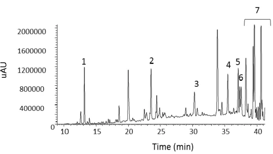
In this work we fully analyzed 11 methanolic leaf extracts of the different plants (in Table 2.1) from Caatinga biome (northeastern Brazil) by HPLC-DAD. High performance liquid chromatography (HPLC) is a technique used in analytical chemistry to separate, identify and quantify the constituents of a mixture, in this case a plant extract. The sample is dissolved in a solvent and introduced under high pressure into the chromatographic column with the stationary phase. This makes the components analysis much faster and better separation of the compounds on the mixture, than the one traditionally used thin layer chromatography (TLC). The solvent is pumped at a constant rate and moves the components of the mixture through the column. These are distributed between the two phases according to their affinities. Substances with higher affinity to stationary phase move more slowly. However substances with little affinity with the stationary phase move faster. The time taken for a compound to be eluted from the column is called retention time and is considered as a characteristic property of a compound under a certain elution program. When leaving the column, the components pass through a detector which gives an electrical signal which is recorded as a series of peaks, constituting a chromatogram per sample analyzed (Bidlingmeyer, 1992; Dong, 2006; Meyer, 2010; Hayes et al., 2014).
Methanolic extracts are composed by a mixture of phenolic compounds, as it can be observed in the HPLC chromatograms (in Fig. 2.2 – 2.12). To see the differences among the plant extracts, phenolic compounds were also quantified by HPLC-DAD-UV, all values were expressed in µg/g dwb (dwb - dry weight biomass). The peaks area was used for measuring the quantities of the compounds, by comparison with several pure compounds, used as external standards. The results of the quantitative analysis by HPLC-DAD are shown in Table 2.3.
VERA CASTRO 43
Fig. 2.3 - HPLC chromatogram (280 nm) of Anadenanthera peregrina (Sp 4) methanolic extract.
Compounds are identified in the figure by numbers: 1-Gallic acid; 2- Quercetin-3-O-rutinoside; 3- Quercetin-3-O-rhammnoside
Fig. 2.2 - HPLC chromatogram (280 nm) of Caesalpinia ferrea (Sp 3) methanolic extract.
Compounds are identified in the figure by numbers: 1- Gallic acid; 2- Ellagic acid; 3- Ellagic acid isomer; 4- Quercitin-3-O-rhamnoside; 5- Kaempferol
VERA CASTRO 44
Fig. 2.4 - HPLC chromatogram (280 nm) of Mimosa tenuiflora (Sp 6) methanolic extract. Compounds are
identified in the figure by numbers: 1-Gallic acid; 2- Ferrulic acid; 3- Quercetin isomer; 4- Quercetin-3-O-rhamnoside; 5- Isorhamnetin; 6- Kaempferol; 7- Kaempferol
Fig. 2.5 - HPLC chromatogram (320 nm) of Sideroxylon obtusifolium (Sp 8) methanolic extract.
Compounds are identified in the figure by numbers: 1- Chlorogenic acid; 2- Quercetin-3-O-rhamnoside; 3- Isorhamnetin
VERA CASTRO 45
Fig. 2.6 - HPLC chromatogram (280 nm) of Schinopsis brasiliensis (Sp 9) methanolic extract. Compounds
are identified in the figure by numbers: 1-Gallic acid; 2- Catechin; 3- Quercetin-3-O-rutinoside; 4- Siringic acid; 5- Quercetin isomer; 6- Ellagic acid;
Fig. 2.7 - HPLC chromatogram (280 nm) of Ziziphus joazeiro (Sp 11) methanolic extract. Compounds are
identified in the figure by numbers: 1- Gallic acid; 3- Catechin; 3- Caffeic acid; 4- Ferulic acid; 5- Quercetin-3-O-rhamnoside; 6- Kaempferol
VERA CASTRO 46
Fig. 2.8 - HPLC chromatogram (280 nm) of Maytenus rigida (Sp 12) methanolic extract. Compounds are
identified in the figure by numbers: 1- Chlorogenic acid; 2- Caffeic acid; 3- Quercetin-3-O-rhamnoside; 4- Isorhamnetin; 5- Kaempferol
Fig. 2.9 - HPLC chromatogram (280 nm) of Capparis flexuosa (Sp 14) methanolic extract. Compounds are
VERA CASTRO 47
Fig. 2.10 - HPLC chromatogram (370 nm) of Erythrina mulungu (Sp 15) methanolic extract. Compounds are
identified in the figure by numbers: 1- Quercetin isomer; 2- Ellagic acid; 3- Quercetin-3-O-rhamnoside
Fig. 2.11 - HPLC chromatogram (280 nm) of Euphorbia tirucalli (Sp 17) methanolic extract. Compounds are identified in the figure by numbers: 1- Gallic acid; 2- Catechin; 3- Siringic acid; 4- Quercetin-3-O-rutinoside; 5- Ellagic acid; 6- Quercetin-3-O-rhamnoside, 7- Kaempferol
VERA CASTRO 48
In recent years, significant attention has been directed towards the identification of plants with biological activity that may be used for human consumption. Phenolic compounds have attracted much attention due to their properties as antioxidant, anti-inflammatory or antitumour agents, among others. Phenolic compounds are aromatic hydroxylated compounds, possessing one or more aromatic rings with one or more hydroxyl groups. They include a large number of subclasses, such as flavonoids, phenolic acids, including hydroxybenzoic acids and hydroxycinnamic acids, stilbenes, lignans, tannins, and oxidised polyphenols, displaying a great diversity of structures. These compounds isolated from fruit, vegetables, and herbs have been extensively studied and their antioxidant potential and bioactive properties are found in all parts of the plants (tree bark, stalks, leaves, fruits, roots, flowers, pods, and seeds) (Ho et al., 2007a; Palacios et al., 2011; Colle et al., 2012; Akomolafe et al., 2013).
As we observe in Table 2.3, all plant extracts present phenolic compounds and the most representative ones are quercetin, catechin, gallic acid and kaempferol. Several studies reported these compounds to possess antioxidant proprieties (Pietta, 2000; Zheng and Wang, 2001; Coskun et al., 2005; Azevedo et al., 2013).
Fig. 2.12 - HPLC chromatogram (280 nm) of Cleome spinosa (Sp 18) methanolic extract. Compounds
are identified in the figure by numbers: 1- Catechin; 2- Ferulic acid; 3- Quercetin-3-O-rhamnoside; 4- Ferulic acid isomer; 5- Kaempferol
VERA CASTRO 49
We observed that quercetin derivatives are present in all extracts. Extract from Sp 9 shows the highest level in quercetin (1485 µg/g dwb) and catechin (471 µg/g dwb) compared with the other plant extracts. Extract from Sp 6 revealed the highest content in kaempferol (503 µg/g dwb). Sp 3 and 9 exhibit both higher levels of gallic acid (95 and 99 µg/g dwb, respectively).
Table 2.3 - Phenolic composition and total phenolic content of plant extracts
Phenolic compounds of methanolic extrats are quantified by HPLC-DAD. Results are expressed in µg/mg dwb. Data represent mean ± SD for three independent experiments.
Species number (Sp)
Gallic acid Chlorogenic
acid Ferulic acid Caffeic acid Ellagic acid
Mean SD Mean SD Mean SD Mean SD Mean SD
3 94,4 0,4 47,4 0,3 4 16,3 0,3 6 44,2 0,3 71,4 0,4 8 12,6 0,2 9 99,9 0,4 28,7 0,3 11 10,0 0,3 14,3 0,3 2,6 0,0 12 15,8 0,0 9,7 0,0 14 15 5,9 0,3 17 33,1 0,2 13,1 0,3 18 33,3 0,2 Species number (Sp)
Catechin Quercetin Isorhamnetin Kaempferol
Total phenolic
content
Mean SD Mean SD Mean SD Mean SD Mean SD
3 59,9 0,4 19,4 0,2 221,0 1,3 4 71,3 0,4 170,5 0,5 258,1 1,1 6 159,4 0,5 89,9 0,4 502,7 0,8 867,7 2,4 8 45,4 0,3 27,6 0,3 85,6 0,9 9 470,9 0,8 1484,9 1,8 13,6 0,0 2097,9 3,3 11 37,2 0,0 74,2 0,4 63,2 0,4 201,4 1,4 12 42,0 0,3 27,1 0,3 33,0 0,3 127,7 1,0 14 63,2 0,4 116,2 0,4 16,8 0,3 196,2 1,1 15 45,9 0,3 51,9 0,6 17 9,0 0,2 39,7 0,3 9,6 0,1 104,6 1,2 18 30,7 0,0 16,4 0,3 15,0 0,2 95,4 0,7