Universidade de Lisboa
Faculdade de Ciências
Departamento de Biologia Animal
Uncovering the effect of low doses of ionizing
radiation in angiogenesis during spinal cord
regeneration
Tiago Filipe Ribeiro Ruas Maçarico
Dissertação de
Mestrado em Biologia Evolutiva e do Desenvolvimento
2014
Universidade de Lisboa
Faculdade de Ciências
Departamento de Biologia Animal
Uncovering the effect of low doses of ionizing
radiation in angiogenesis during spinal cord
regeneration
Tiago Filipe Ribeiro Ruas Maçarico
Dissertação de
Mestrado em Biologia Evolutiva e do Desenvolvimento
Orientado por:
Professora Doutora Susana Constantino
Professora Doutora Gabriela Rodrigues
2014
i
ACKNOWLEDGEMENTS
Firstly, I would like to express my gratitude to both my co-supervisors: Susana Constantino for accepting me as her student and for giving me this opportunity; I am deeply thankful for her teachings and for her guidance in the right direction and Ana Ribeiro, for her patience, for her great support in numerous problems that arose during the course of the work and for the numerous exchanges of insights; I am deeply thankful for her thorough revision of my master's thesis, invaluable comments and excellent expertise.
Secondly, I acknowledge Gabriela Rodrigues for being my internal supervisor, for the inspiration to follow this path and for all the support she gave to me.
I would also like to thank Leonor Saúde for having me in her lab. I am very grateful for her kindness, her enthusiasm and encouraging words.
Thanks to the awesome “Fish” people: to Lara that was always willing to help and give her best suggestions, to Aida for her contagious positive energy and to Sara for her help and kindness.
Thanks to the UAG group: to Dr Augusto, Carolina, Paula, Rita and Adriana for all the discussion and guidance during my work. And a special thanks to Filipa for the patience to listen and discuss my crazy ideas.
Thanks to the development group: to Guida for her patience and kindness, to Susana for all the encouragement words and to Raquel and Zé for the curiosity in this work. A special thanks to Rita for the assistance and interest, and to Sara for always wished me all the best.
I would also like to acknowledge the Bioimaging Unit: Rino, António and Ana for the transmitted knowledge and precious support.
Thanks to Tecnozeb, especially to Sara, Diana and Rita for allowing the behaviour monitoring to happen.
My sincere thanks also go to the Radiotherapy Division of Hospital de Santa Maria, especially to Vera and Inês, for the kindness and for allowing the radiation experiment to happen.
ii I would also like to acknowledge the Histology Unit especially to Tânia and Andreia for all the ideas usefull ideas and interest in this work.
Thanks to Ana Faustino from ISPA for having introduced me in the behavioural experiments and for her kindness.
In addition a thank you to Benedict, Tiago II and Dalila, although their passage by the laboratory was short, they gave me a great help.
Special thanks to my family in particular to my mom for her patience and great support without her I would not done this project. To my uncle (Ti-Tó) for the good mood and interest in my work and to my grandfather for all the encouraging words. Thanks to my friends, to Alex, Margarida, Joana, Stufu and Rodrigo, for the encouragement, moral support and for being there when I needed.
iv
Table of contents
Acknowledgements ...i
Table of contents ...iv
Chapter I - Introduction ... 2
I.1. Spinal cord ... 2
I.1.1. Blood supply in the spinal cord ... 3
I.2. Spinal cord injury ... 5
I.2.1. Response of the vascular system to SCI ... 6
I.3. Experimental models for the study of SCI ... 8
I.3.1. Zebrafish as a regeneration model ... 10
I.4. Blood vessel formation ... 12
I.4.1. Sprouting angiogenesis and angiogenic balance... 13
I.4.2. Angiogenic stimuli – low doses of IR ... 14
I.5. Aims ... 18
1
C
HAPTER
I
2
C
HAPTER
I
-
I
NTRODUCTION
I.1. Spinal cord
The vertebrate spinal cord is a narrow tube of nerve tissue that belongs to the central nervous system (CNS). It is located inside the vertebral column. Although it seems to be well enclosed and protected, it is a very sensitive organ and highly susceptible to injury [19].
The intramedullary region is composed of various types of neuronal and glial cells. The neuronal cells form a complex network that processes information and generates responses; they are responsible for sensory intake and motor response. The glial cells give the support and maintenance required for this tissue, such as electric insulation -oligodendrocytes; immune response - microglia; metabolic exchanges - astrocytes; and covering the central canal, where the cerebrospinal fluid passes - ependymal cells [20].
In mammals histological transverse sections of the spinal cord reveal two distinct regions: the grey matter and white matter. A central region, known as grey matter forms two dorsal horns and two anterior ventral horns (resembles the letter H) is composed of neuron cell bodies, glial cells and interneurons. The peripheral region - white matter - is composed of bundles of myelinated axons and glial cells (Figure 1) [20].
The spinal cord, as the entire CNS, has a high metabolic rate and therefore needs a continuous and tight maintenance in order to maintain homeostasis [3].
Figure 1. Spinal cord intramedullary region. Schematic of a spinal cord transverse section
3
I.1.1. Blood supply in the spinal cord
The blood vessel network has a key role in tissue homeostasis, providing cells with oxygen and nutrients and removing metabolic waste. While this is also the case in the CNS, the spinal cord is not homogeneously vascularized [5, 22]. The grey matter is more vascularized than the peripheral white matter (in human and mouse) as a result of higher metabolic needs [16, 17].
The intramedullary arterial supply comes from external arteries, which in turn enter the spinal cord. The spinal cord vasculature can be divided into two regions: an anterior region that covers two thirds of the spinal cord and a posterior region that covers the remaining third. The anterior region is irrigated by the central artery and the ventral portion of the vasocorona, both in contact with the anterior spinal artery. The posterior region is irrigated by two posterior spinal arteries and the dorsal part of the vasocorona [3, 4, 23] (Figure 2).
This vessel distribution has two flow directions, peripheral blood centripetal from the vasocorona and the posterior spinal arteries and a centrifugal internal blood supply from the central artery. This conformation creates a non-homogeneous distribution of blood vessels, which implies that the spinal cord has zones that do not receive direct blood supply and depend on the overlap of terminal branches. These boundary regions are called “watershed zones” and are particularly vulnerable after injury [3].
Figure 2. Intramedullary blood supply. Schematic of a spinal cord segment showing the
blood supply, grey area is representing grey matter, the remaining area inside the circle represent the white matter (adapted from [23]).
4 The nervous tissue, due to its critical function, is more protected from pathogens and toxins than other tissues. This protection is conferred by a physical barrier between the blood vessels and the nervous tissue – the blood brain barrier (BBB). The BBB, also known as neurovascular unit, is composed of: endothelial cells, tight juntions, pericytes, astrocyte endfeet and basal lamina. The tight junctions, together with adherent junctions and the basal lamina matrix, prevent the passage of macromolecules and cells from the bloodstream, as well as pathogens. Pericytes and astrocytes are involved in the formation of the BBB and also in the control of nutrients and metabolic waste exchange, helping maintaining the nervous tissue homeostasis [4, 24].
Figure 3. Blood brain barrier BBB. Schematic of the neurovascular unit composed of
endothelial cell connected by the apical regions through tight junctions and covered by basal lamina, pericytes and astrocyte endfeet (adapted from [24]).
5
I.2. Spinal cord injury
According to the World Health Organization up to 500 000 people suffer a spinal cord injury each year. SCI in humans frequently leads to paralysis and loss of sensation below the injury site [1, 25, 26]. In addition, people with spinal cord injuries are 2 to 5 times more likely to die prematurely [26].
The causes of spinal cord injury in humans are diverse: trauma can result from contusion, compression, penetration or maceration of the spinal cord and often leads to the formation of cavities and cysts that interrupt the axonal tracks [2, 6]. The SCI pathologic events can be divided into two phases: initial impact and secondary response [3].
First, the traumatic impact leads to the death of many cell types, such as neurons, oligodendrocytes, astrocytes and precursor cells. Subsequently, the tissue damage induces an inflammatory response, which is contained by an accumulation of astrocytes and inhibitory molecules that form the glial scar at the injury region. This glial scar acts as a physical barrier and accumulates molecules that inhibit axon outgrowth, such as myelin associated inhibitors and chondroitin sulphate proteoglycans [2, 6, 27]. Limited axon sprouting does occur, but is blocked by the glial scar barrier and the inhibitory environment generated [2].
Some brain regions were recently shown to have the capacity to regenerate through neural stem cells [28]. However, although there are neural stem cells close to the ependymal canal in the spinal cord, it seems that these cells are in a microenvironment that does not support neurogenesis [29]. As a result, the mammalian spinal cord is unable to replace most of the cells lost with the injury. The secondary injury response follows the initial trauma and further extends in rostral–caudal and medial–peripheral directions. Oligodendrocyte apoptosis leads to the demyelination of spared axons at the periphery that degenerate due to lack of myelin [6, 25].
One of the main events responsible for this response is the disruption of the vascular system that exacerbates cell death and limits the blood supply to spared and repaired tissue. [4].
6
I.2.1. Response of the vascular system to SCI
The disruption of local blood vessels and loss of the BBB are thought to be decisive for the evolution of the destructive events that arise during the secondary injury phase [3].
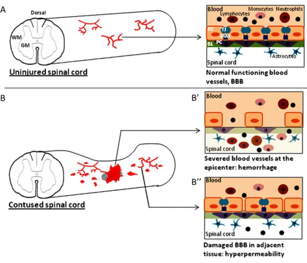
The primary insult results in blood vessel rupture at the injury epicenter that causes a haemorrhagic area leading to tissue damage (Figure 3B’). The haemorrhage happens first in the highly vascularized grey matter and later expands into the peripheral white matter [3, 4].
The initial trauma is followed by a secondary response that further harms the tissue. First, the blood is toxic to nerve tissue, probably due to degradation of haemoglobin that results in the production of free radicals [3, 4]. Moreover, the vessels and BBB disruption lead to an influx of inflammatory cells (neutrophils, T-lymphocytes and macrophages) and toxic molecules (such as calcium and excitatory amino acids) that cause an inflammatory response and the destabilization of tissue homeostasis. These events happen at the injury site but also in the adjacent region, where shear stress leads to blood vessel hyperpermeability and causes more haemorrhages around the injury (Figure 3B’’) [4, 6]. There is also an accumulation of interstitial fluid (oedema) that puts more pressure on the adjacent tissue [4].
Additionally, as the blood flow is reduced in the tissue around the haemorrhage, it leads to a sharp decrease in oxygen and nutrients (glucose) needed for cell metabolism, which in turn leads to ischemia and formation of reactive oxygen species causing more cell death [3, 6].
Healthy blood vessels are crucial for the survival of the damaged tissue and for new cells that may be generated during the repair process. Endogenous repair processes of blood vessels and BBB have been described, but are inefficient and result in malfunctioning (leaky) blood vessels that further aggravate the problem and prevent
tissue repair [4].
Although many repair approaches at the injury site have been tested, such as transplant approaches and inhibition of inhibitors agents, few studies have examined the importance of the vasculature during SCI [2-4]. However, therapies that support the formation of new blood vessels will likely contribute for the functional recuperation of the tissue. The improvement of the vascular support, together with other approaches, may lead to a functional recovery of the multi-facetted nature of
7
spinal cord [4, 6]. However, vessel formation is a complex process regulated by many factors and promoting vascular support in an uncontrolled manner could worsen the problem [3].
Figure 4. Vascular disruption after spinal cord injury. (A) Uninjured spinal cord with intact
blood vessels and blood-brain barrier (BBB). (B) Injured (contused) spinal cord with hemorrhages at the epicenter and in more distance regions. BBB structure in ruptured blood vessels (B’) and leaky blood vessels (B’’). (BL-basal lamina, EC-endothelial cell, JAM-junction adhesion molecules, GM-gray matter, PC-pericyte, TJ-tight junction, WM-white matter) (adapted from [4]).
8
I.3. Experimental models for the study of SCI
Animal models have long been used to investigate the pathophysiological response and repair processes of the spinal cord. They may not have a response to injury as complex as the human but they have several mechanisms that are similar and can help us better understand the repair limitations [30]. Animal models allow us to produce lesions with different degrees of severity and demonstrate a correlation between the severity of the injury and the behavioural response, both sensory and locomotor [31, 32].
There are three main models of SCI: transection, contusion, and compression. Each injury model mimics certain clinical features of mechanical damage and/or post-traumatic events [3, 11, 31].
The transection injury consists in a clean cut. It can be a complete transection or a hemisection, which is useful to compare sides (Figure 4A). The pathological features of this model are restricted to the transected area, making the transection model less clinically relevant [3, 4].
Contusion injuries are performed by a focal force, most commonly applied dorsally (Figure 4B). The pathophysiologic response of this injury spans a wider rostrocaudal extent of the spinal cord; there is a severe vessel disruption that leads to a great inflammatory response and formation of an oedema. This model mimics better the SCI pathology in humans.
The compression injury response is similar to the contusion model when the compression is severe (also called crush in some studies) [12]. The difference to contusion injury is that compression normally involves a lateral compression with forceps and may also affect the lateral side (Figure 4C) [31].
Although rat and mouse models have been widely used in SCI studies, due the similarities to humans, these models do not have the capacity to recover after an injury. In contrast to rodents, some animal models are capable of regeneration during adulthood, such as worms, lizards, salamanders and teleost fish [30, 33, 34]. The study of these animal models can provide clues to new ways to improve the repair process in mammals (including humans) [30].
9
Figure 5. SCI models. (A) Transection injury, (left), lateral hemisections (right), and
complete transection (bottom). (A’) Cell proliferation response is localized at the epicenter. (B) Contusion injury in which some white matter is spared. (B’) Cell proliferation spreads along the rostro-caudal extent. (C) Compression injury resembles the contusion injury in both temporal and proliferative response (C’), but can also affect the lateral white matter and cause a wider rostro-caudal response (adapt from [31]).
10
I.3.1. Zebrafish as a regeneration model
Among the animal models that are capable of spontaneously regenerate we find the zebrafish. Zebrafish is a well known model in developmental biology and has become an attractive organism to study regeneration. This vertebrate has the capacity to regenerate fins, heart and nervous system structures, just to name a few [8, 35]. Adult zebrafish (>4 months) has the capacity to regenerate the spinal cord after trauma [8, 12].
Although this model is not as close to human physiology as rodent models, the basic cytology of the zebrafish spinal cord is relatively similar to the mammalian spinal cord. Yet their functional response is very different, making zebrafish an interesting model to study the mechanisms behind the regenerative process [32, 33].
After SCI zebrafish lose the ability to use the posterior part of the body, having no movement of the caudal fin [32]. Injured zebrafish remain with reduced movement for approximately one week and gradually regain their swimming capacity - within a month in case of strong compression injury [12], or 6 weeks in full transection injury [10, 11].
After a compression injury, the zebrafish spinal cord undergoes apoptotic cell death soon after the lesion, affecting both neural and glial cells. As in mammals, the injury also leads to blood vessel rupture, haemorrhage and influx of inflammatory cells (neutrophils, monocytes and macrophages) (that are useful for myelin debris removal) that further contribute for cell death. However, the apoptotic response reaches a peak at 1 dpi, unlike in mammals where it continues until 8 dpi [12]. Furthermore, the zebrafish spinal cord does not develop a glial scar [9, 12].
The processes behind the regeneration of spinal cord in zebrafish are beginning to be understood. [9]. There is evidence that the zebrafish spinal cord is more permissive for neurogenesis, as new motor neurons are generated from a pool of ependymal cells that are Olig2+ [10, 36]. It has also been shown that severed axons are able to
regrow and an efficient axonal regeneration appears to be correlated with a functional recovery of the fish swimming capacity [8, 13].
Also, since it is a simpler organism, it is possible that it has a higher plasticity. In other words, it might be easier to form new connections that allow different path
11 circuits to adapt and confer functional recovery even though not all affected connections were regenerated [32, 37].
Although the cellular and molecular mechanisms of neuro-regeneration in zebrafish are starting to be uncovered, the process of vascular recovery is still largely unknown.
The blood vessel disruption and malfunctioning contribute for the secondary response to injury in mammals, yet zebrafish seems to overcome this problem and recover motor function. Zebrafish may be able to repair the vascular network and thus support new and spared cells at the injury site [3, 4]. The study of this model organism could help understand how the vascular system can be efficiently restored and how it contributes for spinal cord regeneration. Moreover, genetic and pharmacological tools that report and modulate blood vessels are already available in the zebrafish field [14, 15].
12
I.4. Blood vessel formation
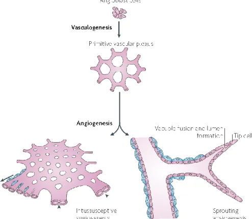
There are two main mechanisms behind the formation of blood vessels: vasculogenesis and angiogenesis. Vasculogenesis is the de novo assembly of blood vessels through recruitment of endothelial precursor cells (EPCs). This process occurs mainly during embryonic development and rarely in adults [38].
Angiogenesis is the formation of new blood vessels from pre-existing ones and can occur through proliferation of endothelial cells, known as sprouting angiogenesis [39]. This process takes place during embryonic development, but also occurs during adulthood in normal physiologic conditions (such as organ growth, wound healing, female reproductive cycle and in the placenta during pregnancy) [40, 41]. Another variant of angiogenesis is splitting angiogenesis or intussusception angiogenesis. As the name suggests, it consists in the splitting of pre-existing vessels to form new ones, through the formation of translumen pillars (Figure 4) [42].
Figure 6. Vasculogenesis and angiogenesis: Vasculogenesis is de novo formation of blood
vessels through differentiation of endothelial precursor cells (EPCs) – Angioblast. Angiogenesis occurs from pre-existing blood vessels. Two types of angiogenesis are often considered: intussusceptive and sprouting angiogenesis. Intussusceptive angiogenesis is the splitting of pre-existing blood vessels through the formation of translumen pillars (arrow head). Sprouting angiogenesis is the formation of vessels through endothelial cell proliferation. Pericytes support both process providing vessel stability (blue cells) (adapted from [43]).
13
I.4.1. Sprouting angiogenesis and angiogenic balance
Angiogenesis is regulated by pro- and anti-angiogenic factors which are in a dynamic balance under physiological conditions (Figure 7) [7]. When this balance is lost it leads to pathologies associated with insufficient angiogenesis (ie. ischemic diseases) or excessive angiogenesis (such as cancer) [41, 44].
When a quiescent vessel senses an angiogenic signal, the process of sprouting angiogenesis starts with proteases, such as matrix metalloproteinases (MMPs), that degrade the surrounding extracellular matrix (ECM) creating openings for endothelial cells to migrate and proliferate [45]. Mural cells detach allowing an activated EC, tip cell, to migrate, in response to guidance signals. Stalk cells trail behind the tip cell and proliferate to allow sprouting elongation and lumen formation. Tip cells anastomose with cells form neighbouring sprouts and a lumen is formed to allow blood flow through the new vessel. Perfusion induces vascular maturation by reestablishment of cell-cell junctions, pericyte maturation and basement membrane deposition [45].
One of the main factors responsible for attracting blood vessels is the vascular endothelial growth factor (VEGF). This factor is a key element that promotes the proliferation and migration of endothelial cells [4, 45].
Many other factors are involved in vessel formation such as FGFs (Fibroblast growth factors), angiopoietins, PDGF (platelet-derived growth factor) and TGF (transforming growth factor)-β that work together with the different VEGF isoforms and regulate vessel proliferation and migration, as well as vessel maturation. Vessel maturation in the CNS requires vessel perfusion, oxygenation and pericyte attachment, but also wrapping by basal lamina and astrocytes feet to form the BBB [4, 39, 46].
Besides vessel maturation and stabilization, a controlled regression of redundant vessels can occur during remodeling. The process of vessel regression starts with
Figure 7. Angiogenic balance. The balance between Pro-angiogenic factors (+) and
14
narrowing and cessation of blood flow leading to endothelial cell apoptosis, pericyte dropout, luminal occlusion and retraction [47, 48].
I.4.2. Angiogenic stimuli – low doses of IR
Radiotherapy has been extensively used as a cancer treatment to kill malignant cells. In this therapy, therapeutic doses of IR are delivered to the tumour area. These doses are known to damage the genetic material inducing cell death and changing the tumour microenvironment [49, 50]. Moreover, the surrounding tissues are exposed to subtherapeutic doses of IR and, although the biological effect of the therapeutic doses is well known, the effect of low doses on healthy tissues still remains largely uncovered.
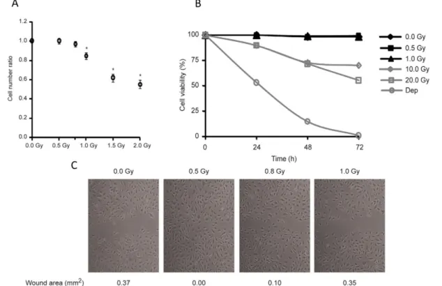
S. Constantino lab has focused on the effect of these low doses of IR on the vasculature, especially on endothelial cells. In vitro assays with human microvascular endothelial cells from lung (HMVEC-L) showed that doses lower than 0,8 Gy did not induce cell cycle arrest or death unlike doses above 1 Gy (Figure 8A, B). Also, confluent monolayers of endothelial cells showed an induced cell migration on doses of 0,5 and 0,8 Gy (Figure 5C) [18].
Figure 8. In vitro effects of low doses IR: Human microvascular endothelial cells from lung
(HMVEC-L) (A)- apoptosis, (dep)- cell death control culture without serum (B) cycle arrest ratio between cell number of irradiated and non-irradiated conditions (C) Confluent monolayers of HMVEC-L (adapted from [18]).
15 Further experiments showed that one of the main molecular mechanisms involved in this effect was the phosphorylation and activation of the receptor 2 of VEGF (VEGFR2). VEGFR2 has been described as a major player in the angiogenic response, being responsible for cellular survival, proliferation, migration and differentiation. This receptor leads to the activation of multiple intracellular proteins that lead to the expression of pro-angiogenic proteins, which in turn will cause an angiogenic response (Figure 6) [51, 52].
The activation of VEGFR2 by IR was shown to be independent of extracellular VEGF. However, the pro-angiogenic response induced by low-dose IR was significantly blocked with a tyrosine kinase inhibitor (TKI) of VEGFR2. Therefore, the phosphorylation of this receptor is one of the most important mechanisms in the angiogenic response induced by low doses of IR [18].
In vivo, the effects of these low doses were evaluated in zebrafish, using a transgenic
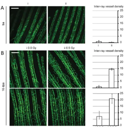
line with EGFP-labelled blood vessels - fli1:EGFP [14, 15]. The effect of low doses of IR on angiogenesis was first assessed in the regeneration of the zebrafish caudal fin. The distal end of the caudal fin was amputated, exposed or not to 0.5 Gy of IR and then allowed to recover. After fin regeneration, the vascular inter-ray density of the caudal fin was increased in fish exposed to IR (Figure 7) [18].
Figure 9. Molecular mechanism: VEGFR2 phosphorylation leads to the activation of multiple
intracellular proteins, which in turn leads to the modulation of the expression of pro-angiogenic factors and an pro-angiogenic response.
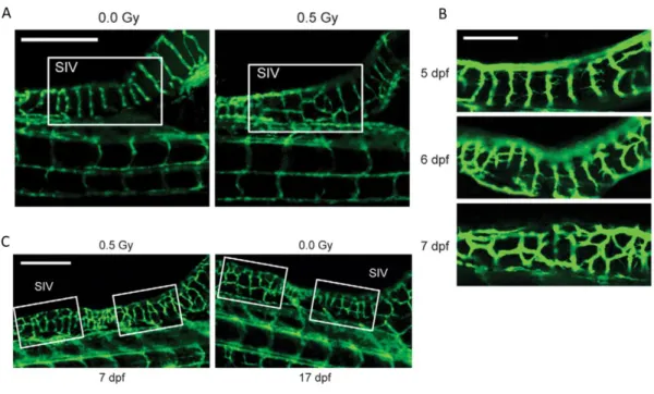
16 The effects of low doses of IR were also tested in zebrafish larvae. Larvae were irradiated for 3 days post fertilization (dpf) with 0.5 Gy. At 7 dpf the sub-intestinal vessels (SIV) in the unirradiated group presented a characteristic pattern of parallel vessels, while in the irradiated group 2/3 of the embryos presented more and irregularly-shaped vessels crossing between the SIV (Figure 8A). However, this irregular pattern of the irradiated group was observed on the unirradiated group on more advanced developmental times (17 dpf) (Figure 8C). Furthermore, endothelial sprouts were observed crossing between SIV in the irradiated group at 5, 6 and 7 dpf (Figure 8B) [18]. In summary, these data show that low doses of IR accelerate angiogenic sprouting during zebrafish development.
Figure 10. Low doses of IR enhances angiogenesis during fin regeneration. Adult zebrafish
caudal fins were amputated at mid-fin level, exposed (i) or not (ii) to 0.5 Gy of IR and then allowed to recover. (A) caudal fin before amputation (ba) (B) 16 days post-amputation (dpa), with and without low-dose IR. Inter-ray vessel density. Scale bars, 250 mm (adapted from [18])
17 These findings suggested that low doses of IR cause a pro-angiogenic response and the molecular analysis points to an activation of pro-angiogenic factors. Furthermore these results suggest that these angiogenesis is functional since new blood vessels presented erythrocytes flowing on the bloodstream, were not haemorrhagic and did not regress over time. However more studies are required to test these blood vessels functionality.
Figure 11. Low doses IR accelerate sprouting angiogenesis in zebrafish development.
Zebrafish embryos exposed or not to 0.5 Gy IR 3 days post-fertilization (dpf). Representative images of Sub-Intestinal Vessels (SIV), (A) a non-irradiated and irradiated zebrafish at 7 dpf; (B) irradiated zebrafish at 5 (top), 6 (middle) and 7 dpf (bottom); (C) irradiated zebrafish at 7 dpf and a non-irradiated zebrafish at 17 dpf. Scale bars, 250 mm (A and C), 100 mm (adapt from [18])
18
I.5. Aims
An angiogenic response after SCI was described in mammals but the new blood vessels are dysfunctional and fail to provide the necessary cell support, further worsening the outcome.
In recent years an increasing number of zebrafish spinal cord regeneration studies have been reported, focusing on the capacity of reestablishment axonal connection and on the capacity to restore the nervous tissue. However, no studies have been conducted on the vascular response that occurs post-injury in the regenerative process.
Our first goal was to characterize the vascular response in a compression injury in adult zebrafish, developed in L. Saúde lab, to mimic the rostro-caudal response that normally occurs in a SCI in humans. This characterization was carried out at several time-points post-injury.
Furthermore, we wanted to investigate whether low doses of IR could stimulate an enhancement or an acceleration of the angiogenic process in the spinal cord, as had been shown in other contexts by S. Constantino Lab. In addition, we aimed to determine if this correlated with an improved functional recovery that could be indicative of the functionality of these blood vessels.
19
C
HAPTER
V
20
C
HAPTER
V
-
R
EFERENCES
1 Barnabé-Heider, F., & Frisén, J. (2008). Stem cells for spinal cord repair. Cell Stem Cell, 3(1), 16–24
2 Thuret S, Moon LDF, Gage FH (2006) Therapeutic interventions after spinal cord injury. Nat Rev Neurosci 7: 628–643.
3 Mautes, A. E. M., Weinzierl, M. R., & Noble, L. J. (2000). Events After Spinal Cord Injury: Contribution to Secondary Pathogenesis, Phy Ther. 673–687. 4 Oudega M (2012) Molecular and cellular mechanisms underlying the role of
blood vessels in spinal cord injury and repair. Cell Tissue Res 349: 269–288. 5 Casella, G. T. B., Marcillo, A., Bunge, M. B., & Wood, P. M. (2002). New
vascular tissue rapidly replaces neural parenchyma and vessels destroyed by a contusion injury to the rat spinal cord. Exp Neurol, 173(1), 63–76.
6 Hagg T, Oudega M (2006) Degenerative and Spontaneous Regenerative Processes after Spinal Cord Injury. J Neurotrauma 23: 263–280.
7 Carmeliet P (2000) Mechanisms of angiogenesis and arteriogenesis. Nat Med. 6: 389–395.
8 Becker CG, Becker T (2008) Adult zebrafish as a model for successful central nervous system regeneration. 26: 71–80.
9 Becker T, Wullimann MF, Becker CG, Bernhardt RR, Schachner M (1997) Axonal Regrowth After Spinal Cord Transection in Adult Zebrafish. 595: 577–595. 10 Reimer MM, Sörensen I, Kuscha V, Frank RE, Liu C, et al. (2008) Motor neuron
regeneration in adult zebrafish. J Neurosci 28: 8510–8516.
11 Fang P, Lin J-F, Pan H-C, Shen Y-Q, Schachner M (2012) A surgery protocol for adult zebrafish spinal cord injury. J Genet Genomics 39: 481–487.
12 Hui SP, Dutta A, Ghosh S (2010) Cellular response after crush injury in adult zebrafish spinal cord. Dev Dyn 239: 2962–2979.
13 Vajn K, Suler D, Plunkett J a, Oudega M (2014) Temporal Profile of Endogenous Anatomical Repair and Functional Recovery following Spinal Cord Injury in Adult Zebrafish. PLoS One 9: e105857.
14 Lawson ND, Weinstein BM (2002) In Vivo Imaging of Embryonic Vascular Development Using Transgenic Zebrafish. Dev Biol 248: 307–318.
15 Bayliss PE, Bellavance KL, Whitehead GG, Abrams JM, Aegerter S, et al. (2006) Chemical modulation of receptor signaling inhibits regenerative angiogenesis in adult zebrafish. Nat Chem Biol 2: 265–273.
21 16 Gillilan, L. A. (1958). The arterial blood supply of the human spinal cord. J.
Comp Neurol, 110(1), 75–103.
17 Attwell D, Laughlin SB (2001) An energy budget for signaling in the grey matter of the brain. J Cereb Blood Flow Metab 21: 1133–1145.
18 Sofia Vala I, Martins LR, Imaizumi N, Nunes RJ, Rino J, et al. (2010) Low doses of ionizing radiation promote tumor growth and metastasis by enhancing angiogenesis. PLoS One 5: e11222.
19. Van De Graaff, K. M. (2001). Human Anatomy.6 ED McGraw-Hill (pp. 384-386) 20 Mescher, A. (2009). Junqueira’s Basic Histology: Text and Atlas, 12th Ed.
Mcgraw-hill. Chapter 9. Nerve Tissue & the Nervous System
21 Bear R, Rintoul D (2014) Nervous System OpenStax-CNX module: m47519: 1–30 22 Martirosyan, N. L., Feuerstein, J. S., Theodore, N., Cavalcanti, D. D.,
Spetzler, R. F., & Preul, M. C. (2011). Blood supply and vascular reactivity of the spinal cord under normal and pathological conditions. J Neurosurg. Spine, 15(3), 238–51.
23 Satran R (1988) Spinal cord infarction. Stroke 19: 529–532.
24 Abbott NJ, Rönnbäck L, Hansson E (2006) Astrocyte-endothelial interactions at the blood-brain barrier. Nat Rev Neurosci 7: 41–53.
25 Ng, M. T. L., Stammers, A. T., & Kwon, B. K. (2011). Vascular disruption and the role of angiogenic proteins after spinal cord injury. Transl Stroke Res, 2(4), 474–91.
26 Sahni, V., & Kessler, J. a. (2010). Stem cell therapies for spinal cord injury. Nat Rev Neurol, 6(7), 363–72.
27 Stichel CC, Müller HW (1998) The CNS lesion scar: new vistas on an old regeneration barrier. Cell Tissue Res 294: 1–9.
28 Bath KG, Lee FS (2010) Neurotrophic factor control of adult {SVZ} neurogenesis. Devel Neurobio: 70: 339–349.
29 Hamilton LK, Truong MK V, Bednarczyk MR, Aumont a, Fernandes KJL (2009) Cellular organization of the central canal ependymal zone, a niche of latent neural stem cells in the adult mammalian spinal cord. Neuroscience 164: 1044–1056.
30 Sharif-Alhoseini M, Rahimi-Movaghar V (2014) Animal Models in Traumatic Spinal Cord Injury. Topics in Paraplegia. InTech.
31 McDonough A, Martínez-Cerdeño V (2012) Endogenous proliferation after spinal cord injury in animal models. Stem Cells Int 2012: 387513.
22 32 Becker CG, Becker T (2006) Zebrafish as a Model System for Successful Spinal Cord Regeneration. Model Organisms in Spinal Cord Regeneration. Wiley-{VCH} Verlag {GmbH} {&} Co. {KGaA}. pp. 289–319.
33 Diaz Quiroz JF, Echeverri K (2013) Spinal cord regeneration: where fish, frogs and salamanders lead the way, can we follow? Biochem J 451: 353–364.
34 Sîrbulescu RF, Zupanc GKH (2011) Spinal cord repair in regeneration-competent vertebrates: adult teleost fish as a model system. Brain Res Rev 67: 73–93.
35 Gemberling M, Bailey TJ, Hyde DR, Poss KD (2013) The zebrafish as a model for complex tissue regeneration. Trends Genet 29: 611–620.
36 Guo Y, Ma L, Cristofanilli M, Hart RP, Hao A, et al. (2011) Transcription factor Sox11b is involved in spinal cord regeneration in adult zebrafish. Neuroscience 172: 329–341.
37 Fleisch VC, Fraser B, Allison WT (2011) Investigating regeneration and functional integration of CNS neurons: lessons from zebrafish genetics and other fish species. Biochim Biophys Acta 1812: 364–380.
38 Davis GE, Stratman AN, Sacharidou A, Koh W (2011) Molecular Basis for Endothelial Lumen Formation and Tubulogenesis During Vasculogenesis and Angiogenic Sprouting. International Review of Cell and Molecular Biology. Elsevier {BV}. pp. 101–165.
39 Potente M, Gerhardt H, Carmeliet P (2011) Basic and therapeutic aspects of angiogenesis. Cell 146: 873–887.
40 Carmeliet P (2003) Angiogenesis in health and disease. Nat Med 9: 653–660. 41 Cao Y, Hong A, Schulten H, Post MJ (2005) Update on therapeutic
neovascularization. Cardiovasc Res 65: 639–648.
42 Burri PH, Hlushchuk R, Djonov V (2004) Intussusceptive angiogenesis: its emergence, its characteristics, and its significance. Dev Dyn 231: 474–488. 43 ten Dijke P, Arthur HM (2007) Extracellular control of TGFbeta signalling in
vascular development and disease. Nat Rev Mol Cell Biol 8: 857–869.
44 Carmeliet P (2005) Angiogenesis in life, disease and medicine. Nature 438: 932–936.
45 Gerhardt H (2008) {VEGF} and Endothelial Guidance in Angiogenic Sprouting. {VEGF} in Development. Springer New York. pp. 68–78.
46 Adair TH, Montani J-P (2010) Angiogenesis. Colloq Ser Integr Syst Physiol From Mol to Funct 2: 1–84.
23 47 Korn C, Augustin HG (2012) Born to die: blood vessel regression research
coming of age. Circulation 125: 3063–3065.
48 Pries AR, Reglin B, Secomb TW (2005) Remodeling of blood vessels: responses of diameter and wall thickness to hemodynamic and metabolic stimuli. Hypertension 46: 725–731.
49 Barcellos-Hoff MH, Park C, Wright EG (2005) Radiation and the microenvironment - tumorigenesis and therapy. Nat Rev Cancer 5: 867–875. 50 Connell PP, Kron SJ, Weichselbaum RR (2004) Relevance and irrelevance of
DNA damage response to radiotherapy. DNA Repair (Amst) 3: 1245–1251. 51 Domingues I, Rino J, Demmers J a a, de Lanerolle P, Santos SCR (2011)
VEGFR2 translocates to the nucleus to regulate its own transcription. PLoS One 6: e25668.
52 Cross MJ, Dixelius J, Matsumoto T, Claesson-Welsh L (2003) VEGF-receptor signal transduction. Trends Biochem Sci 28: 488–494.
53 Becker CG, Lieberoth BC, Morellini F, Feldner J, Becker T, et al. (2004) L1.1 is involved in spinal cord regeneration in adult zebrafish. J Neurosci 24: 7837– 7842.
54 Hama H, Kurokawa H, Kawano H, Ando R, Shimogori T, et al. (2011) Scale: a chemical approach for fluorescence imaging and reconstruction of transparent mouse brain. Nat Neurosci 14: 1481–1488.
55 Stewart A, Cachat J, Wong K, Gaikwad S, Gilder T, et al. (2010) Homebase behavior of zebrafish in novelty-based paradigms. Behav Processes 85: 198– 203.
56 Fontaine E, Lentink D, Kranenbarg S, Müller UK, van Leeuwen JL, et al. (2008) Automated visual tracking for studying the ontogeny of zebrafish swimming. J Exp Biol 211: 1305–1316.
57 Van Ham T, Kokel D, Peterson R (2012) Apoptotic Cells Are Cleared by Directional Migration and elmo1- Dependent Macrophage Engulfment. Curr Biol 22: 830–836.
58 Xiaowei H, Ninghui Z, Wei X, Yiping T, Linfeng X (2006) The experimental study of hypoxia-inducible factor-1alpha and its target genes in spinal cord injury. Spinal Cord 44: 35–43.
59 Cao R, Jensen LDE, Söll I, Hauptmann G, Cao Y (2008) Hypoxia-induced retinal angiogenesis in zebrafish as a model to study retinopathy. PLoS One 3: e2748.
24 60 Hui SP, Sengupta D, Lee SGP, Sen T, Kundu S, et al. (2014) Genome wide expression profiling during spinal cord regeneration identifies comprehensive cellular responses in zebrafish. PLoS One 9: e84212.
61 White RM, Sessa A, Burke C, Bowman T, LeBlanc J, et al. (2008) Transparent Adult Zebrafish as a Tool for In Vivo Transplantation Analysis. Cell Stem Cell 2: 183–189.
62 Eichmann, A. (2005). Neural guidance molecules regulate vascular remodeling and vessel navigation. Genes & Development, 19(9), 1013–1021.
63 Tysseling VM, Mithal D, Sahni V, Birch D, Jung H, et al. (2011) SDF1 in the dorsal corticospinal tract promotes CXCR4+ cell migration after spinal cord injury. J Neuroinflammation 8: 16.
64 Mizoguchi T, Verkade H, Heath JK, Kuroiwa A, Kikuchi Y (2008) Sdf1/Cxcr4 signaling controls the dorsal migration of endodermal cells during zebrafish gastrulation. Development 135: 2521–2529.
65 van Raamsdonk W, Maslam S, de Jong DH, Smit-Onel MJ, Velzing E (1998) Long term effects of spinal cord transection in zebrafish: swimming performances, and metabolic properties of the neuromuscular system. Acta Histochem 100: 117–131.
![Figure 1. Spinal cord intramedullary region. Schematic of a spinal cord transverse section (adapted from [21])](https://thumb-eu.123doks.com/thumbv2/123dok_br/15649241.1058652/10.892.266.638.854.1060/figure-spinal-intramedullary-region-schematic-transverse-section-adapted.webp)
![Figure 2. Intramedullary blood supply. Schematic of a spinal cord segment showing the blood supply, grey area is representing grey matter, the remaining area inside the circle represent the white matter (adapted from [23])](https://thumb-eu.123doks.com/thumbv2/123dok_br/15649241.1058652/11.892.311.596.727.1052/figure-intramedullary-schematic-segment-showing-representing-remaining-represent.webp)
![Figure 3. Blood brain barrier BBB. Schematic of the neurovascular unit composed of endothelial cell connected by the apical regions through tight junctions and covered by basal lamina, pericytes and astrocyte endfeet (adapted from [24])](https://thumb-eu.123doks.com/thumbv2/123dok_br/15649241.1058652/12.892.318.578.408.751/schematic-neurovascular-composed-endothelial-connected-junctions-pericytes-astrocyte.webp)
