Mariana Antas Rebocho da Costa
Licenciatura em Biologia Celular e MolecularUncovering the role of blood vessels during
spinal cord regeneration in zebrafish
Dissertação para obtenção do Grau de Mestre em Genética Molecular e Biomedicina
Orientador:
Doutora Ana Ribeiro, Instituto de Medicina
Molecular – João Lobo Antunes
Co-orientador:
Professora Doutora Leonor Saúde, Instituto de
Medicina Molecular – João Lobo Antunes
Presidente: Professora Doutora Alexandra Fernandes
Arguente: Professor Doutor Sérgio Dias
Uncovering the role of blood vessels during spinal cord regeneration in zebrafish
2
0
1
Mariana Antas Rebocho da Costa
Licenciatura em Biologia Celular e MolecularUncovering the role of blood vessels during spinal cord
regeneration in zebrafish
Dissertação para obtenção do Grau de Mestre em Genética Molecular e Biomedicina, Faculdade de Ciências e Tecnologia, Universidade Nova de Lisboa
Orientadores:
Ana Ribeiro e Leonor Saúde
Uncovering the role of blood vessels during spinal cord
regeneration in zebrafish
Copyright © Mariana Antas Rebocho da Costa, Faculdade de Ciências e Tecnologia, Universidade Nova de Lisboa
i
Acknowledgments
Bem, por onde começar?
Acaba um assim o ano da tese de Mestrado. Aquele que todos os alunos de mestrado temem mas no qual, no fundo, têm uma expectativa e entusiasmo imensos, já que para muitos de nós é a primeira longa experiência de trabalho num grupo de investigação no qual temos de desenvolver um projecto que vai ser o nosso “bebé”. E tal como um bebé, este projecto precisou de muito carinho, paciência, dedicação e tempo (muitooo tempo). Mas tudo acaba, e com a escrita desta dissertação, está (quase) finalizado este capítulo na vida desta estudante de Mestrado e é hora de agradecer a todas as pessoas que fizeram com que esta experiência pudesse acontecer ou que apenas deram o seu tempo e apoio total ao longo deste ano.
Antes de tudo, queria agradecer à Professora Leonor Saúde, a melhor chefe que alguém pode ter. Desde ter aceite no seu grupo está miúda que não sabia muito bem se queria ou iria gostar de fazer investigação, às reuniões semanais para discutir os resultados que podiam não ser fantásticos, mas que com uma pitada de otimismo eram novas possibilidades e incentivavam a manter a cabeça erguida e não desmotivar. Um grande obrigada por tudo!
Quero também agradecer imenso à minha orientadora, Ana Ribeiro, por me ter ensinado todas as técnicas essenciais para este projecto. Foste uma professora fantástica (e a Alice também, que tecnicamente estava presente nesta altura)! Obrigada por me teres apoiado ao longo deste ano, de me teres ajudado e dado uns empurrõezinhos quando precisei (principalmente em partes durante a escrita desta dissertação, quando eu não sabia muito bem por onde pegar). Obrigada por nestas últimas semanas teres sido um apoio constante e uma guia e teres tornado possível a entrega desta tese a tempo (não sei o que faria sem os teus feedbacks tão rápidos)! OBRIGADA!
ii
das minhas linhas e a fazer os screens quando eu já não sabia se os peixes eram verdes ou azuis ou quando tinha tanta coisa para fazer que o que me apetecia mesmo encostar a um cantinho e não me mexer. Obrigada por seguires as minhas maluquices e implementares comigo o dia mensal da batata frita, porque na verdade toda a gente precisa de um diazinho (ou mais) de comida de conforto. Obrigada principalmente por nestas duas semanas de stress intensivo teres tentado quebrar os momentos de tensão de escrita a desoras, és fantástica (but u know that ). Quero agradecer ainda à Filipa por todo o apoio, sugestões e dicas como “veterena” de MGMB e por a certa altura ter sido ao mesmo tempo minha professora em algumas técnicas e minha aluna em SCI, foi engraçado, és uma boa professora e espero ter sido uma boa professora para ti (pelo menos lesionas que é uma maravilha! Eheheh). Obrigada ainda à Carolina. Desejo-te imensa sorte no teu mestrado! (para o ano és tu assim)
Agradeço também à equipa de Bioimaging do IMM por todo o apoio e ajuda que me deram ao longo de todo este ano, principalmente ao António Temudo que me deu a minha primeira (e única, até agora) aula de bioimaging e me incentivou a utilizar o equipamento maravilhoso (quando lhe apetece) que é o Lighsheet. Agradeço ainda à Anna Pezzarossa pela ajuda e contributo que deu para este trabalho.
Quero agradecer à Mariana (ainda não oficialmente bioimaging, but one day!) por ser aquela amiga que eu não estava à espera de encontrar, por ser a minha companheira durante todo este ano no iMM e a pessoa com quem eu posso contar e falar sem me importar de dizer as maiores baboseiras à face da terra. U DA GURL! Quero também agradecer à Dalila que, desde o início, me deu dicas sobre o que esperar deste ano e para escrever esta dissertação, que me ajudou e apoio ao longo de todo este processo e que foi outra amizade com a qual eu não contava. Obrigada meninas! No fundo, somos a #iMMMScrew!
Quero agradecer aos meus amigos e família que, embora não tenham estado envolvidos directamente no projecto em termos laboratoriais, foram o meu apoio exterior. Às minhas meninas, Ana, Catarinas e Joana, obrigada por todo o apoio, por terem ouvido os desabafos, por serem a melhor distração com os nossos lanches e as nossas sessões de filmes de terror. Desculpem não ter estado tão presente nos últimos tempos, mas a “época final” de uma tese faz destas coisas. De qualquer das formas um enorme obrigada! (e viva o abalam ou o flower power, não sei bem, são coisas um bocado contraditórias, não?).
Obrigada aos amigos do mesmo percurso académico, à Laura, por estar presente desde o primeiro dia de faculdade, literalmente desde que nos conhecemos somos amigas. És a melhor sabes? Obrigada ao Magalhães pelas parvoíces e pelas discussões de arte. A estas duas pérolas de LBCM, obrigada por estarem sempre a uma mensagem de distância se for necessário. À Rita e ao Martins, colegas de licenciatura e de mestrado, obrigada por estes últimos dois anos de companheirismo, de parvoíces e, agora, de stress conjunto por termos de estar a escrever esta dissertação. Está quase a acabar!
iii
não só nestes anos de mestrado mas desde sempre! Nada disto era possível sem o vosso apoio incondicional, incentivo e, sim, não era possível sem a vossa preocupação (embora muitas seja vezes excessiva, convenhamos). Um Obrigado Gigante!
E agora sim, o último obrigado vai mesmo para o meu gato que é um fofo e eu não o ia deixar fora disto, obviamente!
v
Abstract
The spinal cord is the region of the central nervous system responsible for the bidirectional relay of information between the brain and the rest of the body. For this reason, damages to the spinal cord can result in devastating consequences. Spinal cord injury (SCI) occurs due to a physical trauma and causes loss of motor and sensitive function. Additionally, the initial trauma provokes the disruption of the blood-spinal cord barrier (BSCB). This results in the leakage of blood to the tissue, further damaging the spinal cord. In mammals, like humans and mice (Mus musculus), endogenous attempts to repair the resulting damage occur, however, these attempts are mostly unsuccessful due to the present of growth-inhibitory molecules and structures. As such, no significant recovery is accomplished. By contrast, zebrafish (Danio rerio) are able to regenerate their spinal cord and previous work from our lab showed that, during regeneration, the injured tissue revascularizes and that blood flow is observed in these vessels.
In this work, we followed the recovery of the BSCB during spinal cord regeneration in zebrafish at different timepoints after injury. Our results showed that the reestablishment of the BSCB occurred between 3 dpi and 7 dpi, indicating that the new blood vessels rapidly become functional in zebrafish. In addition, in order to study the importance of revascularization after SCI, we attempted to inhibit the angiogenic process that occurs during spinal cord regeneration. Our preliminary results suggest that the inhibition of angiogenesis results in impaired motor function. However, the cellular and molecular mechanisms involved are not yet understood.
These results allow a better understanding of the regenerative process in zebrafish and may provide clues regarding the fundamental differences that exist between this animal model and mammals.
vii
Sumário
A medula espinhal é a região do sistema nervoso central responsável pela troca bidirecional de informação entre o cérebro e o resto do corpo. Desta forma, qualquer dano que afete a medula resulta em consequências devastadoras para o indivíduo. As lesões vertebro-medulares (LVM) são causadas por um trauma físico e têm como consequências a perda da função motora e sensorial. Este trauma pode ainda levar à disrupção da função da barreira hematoencefálica (BHE) da medula, provocando uma hemorragia que promove a deterioração do tecido nervoso. Nos mamíferos, como por exemplo em humanos e em murganho (Mus musculus), ocorre uma tentativa endógena de reparação dos danos causados. No entanto, devido à existência de substâncias e estruturas que inibem estas tentativas, nenhuma melhoria significativa ocorre a longo prazo. Por outro lado, o peixe-zebra (Danio rerio) é capaz de regenerar a medula espinhal após uma lesão. Estudos feitos pelo nosso laboratório indicam também que, durante o processo regenerativo, ocorre uma revascularização do tecido lesado e, nestes vasos, é ainda possível observar fluxo sanguíneo.
Neste trabalho, estudámos o restabelecimento da BHE em peixe-zebra, utilizando amostras com diferentes dias após a lesão. Os resultados obtidos apontam para a recuperação da BHE entre o terceiro e o sétimo dia após a lesão, indicando que os vasos recentemente formados rapidamente se tornam funcionais. Adicionalmente, tentámos inibir a formação de novos vasos durante o processo regenerativo de modo a estudar a importância da revascularização após uma LVM. Os resultados preliminares indicam que a inibição da revascularização parece resultar numa diminuição da função motora. No entanto, os mecanismos celulares e moleculares envolvidos neste processo não são claros.
Os resultados obtidos neste trabalho contribuem para uma melhor compreensão do processo regenerativo em peixe zebra e poderão fornecer informações e pistas sobre as diferenças existentes entre o peixe-zebra e os mamíferos.
Palavras-chave: Medula Espinhal, Lesões Vertebro-Medulares, Barreira Hematoencefálica,
ix
Table of Contents
Acknowledgments ... i
Abstract ... v
Sumário ... vii
Table of Contents ... ix
Figure index ... xi
Abbreviation and Symbols ... xiii
Chapter 1. Introduction ... 1
1.1 Spinal Cord ... 1
1.1.1 Function, structure and composition ... 1
1.1.2 Spinal cord vasculature ... 2
1.1.2.1 Vascular structure and blood supply ... 2
1.1.2.2 Blood - Spinal Cord Barrier ... 3
1.2 Spinal cord injury ... 4
1.2.1 General description and types of spinal cord injury ... 4
1.2.2 Epidemiology ... 5
1.2.3 Physiological events after spinal cord injury... 5
1.2.3.1 Blood - Spinal Cord Barrier disruption ... 7
1.2.4 Complications and current treatments ... 8
1.2.5 Spinal cord injury experimental models ... 8
1.2.5.1 Zebrafish as a spinal cord injury model ... 10
1.3 Blood vessel formation ... 11
1.3.1 Sprouting angiogenesis ... 11
1.3.1.1 Main cellular and molecular players ... 12
1.3.1.2 Angiogenesis during spinal cord injury ... 13
1.4 Aims of the study ... 15
Chapter 2. Methods & Materials ... 17
2.1 Animal Model ... 17
2.1.1 Zebrafish Husbandry ... 17
2.1.2 Zebrafish lines ... 17
2.2 Spinal cord injury ... 18
2.3 Inhibition of angiogenesis - Heat-shock Protocols ... 19
2.3.1 Heat-shock protocols in larvae for imaging ... 19
2.3.2 Heat-shock protocols in adults for imaging ... 19
2.4 Rhodamine Injection ... 20
2.5 Spinal cord extraction ... 20
2.6 Immunohistochemistry... 21
2.7 Whole spinal cord clearing ... 21
x
2.9 Image Acquisition & Analysis ... 22
Chapter 3.Results ... 25
3.1 Reestablishment of the Brain - Spinal Cord Barrier ... 25
3.1.1 Spinal cord vasculature after injury ... 25
3.1.2 Quantification of the Reestablishment of the BSCB ... 27
3.2 Inhibition of angiogenesis - Heat-shock treatment ... 28
3.2.1 Transgenic line tests – without Heat-shock ... 29
3.2.2 Heat-shock at 37ºC ... 30
3.2.2.1 37ºC Heat-shock tests in embryos ... 30
3.2.2.2 37ºC Heat-shock in spinal cord injured adults ... 31
3.2.3 Heat-shock at 34ºC ... 35
3.2.3.1 34ºC Heat-shock tests in embryos ... 35
3.2.3.2 34ºC Heat-shock in spinal cord injured adults ... 37
3.2.3.3 Image analysis and quantification ... 41
3.2.4 Motor function recovery assay ... 42
Chapter 4. Discussion ... 47
4.1 Reestablishment of the Blood - Spinal Cord Barrier ... 47
4.2 Inhibition of angiogenesis ... 49
4.2.1 Heat-shock protocols ... 49
4.2.2 Motor Function Recovery Assay ... 53
4.3 Concluding remarks ... 54
Bibliography ... 55
Attachments ... 59
1.Suplementary Figures ... 59
xi
Figure index
Figure 1.1 – Central nervous system…...1
Figure 1.2 – Spinal cord transverse section...1
Figure 1.3 – Spinal cord vasculature and blood supply...3
Figure 1.4 – Representation of the blood - spinal cord barrier...4
Figure 1.5 – Events after spinal cord injury...6
Figure 1.6 – Blood - Spinal cord barrier disruption after spinal cord injury...7
Figure 1.7 – Spinal cord injury models...9
Figure 1.8 –Mechanisms of blood vessel formation...11
Figure 1.9 – VEGF receptors expressed by endothelial cells and VEGF specificity...11
Figure 1.10 –Vascular and functional recovery after spinal cord injury...14
Figure 2.1 – SCI material and set-up...18
Figure 2.2 – Spinal cord injury procedure...18
Figure 2.3 – Rhodamine injection procedure...20
Figure 2.4 – Light sheet microscopy acquisition...23
Figure 3.1 – Representative images of Sham and 1, 3, 5, 7 and 14 dpi of Tg (kdrl:EGFP) spinal cords...26
Figure 3.2 – Tg (kdrl:EGFP) 30 dpi samples...27
Figure 3.3 – Ratio rhodamine/GFP per timepoint...28
Figure 3.4 – Transgenic line tests without heat-shock...29
Figure 3.5 – Spinal cord injuries at 7 dpi and 14 dpi of Tg (hsp70l:sflt1)...29
Figure 3.6 – Heat-shock at 37ºC in embryos...30
Figure 3.7 – Representation of the HS 37ºC (-1, 5 - 7) dpi protocol and representative Tg (hsp70l:sflt1) samples obtained...32
Figure 3.8 – Representation of the HS 37ºC (4 - 7) dpi and representative Tg (hsp70l:sflt1) and Tg (kdrl:EGFP) samples obtained...33
Figure 3.9 – Representation of the HS 37ºC (4 - 14) dpi protocol and Tg (hsp70l:sflt1) samples obtained... ...34
Figure 3.10 – Heat-shock at 34ºC in embryos...36
Figure 3.11 – Representation of the HS 34ºC (4 - 7) dpi continuous protocol...37
Figure 3.12 - Tg (hsp70l:sflt1) samples obtained with the HS 34ºC (4 - 7) dpi protocol...38
Figure 3.13 - Tg (kdrl:EGFP) samples obtained with the HS 34ºC (4 - 7) dpi protocol...39
Figure 3.14 – Representation of the HS 34ºC (2 - 14) dpi protocol and Tg (hsp70l:sflt1) samples obtained... ...40
Figure 3.15 – Macro analysis steps...41
Figure 3.16 – Analysis of Tg(kdrl:EGFP) and Tg (hsp70l:sflt) injured and caudal acquisitions...41
xii
Figure 3.18 – Heat-shock tracking protocol results...44
Figure 3.19 – Normalized heat-shock tracking protocol results...45
xiii
Abbreviation and Symbols
Abbreviations
Ang - AngiopoetinBSCB - Blood-Spinal Cord Barrier CFP - Cerulean fluorescent protein CNS - Central nervous system CSF - Cerebrospinal fluid dpi - Days post injury ECs - Endothelial cells
EGFP or GFP - (Enhanced) Green fluorescent protein FGF - Fibroblast growth factor
h - hour
HS - heat-shock
hpf - Hours post fertilization ISVs - Intersomitic vessels KDRL – VEGFR-2 like
PDGF-β - Platelet-derived growth factor PFA - Paraformaldehyde
qPCR – Quantitative polymerase chain reaction SCI - spinal cord injury
sFLT1 - soluble form of VEGFR-1 TGF - Transforming growth factor
VEGF - Vascular endothelial growth factor
VEGFR-1 (also known as FLT1) - Vascular endothelial growth factor receptor 1 VEGFR-2 (also known as KDR) - Vascular endothelial growth factor receptor 2
1
Chapter 1. Introduction
1.1 Spinal Cord
1.1.1 Function, structure and composition
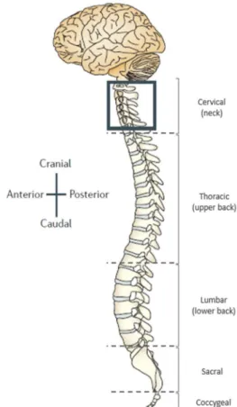
The spinal cord is a narrow tube composed of nervous tissue and is a major component of the central nervous system (CNS). It contacts with the brain through an orifice in the skull, the foramen magnum, and is protected by the vertebral column (Figure 1.1), extending through the spinal canal of each vertebra. Additionally, the spinal cord is also protected by the meninges, three membranes of connective tissue that directly cover the CNS, and by the cerebrospinal fluid (CSF) (Marcus et al., 2014; Van de Graaff, 2011). The spinal cord has two main functions: the conduction of impulses, processing sensory information and providing bidirectional relay between the brain and remaining organs and tissues; and the integration of reflexive involuntary movements, with different nerve pathways than those initiated voluntarily by the brain (Marcus et al., 2014; Van de Graaff, 2011).Transverse cuts of the spinal cord allow the identification of two distinct areas, the white matter and the grey matter (Figure 1.2). The white mater is the outermost part of the spinal cord, consisting of glial cells and bundles of myelinated axons of sensory and motor fibers, running to and from the brain, respectively. The grey matter is the innermost part of the spinal cord and consists of cell bodies and
Figure 1.2 – Spinal cord transverse section. Representation of the grey matter and white matter regions of the spinal cord, as well as the dorsal and ventral horns and the central canal (adapted from OpenStax Anatomy & Physiology, Rice University, 2013)
Figure 1.1 – Central nervous system. Representation of the CNS regarding the body axis. The vertebral column
surrounds and protects the spinal cord (as seen in boxed
area) and can be divided 5 different levels: cervical,
thoracic, lumbar, sacral and coccygeal (Adapted from
2
synapses, neuroglia and unmyelinated interneurons. These two regions are organized in a butterfly-like shape, in which the grey matter is surrounded by the white matter, with two dorsal horns and two ventral horns (Marcus et al., 2014; Mescher, 2013; Van de Graaff, 2011).
Although it is the least complex of the CNS elements, the spinal cord is still composed of various cell types with specific functions and distinct distribution. One of the main cell types are neurons, which form a complex network that receives information, like sensory intakes, and generates motor responses. As previously mentioned, the cell bodies and the axons of neurons in the spinal cord have distinct locations, the grey matter and the white matter, respectively. Different types of glial cells provide the support and maintenance needed for the nervous tissue survival, such as oligodendrocytes, astrocytes, microglia and ependymal cells. Oligodendrocytes produce myelin sheaths, which wrap around axons and give electrical insulation, allowing the efficient transmission of the electric impulse. Astrocytes, the most numerous type of glial cells, are responsible for not only regulating metabolic exchanges but also directly influencing the metabolism of surrounding cell types. Microglia are motile antigen-presenting cells of the CNS, responsible for the immune surveillance of these tissues. Finally, ependymal cells line the central canal of the spinal cords, are responsible for the circulation of the CSF and are a source of adult neural stem cells. (Marcus et al., 2014; Meletis et al., 2008; Mescher, 2013).
1.1.2 Spinal cord vasculature
The spinal cord, as the rest of the CNS, is a highly energy-demanding tissue. It is, therefore, of special importance the existence of an organized and tightly controlled vascular system, in order to provide cells with oxygen and nutrients and remove metabolic waste, maintaining the homeostasis of the tissue (Attwell and Laughlin, 2001; Martirosyan et al., 2011).
1.1.2.1 Vascular structure and blood supply
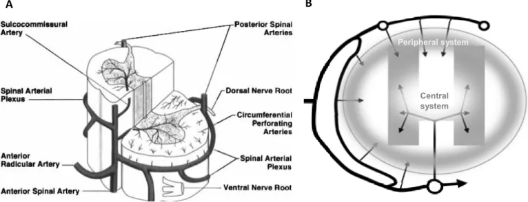
3
Although there is no connection between the capillary beds of the central and peripheral systems, in an intermediate zone there is an overlap of their terminal branches, being indirectly supplied by one system or the other. As the blood flow in each system is different, with a centrifugal flow in the central system and a centripetal flow in the peripheral system (Figure 1.3 B), a watershed zone is established. These zones are characterized by the inexistence of direct blood supply and are, therefore, dependent on the overlapping vasculature. In case of interruption of the blood supply, as occurs in spinal cord injuries (SCI), these zones are particularly vulnerable (Martirosyan et al., 2011; Mautes et al., 2000).
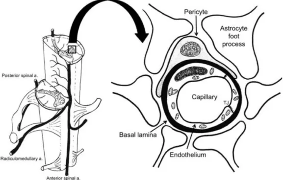
1.1.2.2 Blood - Spinal Cord Barrier
As arteries get thinner and blood vessels reach the intramedullary territory, the blood - spinal cord barrier (BSCB) is established (Figure 1.4). This vascular specialization occurs at the capillary level and involves pericytes, basal lamina, and astrocytes. Capillaries are the smallest blood vessels in diameter, with just one endothelial cell (EC) of thickness, and lack smooth muscle, having instead pericytes with stabilizing and contractile functions. Unlike the capillaries that exist in the peripheral circulation, those of the BSCB are characterized by the absence of cell membrane fenestration and the existence of tight junctions between neighbouring ECs. These cells are then surrounded by a continuous basal lamina, where pericytes are attached. Surrounding the outer surface of the capillaries, astrocytic foot processes are essential to the development and maintenance of the barrier mechanism, modulating the properties of the other BSCB components via secretory mechanisms (Bartanusz et al., 2011; Mautes et al., 2000; Van de Graaff, 2011).
The BSCB, through the existence of physical and molecular barriers, regulates the transport of molecules into the nervous tissue and restricts the contact of potentially toxic agents, such as metabolic
A
Figure 1.3 – Spinal cord vasculature and blood supply. (A) General representation of the main arteries
irrigating the spinal cord and (B) of the blood supply and flow (arrows) to the peripheral and central systems of
the spinal cord. (A –Cheshire et al., 1996; B – Adapted from Mautes et al., 2000)
B
Central system
4
waste or pathogens, with the spinal cord. This way, a stable microenvironment, necessary for the normal neuronal function, is established (Bartanusz et al., 2011; Mautes et al., 2000).
1.2 Spinal cord injury
Although the spinal cord is protected by the vertebrae and the meninges, it is a relatively soft and fragile tissue, with very little collagen and fibrous components and, as such, is especially susceptible to damage directly inflicted to the vertebral column (Mescher, 2013).
1.2.1 General description and types of spinal cord injury
Spinal cord injury is a highly disabling injury and is defined as a damage to the spinal cord, which can result from a contusion, compression, laceration or maceration, causing a temporary or permanent change in its function, bellow the injury site, like loss of sensation and partial or total paralysis, and it may also affect the performance of multiple organs (Ahuja et al., 2017; Kang et al., 2018, Thuret et al., 2006).
It can be divided in two categories: traumatic SCI, that occurs due to an external physical impact, like falls, motor vehicle accidents, sport-related accidents, violence, etc.; or non-traumatic SCI, which can be the result of acute or chronic illnesses, such as vertebral spondylosis, tumorous compression, infections, vascular ischemia and congenital or degenerative diseases (Ahuja et al., 2017; Kang et al., 2018).
5
1.2.2 Epidemiology
According to data provided by the World Health Organization (WHO) in 2013, between 250 000 and 500 000 people world-wide suffer from SCI each year, with the most common cause being falls, followed by motor vehicle crashes and violence, all of them preventable.
Recent information indicates that, with the rise in human activity, the incidence of traumatic SCI also increased, being estimated that it varies from 13.019 cases per million individuals to 163.420 cases per million individuals. Most SCI patients are male (79,8%) and the age profile of SCI patients has a bimodal distribution, with most cases happening in patients with 15 to 29 years of age and, although with a smaller number of cases but growing, in patients with more than 50 years of age. (Ahuja et al., 2017; Kang et al., 2018).
The mortality of SCI patients, although having improved over time, is still 1,5 times to 5 times higher to that of the general population. The survival, longevity and quality of life of SCI patients are positively correlated with less severe injuries and SCI affecting lower levels of the spinal cord like at the lumbar or sacral level, as opposed to more severe injuries and those affecting higher levels, like lesions in the cervical and high thoracic regions (Chamberlain et al., 2015, Kirshblum et al. 2011).
1.2.3 Physiological events after spinal cord injury
The events that occur after a traumatic spinal cord injury can be temporally divided into acute (< 48 hours), subacute (> 48 hours to 14 days), intermediate (14 days to 6 months) and chronic (> 6 months) phases (Figure 1.5), and physiologically divided into primary and secondary injuries (Ahuja et al., 2017).
The primary injury happens immediately after a physical trauma to the vertebral column, leading to the compression or transection of the spinal cord tissue. It instantly causes neural cell death, axon damage and demyelination, which results in an immediate loss of motor and sensory function. The disruption of the BSCB also ensues, causing a severe haemorrhage at the injury site and exposing the spinal cord to immune cells and pro-inflammatory molecules. As a consequence, swelling of the tissues occurs, further compressing the injured spinal cord. These events describe the acute phase of the injury (Figure 1.5 A) and, over time, lead to the spread of the damage to adjacent areas, resulting in a secondary injury. The consequent cell death and axonal retraction, the absence of a functional microvasculature, the presence of pro-apoptotic, of pro-inflammatory and of cytotoxic molecules and the resulting activation of immune cells, all aid in the maintenance of an acute inflammatory response that is described as a subacute phase of the injury (Figure 1.5 B) (Ahuja et al., 2017; Oudega, 2012; Tewari et al.,2010).
6
and vascular repair (Oudega, 2012) and by the expression of regeneration associated genes (Tetzlaff et al. 1991) and remodelling of the neural circuitry (Dietz et al., 2009). However, and even if in some cases some functional recovery is observed years after the injury, these self-repair attempts are mostly unsuccessful due to the development of growth-inhibitory structures, like a scar, with reactive astrocytes, that surrounds and isolates fluid filled cystic cavities where cell death occurred. In addition, the presence of growth-inhibitory molecules, like myelin debris and other molecules released by degenerating oligodendrocytes, and lack of functional vasculature further hinder the self-repair process (Ahuja et al., 2017; Oudega, 2012).
Figure 1.5 – Events after spinal cord injury. Main events that occur, after a mechanical injury to the spinal cord. (A) In the acute phase, death by necrosis and apoptosis, axonal disruption, swelling of the tissue, BSCB disruption and severe
haemorrhage are observed and the beginning of the inflammatory response occurs. (B) In the subacute phase, an increased
inflammatory response occurs due to the production/release of pro-inflammatory molecules and the invasion of immune
cells, further damaging the tissue. (C) The intermediate to chronic phase is marked by the presence of a cystic cavity
surrounded by a glial scar, composed of reactive astrocytes, that restricts endogenous attempts at a functional recovery.
(Ahuja et al., 2017)
C
7 1.2.3.1 Blood - Spinal Cord Barrier disruption
The disruption of the BSCB is a major event after SCI (Figure 1.6). As previously mentioned, the destruction of blood vessels at the injury site leads to the consequent contact between nervous tissue and toxic blood components, further increasing cell death. Additionally, the shear stress from the initial trauma may damage blood vessels in adjacent areas, leaving them hyperpermeable, allowing the passage
A
B
B’
B’’
C
Figure 1.6 – Blood - Spinal cord barrier disruption after spinal cord injury. (A) BSCB in a healthy spinal cord. Blood and nervous tissue are physically separated by a vascular specialization, maintaining the homeostasis of the spinal cord
tissue. (B) Disruption of the BSCB after a physical trauma. Blood vessels are severed at the injury site (B’), causing
haemorrhage and, consequently, loss of homeostasis and cell death. At the periphery of the injury (B’’), the BSCB is damage
and blood components leak to the tissue. (C) In a chronically contused spinal cord, no new blood vessels are formed at the
injury site and BSCB in peripheral areas continue to be hyperpermeable. WM – white matter, GM – grey matter, EC-
endothelial cells, TJ – tight junctions, PC – pericytes, BL – basal lamina, JAM - junction adhesion molecules (Oudega,
2012)
Damaged BSCB in adjacent tissue: Hyperpermeability Normal functioning blood vessels:
BSCB
Severed blood vessels at the epicentre: haemorrhage
No new blood vessels in the epicentre; leaky blood vessels in
8
of immune cells and toxic molecules, further damaging the spinal cord tissue. Through time, an angiogenic response fails to take place. As a consequence, no new blood vessels are formed in the epicentre of the injury, impairing the normal blood supply to this area. In adjacent regions, new and old blood vessels remain leaky, being a continuous source of damage to the already hurt tissue (Figure 1.6 C) (Oudega, 2012). The leakiness of the peripheral vessels may result from the detachment of pericytes from the blood vessels and their migration to the injury site, where they differentiate into fibroblast-like cells and contribute for the formation of the stromal component of the glial scar (Goritz et al., 2011).
1.2.4 Complications and current treatments
Besides the immediate loss of sensation and of motor function, many other complications can arise from a spinal cord injury. In most cases, patients suffer from spasticity (involuntary muscle contractions), pressure ulcers, excessive neuro-inflammation and consequent neuropathic pain. Bladder control is affected and, in many cases, bowel control is also affected. In more severe SCI cases, like in cervical and high thoracic lesions, autonomous nervous function can be affected, resulting in loss of core temperature control, cardiovascular and respiratory complications. Furthermore, SCI patients, due to the seriousness of this type of injury and the future obstacles they face, are especially susceptible to experience anxiety and develop depression (Ahuja et al., 2017; Hagen, 2015).
Although there is no effective treatment for SCI, neuroprotective interventions can be applied to minimize loss of neural tissue and improve quality of life. It is therefore important to provide efficient and effective medical care right after injury. Non-pharmacological treatments involve the surgical decompression of the spinal cord right after injury, rehabilitation to prevent muscle waste and, in cases of subacute phase SCI or chronic motor-incomplete SCI, to recover as much function as possible, functional electrical stimulation. Pharmacological approaches involve the use of medication to reduce the inflammation and swelling of the spinal cord, control pain, manage spasticity and improve bowel and bladder function. (Ahuja et al., 2017; Baptiste and Fehling, 2007; Cristante et al., 2012) Cell transplantation therapies, like of neural stem cells, present a interesting method to improve sensory and motor function in chronic SCI patients, with some studies in phase I human clinical trials (Curtis et al., 2018; Thuret et al., 2006). However, a better understanding regarding the mechanisms involved to promote the potential functional recovery is still needed as well as further investigation about their safety and risks in human applications (Assinck et al., 2017).
1.2.5 Spinal cord injury experimental models
9
Three different models for SCI, that mimic specific clinical features, have been established: the transection model, the contusion model and the compression model (Figure 1.7). The transection model (Figure 1.7 A) includes all forms of lacerations, from small incisions and dorsal or lateral hemisections to complete cuts of the spinal cord, and has a more contained area of damage. Despite not being a very common type of SCI in humans, this model is used in studies where direct axonal growth, through the glial scar, is of interest. The contusion model (Figure 1.7 B) is produced by a focal impact on the spinal cord, most commonly dorsal, giving rise to an anatomically incomplete injury with a rim of spared white matter, the formation of cavities inside the spinal cord and with spread of the damage to areas adjacent to the injury. Contusions are the most common type of SCI in humans and therefore, this model is of particular relevance in terms of human SCI pathology. It is experimentally performed using a weight drop method or an impactor. The compression model (Figure 1.7 C) has the same pathophysiological consequences as the contusion model, however, rather than a focal force, a lateral or dorsal force is generally applied with the use of forceps, clips or specialized apparatus. Compression models have a broader dorsal and rostral impact and can have a bigger effect on lateral white matter areas than contusion models. The severity of this type of injury can vary depending on the instrument used and the duration of compression (McDonough, 2012; Oudega, 2012).
Mammal models like rat (Rattus norvegicus) or mouse (Mus musculus) are valuable SCI models as they emulate the locomotor and sensory deficits that happen in humans after a SCI, not being able to recover to their initial state. Although larger animals and non-human primates have more similarities with humans regarding the physiological response to SCIs, rodent models are better suited for preliminary studies since they are relatively smaller and cheaper to maintain, have better understood anatomy and genome, bigger availability of genetic tools, and less regulatory requirements and ethical restrictions. In contrast with these models, animals that are capable of regeneration during adulthood are used in order to understand and compare the pathophysiological, cellular and molecular differences during SCI and provide hints to improve the repair process in non-regenerating animals. An example of Figure 1.7 –Spinal cord injury models. Main types of experimentally induced spinal cord injuries models, (A)
transection model, (B) contusion model and (C) compression model, and the respective extent of damage, seen
10
an animal model with regenerative abilities is the zebrafish (Danio rerio) (Sharif-Alhoseini et al., 2017; Steward and Willenberg, 2017).
1.2.5.1 Zebrafish as a spinal cord injury model
Zebrafish, a small teleost fish, is a well known developmental model due to characteristics like having external fertilization and development, being optically transparent until early adulthood and having a rapid development, with the completion of embryogenesis after 5 days post fertilization. Furthermore, only one couple can produce a high number of offsprings (100-300 embryos), providing statistically significant sample size and facilitating the maintenance of zebrafish lines. Since these fish exhibit a high degree of similarities with mammals regarding molecular pathways and mechanisms, and due to the variety of genetic tools and mutant and transgenic lines currently available, zebrafish present an interesting and less expensive model to use in preliminary studies. (Kari et al., 2007)
As previously mentioned, zebrafish has regenerative abilities, being able to regenerate fins, jaw, heart, pancreas, liver, kidney and, of course, CNS structures, like the spinal cord (Gemberling et al., 2013). Although the result after SCI in vastly different to that of mammals, the spinal cord cellular architecture is relatively similar and therefore useful to study the regenerative process (Dias Quiroz and Echeverri, 2013; Hui et al., 2010)
Transection and compression models of spinal cord injury can be performed in zebrafish (Becker and Becker, 2008; Fang et al., 2012). After SCI, loss of swimming behaviour is observed mainly due to the paralysis of the posterior portion of the body, bellow the injury site. In compression models, very little movement is observed 3 days after the injury, when compared to unlesioned fish. Movement progressively increases after 15 days and an almost complete recovery of swimming ability is observed 1 month after injury. In a complete transection of the spinal cord, 42 days after injury, axonal projections of brainstem neurons with spinal projection are seen beyond the injury site and fish show almost complete recovery of their normal swimming behaviour (Becker and Becker, 2008; Hui et al., 2010).
Pathophysiological analysis show that, after a compression injury, adult zebrafish spinal cord experiences cell death of both neurons and glial cells, rupture of blood vessels, with consequent haemorrhage and release of immune cells and pro-inflammatory molecules to the spinal cord. This follows what is observed in mammals. However, and in contrast to the SCI response in mammals, apoptotic cell death and macrophage infiltration reach a peak earlier, at 1 day and 2-3 days after injury, respectively, decreasing afterwards. Furthermore, the formation of a glial scar is not observed in zebrafish (Ghosh and Hui, 2018; Hui et al., 2010).
11
indicates a permissiveness of the environment of the adult zebrafish spinal cord for axonal regeneration and a plasticity in terms of cell differentiation and network integration (Becker and Becker, 2008; Becker and Becker, 2014; Hui et al., 2010; Reimer et al., 2008).
Despite these findings, not many studies focus on the vascular recovery during spinal cord regeneration in zebrafish, even though vascular integrity is essential in the maintenance of homeostasis and its disruption contributes to the spread of the secondary injury in mammals, as previously mentioned. Information regarding the recovery of the vasculature after SCI could be essential in establishing new players and/or mechanisms that, together with other biological or pharmacological strategies, could be applied to efficiently restore the malfunctioning vasculature in mammal SCIs (Oudega, 2012)
1.3 Blood vessel formation
The two main processes from which new blood vessels arise are called vasculogenesis and angiogenesis (Figure 1.8). Vasculogenesis mainly occurs during development and is defined as a process where epithelial precursor cells, also known as angioblasts, differentiate and join to form a primitive vascular network (de novo formation). Angiogenesis is the development of new blood vessels from pre-existing ones and can be divided in sprouting angiogenesis with proliferation of ECs, and in intussusceptive angiogenesis, where a split in pre-existing blood vessels occurs (Kolte et al., 2015).
1.3.1 Sprouting angiogenesis
12
metalloproteinases; (ii) ECs proliferation and migration into the connective tissue, with specialization into tip and stalk cells; (iii) contact of ECs and cord formation; (iv) lumen formation and (v) establishment of anastomosis to establish functional capillary loops, with synthesis of new basement membrane and recruitment of pericytes. (Kolte et al., 2015; Ribatti and Crivellato, 2012).
1.3.1.1 Main cellular and molecular players
The angiogenic process is regulated by pro- and anti-angiogenic factors that exist in a dynamic balance, in physiological conditions. ECs can remain quiescent for years. However, in response to tissue damage or oxygen and nutrient deprivation, the molecular angiogenic balance is disturbed and angiogenic sprouting is initiated. This process requires ECs to take different roles and morphologies, giving rise to tip cells, which primarily migrate, and stalk cells, that mainly proliferate. Tip cells extend numerous filopodia and respond to angiogenic stimuli, guiding the new branch vessel. Stalk cells are responsible for the formation of tubes, branches and of the nascent vascular lumen. After the formation of new blood vessels, tip cells adopt a lumenized, immobile phenotype that promotes vessel integrity and stabilization, not responding to angiogenic clues. (Kolte et al., 2015; Ribatti and Crivellato, 2012)
The main regulator of the angiogenic process is the vascular endothelial growth factor (VEGF) pathway (Figure 1.9). Tip cell migration is driven by a gradient of VEGF, drifting away from the parent blood vessel, while stalk cells proliferate and lumenize as a response to VEGF concentration. Tip cells filopodia express VEGF receptor 2 (VEGFR-2, also known as KDR or FLK1), a tyrosine kinase receptor that positively responds to VEFG and activates a angiogenic cascade. VEGFR-1 (also known as FLT1), can have a membranar (mFLT1) or soluble form
(sFLT1) and is mainly expressed in stalk cells. It is involved in guidance of tip cells, preventing their outward migration through the reduction of VEGF availability, and limiting tip cell formation, therefore having an anti-angiogenic effect. Interference with these receptors can result in angiogenic defects, such as increase of sprouting and vascularization in case of loss of VEGFR-1 and defects of sprouting in cases of blockade of VEFGR-2. The opposite effects are seen in case of increase of each receptor (Chappel and Bautch, 2010; Chappel et al., 2012; Matsuoka et al., 2016; Ribatti and Crivellato, 2012).
Other pathways also have an important contribute to the angiogenic process, such as the Notch pathway, and other factors, like the fibroblast growth factor (FGF), angiopoetin (Ang), platelet-derived Figure 1.9 – VEGF receptors expressed by endothelial cells and VEGF specificity. Differents types of VEGFs, including placenta growth factor (PIGF) bind in a specific manner to different VEGFR, having distinct downstream
13
growth factor (PDGF)-β and transforming growth factor (TGF) work together with VEGFs and regulate vessel proliferation, migration and maturation (Chappel and Bautch, 2010; Chappel et al., 2012; Kolte et al., 2015; Ribatti and Crivellato, 2012; Ten Dijke and Arthur, 2007).
1.3.1.2 Angiogenesis during spinal cord injury
As previously mentioned, after SCI in mammals, an attempt at vascular repair occurs. However, no blood vessels are formed at the injury site and, in areas adjacent to the injury, the newly formed blood vessels are not functional, allowing the leakage of their contents to the spinal cord tissue, further damaging it. The use of different strategies to promote the revascularization of the spinal cord after injury, like the use of biomaterials coupled with pro-angiogenic molecules (Haggerty et al., 2018; Rao et al., 2018), the delivery of pro-angiogenic factors to the injury site (Yu et al., 2016) or use of microRNAs (Hu et al., 2015), have resulted in an improvement of motor function, pointing to the importance of tissue revascularization for spinal cord regeneration.
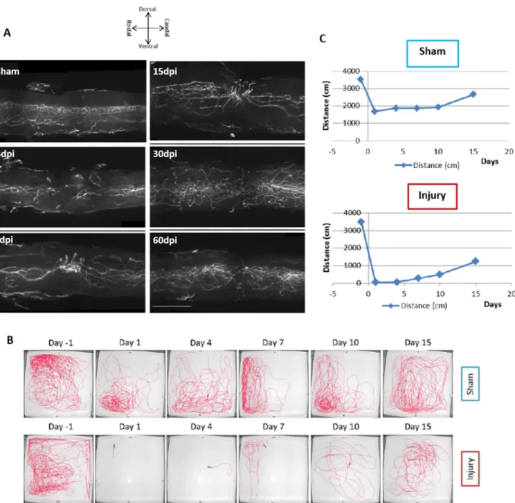
In zebrafish, the recovery of the vasculature seems to follow the regenerative process of the spinal cord. Previous work done in our lab (Figure 1.10), using the zebrafish compression model, showed that, since injury and until 15 days post injury (dpi), there is an increase of blood vessels at the injury site, followed by a decrease in number and a vascular reorganization seen at 30 and 60 dpi. It is worth to note that these images represent just a portion of the spinal cord vasculature (160 µm), due to restraints from the method of image acquisition used. Additionally, blood flow, through cardiac injection of a fluorescence compound at 7 dpi, was observed, indicating that, at this timepoint, the BSCB is reestablished. A functional swimming assay, done 1 day before injury and until 15 dpi, confirms an improvement of swimming ability after injury. These results could potentially indicate a correlation between the repair of the vasculature and the recovery of motor function that occurs during the regenerative process of the spinal cord (Maçarico, 2014).
14
Figure 1.10 – Vascular and functional recovery after spinal cord injury. (A) Spinal cord vasculature was observed
at different time points, (3, 7, 15, 30 and 60 dpi).Until 15dpi, an increase of blood vessels at the injury site is observes.
At 30 and 60 dpi, a reorganization of the vessels at the injury site is seen. (B and C) Funtional swimming assays in
15
1.4 Aims of the study
In this study, we took advantage of light sheet fluorescence microscopy to revisit the dynamics of the vasculature recovery after SCI to allow the observation of the full vasculature at the injury area, as previous work was only able to obtain portions of the target vasculature.
As previously mentioned, the vascular response of mammals, like humans, rat and mouse, and that of zebrafish to a SCI is quite distinct, with zebrafish being able to form new blood vessels at the injury site, in contrast to what happens in mammals. In addition, in mammals, blood vessels from areas adjacent to the injury become malfunctional, leaking blood to the spinal cord tissue. In zebrafish, although previous work from our lab showed that, at least at 7 dpi, the BSCB is reestablished, it was not known at what time after injury did the newly formed blood vessels regain their function. Therefore, in this work, the blood vessels of the previously mentioned spinal cords were observed in order to pinpoint the period of reestablishment of the BSCB. This was possible through the cardiac injection of a fluorescent compound, before spinal cord extraction, at defined timepoints.
17
Chapter 2. Methods & Materials
2.1 Animal Model
2.1.1 Zebrafish Husbandry
Adult zebrafish (Danio rerio) were kept in standard Tecniplast rack systems and were maintained at 28ºC, in standard pH and conductivity conditions (Westerfield, 2000). During the week, adult zebrafish were fed two meals of only dry food (SPAROS, Portugal) or dry food and a live feed, decapsulated Artemia (ZM Systems), when available. On the weekends and holidays, only a single dry food meal was given.
The surgical procedures involving adult zebrafish were done or supervised by users licenced by the Direcção Geral de Alimentação e Veterinária (DGAV). All the experiments involving animals were approved by the Animal User and Ethical Committees at Instituto de Medicina Molecular – João Lobo Antunes, in accordance with directives from DGAV (PORT 1005/92).
Fish with SCI were kept in individual 1L breeding tanks (external tank and internal tank with perforated bottom) (Tecniplast), in baths at 28ºC or at heat-shock temperature, in 1x Embryo Medium (reverse osmosis water with 5 mM NaCl; 0,17 mM KCl; 0,33 mM CaCl2٠2H2O; 0,33 mM
MgSO4٠7H2O), and were only fed once a day, with decapsulated Artemia, after the second day of injury.
The temperature of the bath was continuously monitored to avoid increase or decrease of set temperature. To avoid ammonia peaks and development of fungi or wound infections, the Embryo Medium and external tanks were changed daily.
For breeding, male and female zebrafish were placed in 1L breeding tanks before the end of the day, with divider. In the next morning, the divider was removed to initiate mating. Sometime later, embryos were collected and rinsed, first with reverse osmosis water and afterwards with 1x Embryo Medium with Methylene Blue (0.97 µM) (Sigma Aldrich) and were placed in petri dishes with 1x Embryo Medium with Methylene Blue. Embryos were then placed in the incubator at 28ºC until the start of procedures. Embryonic stages were confirmed according to Kimmel et al., 1995.
2.1.2 Zebrafish lines
In this study, the following zebrafish lines were used: Wildtype AB strain established in the fish facility at iMM-JLA, Tg(kdrl:EGFP)s843 (Jin et al., 2005) and Tg(kdrl:EGFP)s843(hsp70l:sflt1;
cryaa:cerulean)bns80 (Matsuoka et al., 2016). Wildtype AB zebrafish were only used as breeders with the
18
increase of temperature, also known as heat-shock treatment. This construct also contained genetic information for a cyan fluorescent protein (CFP), cerulean, whose expression was driven by the cryaa promotor, labelling the eye lens.
2.2 Spinal cord injury
Adult zebrafish (5 - 9 months-old) were anesthetised by immersion in 0,016% (w/v) Tricaine-S solution. The fish were placed, left side up, on a moulding clay support covered with chromatography paper (GE Healthcare), on top of a cooling pad, as shown in Figure 2.1.
After removal of the scales, a longitudinal incision, parallel to the first light dorsal stripe, was made halfway between the base of the skull and the dorsal fin using surgical scissors (Vannas-Tübingen Spring Scissors - Straight, FST). The vertebral column and the spinal cord were exposed, and the spinal cord was compressed dorsoventrally using forceps (Dumont #55, FST) (Figure 2.2). After gently removing the forceps and bringing together the limits of the incision, the wound was sealed with Vetbond (3M) and the fish were allowed to recover in individual tanks. Sham injury fish were obtained following the same procedure without the compression of the spinal cord. (Fang et al., 2012) All surgical material was sterilized with 75% ethanol between procedures.
If at any point before the end of the experiment the injured fish showed abnormal behaviour or signs of decay after the injury procedure, the fish were euthanized using a lethal dose (>1500 mg/L) of Tricaine-S solution (MS222, Western Chemical Inc.).
A
B
C
19
2.3 Inhibition of angiogenesis - Heat-shock Protocols
2.3.1 Heat-shock protocols in larvae for imaging
To confirm promoter activation of the hsp70l:sflt1 transgenic line after heat-shock and to assess the inhibitory effect of sflt1 overexpression on angiogenesis, the development of the intersomitic vessels (ISVs) was followed.
Embryos at 17-somite stage, before the development of these vessels (Isogai et al., 2003), were subjected to 1 hour (h) heat-shock, in pre-warmed embryo medium, at 37ºC (Matsuoka et al., 2016) and were observed at, approximately, 36 hours post fertilization (hpf).
Heat-shock at 34ºC was also done, with several periods of heat-shock: 30 minutes, 1h, 1h30, 2h, 4h, 6h, 8h, 10h,16h, 18h and 1 day. Heat-shock was induced at 17-somite stage and, once again, the embryos were observed at approximately 36hpf.
In addition, the development of the vasculature at standard temperature (28ºC) was likewise monitored at 36 hpf.
2.3.2 Heat-shock protocols in adults for imaging
Several heat-shock protocols for adult zebrafish were tested. Heat-shock protocols at 37ºC had a duration of 2h, twice a day, until 7 dpi with the difference between them being the day of the beginning of the protocol.
The following protocols were tested: heat-shock right after injury – HS 37ºC (0 - 7) dpi; heat-shock beginning at 3 dpi – HS 37ºC (3 - 7) dpi; heat-shock beginning at 4 dpi – HS 37ºC (4 - 7) dpi; and heat-shock beginning at 5 dpi, with an extra day of heat-shock before injury – HS 37ºC (-1, 5 - 7) dpi. In addition to these, an extended heat-shock protocol was tested, beginning at 4 dpi and ending at 14 dpi – HS 37ºC (4 - 14) dpi.
Figure 2.2 – Spinal cord injury procedure. (A) After scale removal, a clean longitudinal incision was done, (B)
the spinal cord was gently exposed using forceps and (C) compressed dorsoventrally. (Adapted from Fang et
20
At 34ºC, the following protocols were tested: 6h heat-shock, twice a day, starting at 2 dpi – HS 34ºC 6h (2 - 7) dpi; and continuously at 34ºC, beginning at 2 dpi – HS 34ºC cont. (2 - 7) dpi. An extended version of the continuous protocol was also tested, starting at 2 dpi and ending at 14 dpi – HS 34ºC cont. (2 - 14) dpi.
2.4 Rhodamine Injection
Injured adult zebrafish were anesthetised by immersion in 0,016% (w/v) Tricaine-S solution. The fish were placed, ventral side up, in a foam support and the heart was exposed using forceps and surgical scissors (Figure 2.3 A). Tetramethylrhodamine dextran 10 kDa (3µg/µl), (ThermoFisher Scientific, D1824), here referred to as rhodamine, was injected directly in the heart using needles made from borosilicate capillaries (World Precision Instruments, Inc.) and inserted into an aspirator tube (Sigma, A5177). The procedure was done in an Olympus MVX10 microscope and the injection was followed using fluorescence. The injection was then confirmed by inspecting the presence of rhodamine inside the caudal fin blood vessels (Figure 2.3 B).
2.5 Spinal cord extraction
The anesthetised fish were euthanized through decapitation using a scalpel blade and the vertebral column was dissected in cold 1x Phosphate Buffer Saline (1x PBS) (1.37mM NaCl, 0.27mM KCl, 1mM Na2HPO4·7H2O, 0,2mM KH2PO4). The samples were fixed using 4% paraformaldehyde
(PFA) at 4ºC overnight. After fixation, the spinal cord was isolated from the vertebrae.
B
B.2
A
Figure 2.3 – Rhodamine Injection Procedure. (A) After anaesthesia, the fish’s heart was exposed (arrow) and
rhodamine was injected directly into the blood stream. (B) The injection’s success was confirmed by the presence
of rhodamine inside the caudal tail blood vessels. B.1 is a close up of the selected area of image B.
21
2.6 Immunohistochemistry
Two immunohistochemistry protocols for whole spinal cord were tested, with the main difference between them being the composition of the blocking solution. In both protocols, the primary and secondary antibodies used were the same: anti-GFP rabbit primary antibody (ThermoFisher Scientific, A6455) and anti-rabbit Alexa 488 goat secondary antibody (ThermoFisher Scientific, A-11034).
In the first protocol, spinal cords were incubated in Blocking Goat Serum (BGS) solution (1x PBS; 10% Goat Serum; 0,1% Triton X-100) for 2 days at 4ºC and then incubated with anti-GFP primary antibody solution (BGS with 2µl/ml of primary antibody) for another 2 days. The samples were twice washed in PBST (1x PBS; 0,1%(v/v) Triton X-100) and left in the roller during the day, and then incubated with secondary antibody solution (BGS with 1µl/ml of secondary antibody) for 2 days. Finally, the samples were twice washed in PBST.
For the second protocol, spinal cords were incubated in a different blocking solution (1x PBS; 1%(w/v) Bovine Serum Albumin (BSA); 1%(v/v) Dimethyl sulfoxide (DMSO); 0,05%(v/v) Triton X-100) for 2 days at 4ºC and then incubated with anti-GFP primary antibody solution (1x PBS; 1%(w/v) BSA; 0,1%(v/v) Triton X-100; 2µl/ml primary antibody) for another 2 days. The samples were twice washed in PBST (1x PBS; 0,1%(v/v) Triton X-100) and left in the roller during the day, and then incubated with secondary antibody solution (1x PBS; 1%(v/v) BSA; 0,1%(v/v) Triton X-100; 1µl/ml secondary antibody) for 2 days. Finally, the samples were twice washed in PBST.
2.7 Whole spinal cord clearing
The clearing protocol was adapted from the Scale protocol from Hama et al.,2015. Whole spinal cords were subjected to a clearing protocol before light sheet fluorescence microscopy acquisition. Samples were placed in Scale A2 (4M urea; 10%(w/v) glycerol; 0,1%(v/v) Triton X-100) for 3 days, at 4ºC. The samples were then incubated in Scale B4 (8M urea; 0,1%(v/v) Triton X-100) at 37ºC for 1 day and, afterwards, switched back to Scale A2 for a minimum of 3 days. At least a day before acquisition, the samples were incubated in Scale S4 (4M urea; 40%(w/v) Sorbitol; 10%(w/v) glycerol; 0,2%(v/v) Triton X-100; 15%(v/v) DMSO).
2.8 Motor Function Recovery Assay
22
injury (1 dpi). From 2 dpi, the fish were subjected to a continuous heat-shock at 34ºC and their swimming ability was again analysed at 7 dpi, 14 dpi and 21 dpi. For each measurement, zebrafish were individually placed in a 35cm x 35cm tank filled with system water, illuminated from below. The trials were recorded using a camera placed above the tank and the tracking was done using Ethovision software (Noldus, Wageningen, The Netherlands). Each fish was allowed to freely explore the tank for 15minutes – Open Field Test (Stewart et al., 2012)- of which 5 minutes were done so the animal could acclimatise to the tank and the final 10 minutes to record the fish’s movement. After each trial, the water of the tank was changed to avoid exposure of the next fish to stress hormones from previous ones. Different people were responsible for the injury and tracking procedure to ensure that the person recording the fish did not know their genotype. After the end of the experiment, the results of injured zebrafish were divided into the corresponding groups and their swimming distance for each fish per timepoint was analysed using Prism software (GraphPad Software). Swimming distances from both lines, for each tracking timepoint, were statistically compared using a two-way ANOVA multiple comparisons test (alpha of 0.05, with 36 degree of freedom (df)), with a Sidak test for multiple comparison corrections.
2.9 Image Acquisition & Analysis
Transmitted light and fluorescence images from adult zebrafish and from control and heat-shocked embryos were acquired either using a Zeiss AxioZoom V16 microscope (Carl Zeiss MicroImaging) with a PlanNeoFluar Z 1x objective, with GFP (excitation: 450 – 490 nm, emission: 500 – 550 nm) and RFP (Red Fluorescent Protein; excitation: 559 – 585 nm, emission: 600 – 690 nm) filter sets, or an Olympus MVX10 microscope (Olympus) with an Olympus MVPlapo 1x objective, and with GFP (excitation: 460 – 480 nm, emission: 495 – 540 nm), RFP (excitation: 535 – 555 nm, emission: 570 – 625 nm) and CFP (excitation: 425 – 445 nm, emission: 460 – 510 nm) filter sets. Both microscopes were equipped with monochromatic AxioCam MRm cameras (Carl Zeiss MicroImaging). The software used was ZEN 2012 Blue Edition (Carl Zeiss MicroImaging).
23
and the injected rhodamine were excited with 488nm and 561nm wavelength laser units, respectively. Emitted light was directed to GFP (emission: 505 – 545 nm) or Cy3 (emission: 575 – 615nm) filter sets to reduce unspecific signal, before reaching the primary camera. With this method, thin optical slices were made using double-sided illumination, with left and right adjustable light sheets, and a z-stack of the complete target area of the cleared sample was obtained (Weber et al., 2014).
Preliminary images from spinal cord sample obtained in the motor functional recovery assay were preliminarily acquired using confocal laser point-scanning microscopy with a Zeiss LSM 880 (Carl Zeiss MicroImaging) equipped with a 25x LCI Plan-Neofluar objective, Argon (488 nm), DPSS 561-20 (561nm) laser units and Green (excitation : 450 nm – 490 nm; emission: 500 nm – 550 nm) and Red (excitation: 533 nm – 558 nm; emission: 570 nm – 640 nm) filter sets.
Maximum intensity orthogonal projections of the acquired z-stack images were done using ZEN 2012 Blue Edition and stitching of the tiles was done manually using Photoshop (Adobe).
Image analysis for heat-shock samples was done using a custom macro for FIJI software (Schindelin et al., 2012), written by Anna Pezzarossa from Edgar Gomes Lab, at iMM-JLA Lisbon. It required a .czi file of the acquired spinal cord z-stack as input and, when running, selection of regions of interest (ROI) to analyse. The following parameters were analysed: branch length and tortuosity (branch length/ euclidian distance).
A
C
B
Figure 2.4 – Light sheet microscopy acquisition. Representation of (A) natural position of the spinal cord and
body axis; (B) spinal cord placement for light sheet acquisition and coordinates (x,y,z), and (C) direction of
z-stack acquisition and corresponding body axis according to the spinal cord placement; D – dorsal, V – ventral, R
24
25
Chapter 3.Results
3.1 Reestablishment of the Brain - Spinal Cord Barrier
Work done previously in our lab showed that new blood vessels are formed at the injury site during spinal cord regeneration in zebrafish. However, it was important to obtain information regarding their functionality as this could be one of the main differences between zebrafish and mammals, whose remaining blood vessels are not functional, further damaging the spinal cord tissue.
In order to observe the reestablishment of the BSCB, Tg (kdrl:EGFP) zebrafish at 1, 3, 5, 7, 14 and 30 dpi were subjected to cardiac injection of rhodamine. If these new blood vessels were functional, rhodamine would be retained due to the reestablishment of the BSCB. If not, rhodamine would be leaked to the surrounding tissue. Sham injuries were also done to ensure that the surgical procedure of exposing the spinal cord did not influence the overall vasculature. Spinal cord samples were isolated, fixed and subjected to a whole mount immunohistochemistry protocol, followed by a clearing protocol, in order to observe the vasculature and its overall recovery and gain of function through time, using light sheet microscopy.
3.1.1 Spinal cord vasculature after injury
26
Figure 3.1 – Representative images of Sham and 1, 3, 5, 7 and 14 dpi of Tg (kdrl:EGFP) spinal cords.
The images show an increase blood vessels (GFP) at the injury site, starting at 3dpi, and an increase in their
27
Throughout the analysed timepoints, a perceptible increase of rhodamine at the injury site occurs between 3 dpi and 7 dpi. This indicated that the major gain of function of the new blood vessels and, consequently, the reestablishment of the BSCB happened between these timepoints.
3.1.2 Quantification of the Reestablishment of the BSCB
Although a perceptible difference in the presence of rhodamine inside the newly formed blood vessels is seen, throughout the timepoints, it was important to quantify these results and confirm their significance. For this, a simple analysis of the orthogonal projection for Sham injury and 1, 3, 5, 7 and 14 dpi samples was done, firstly using Fiji and then Prism software. A threshold was defined for both channels of each sample and the area of the positive pixels measured, obtaining the percentage (%) of the positive area for each channel. It is important to note that blood vessels in injured areas are much brighter than those in adjacent areas, independently of the immunohistochemistry protocol used (although spinal cords subjected to the second protocol presented a more uniform GFP signal than those subjected to the fist protocol (images not shown)). Therefore, the threshold was manually set as to include fainter blood vessels, without losing definition of brighter ones. Samples were then analysed for the amount of rhodamine and GFP in the samples (ratio %red / %green), and the results (mean ± standard deviation) were plotted (Figure 3.3). Sham injuries, as the vasculature was not affected, had an almost perfectly matched % of rhodamine and GFP, having a ratio of 0.998 ± 0.0014. One day after injury, a ratio of 0.214 ± 0.038 was observed (although setting the threshold in these samples was more difficult due to the existence of leaked rhodamine in the tissues). At 3 dpi, a ratio of 0.342 ± 0.094, was obtained; over time, this ratio increased, being 0.496 ± 0.066 at 5 dpi and 0,706 ± 0.039 at 7 dpi. At 14 dpi, the ratio changed only slightly, being 0.738 ± 0.125.
Figure 3.2 – Tg (kdrl:EGFP) 30 dpi samples. The injury site was still visible, now with less blood vessels
(GFP). The vasculature was able to retain rhodamine. Scale bar: 100 µm
28
The results for each timepoint were then compared, using an unpaired T-test statistical test with a correction for multiple comparisons using the Holm-Sydak method, with a p value of < 0.05, to measure statistically significance. Differences between 3 dpi and 5 dpi samples (P value = 0.016) and between 5 dpi and 7 dpi (P value = 0.0003) were found to be statistically significant but not those between 1 dpi and 3 dpi (P value = 0.076) and between 7 dpi and 14 dpi (P value = 0.614). This goes in accordance to what was previously mentioned and supports the idea that the BSCB reestablishment, with the timepoints tested, occurred mainly between 3 dpi and 7 dpi.
3.2 Inhibition of angiogenesis - Heat-shock treatment
In order to assess the importance of the angiogenic response observed during spinal cord regeneration, a genetic method was chosen to inhibit angiogenesis, as previous attempts with pharmacological methods were not successful. The transgenic Tg (kdrl:EGFP)(hsp70l:sflt1, cryaa:cerulean) zebrafish line was used, hereby abbreviated Tg (hsp70l:sflt1) (Matsuoka et al, 2016). This transgenic line has GFP labelled blood vessels due to the GFP driven expression by the kdrl promotor, which is a known receptor of the VEFG pathway and is present in ECs. This line also has a genetic construct that allows temperature inducible activation of the hsp70l promotor that drives the expression of sflt1. sFLT1 is the soluble form of a receptor also involved in VEGF signalling, and therefore in angiogenesis. However, and in contrast to KDRL, sFLT1 acts as a decoy receptor, controlling the availability of VEFG and restricting blood vessels formation. Due to this, this transgenic line can be used as a tool to inhibit the angiogenic process during regeneration.
Figure 3.3 – Ratio rhodamine/GFP per timepoint. Mean ± standard deviation values regarding the ratio
%rhodamine/%GFP, for Sham injury and days post injury timepoints, were calculated and plotted. * - Statistically