Universidade de Lisboa
Faculdade de Medicina Dentária
The Effect of Platelet Rich Fibrin in Oral Surgery:
A Literature Review
Beatriz Mancebo Vieira Pedro
Dissertação
Mestrado Integrado em Medicina Dentária
2017
Faculdade de Medicina Dentária
The Effect of Platelet Rich Fibrin in Oral Surgery:
A Literature Review
Beatriz Mancebo Vieira Pedro
Dissertação orientada
Pelo Prof. Doutor João Caramês
e Prof.ª Doutora Helena Francisco
Mestrado Integrado em Medicina Dentária
I
A quem sempre acreditou em mim e, mesmo longe, esteve ao meu lado.
II
Chegar ao final desta etapa e concretizar este sonho não teria sido possível sem o apoio incondicional de pessoas fantásticas que cruzaram o meu caminho e que me enriqueceram tanto a nível pessoal como profissional.
A todos, o meu profundo e sincero obrigada,
Ao Professor Doutor João Caramês, uma referência para mim no ramo da Cirurgia Oral, por ter aceite o meu convite para orientar esta dissertação. É, sem dúvida, um exemplo a seguir no que respeita aos seus valores como pessoa, à sua dedicação à profissão e à sua vontade sincera em ajudar quem o procura.
À Professora Doutora Helena Francisco, pela orientação cuidadosa e amável que sempre me prestou e por todas as horas dedicadas a este trabalho. Foi essencial na elaboração e conclusão desta dissertação.
A todos os colegas, docentes, e funcionários da Faculdade de Medicina Dentária da Universidade de Lisboa, que deixaram marca no meu percurso académico pelos ensinamentos e amizade que levo no coração.
Aos meus pais, Filomena e Leonel Pedro, porque a eles devo tudo o que sou hoje! Por serem a minha fonte diária de inspiração e verdadeiros alicerces todos os dias da minha vida. Porque sem eles não tinha tido coragem de embarcar nesta aventura e acreditar que posso fazer sempre mais e melhor. Não existem palavras para vos dizer a importância que têm para mim.
À minha irmã, Bárbara Pedro, a outra metade de mim. Por todos os momentos que passámos juntas, por me apoiar a cada passo que dou e por partilhar comigo a cumplicidade que nos caracteriza. Obrigada por todos os momentos desde o mais silencioso à maior gargalhada.
III
enorme a distância de casa, pelas palavras de conforto e admiração e pelo sorriso que me permites mostrar todos os dias menos bons.
A toda a minha família, agradecer-vos simplesmente por fazerem de mim uma pessoa melhor e feliz e por me deixarem contar sempre convosco para o bem e para o mal.
Por último, mas não menos importante, aos meus amigos, por me mostrarem que a distância nunca afastará as pessoas: sempre as tornará mais unidas no nosso coração. Levo-vos a todos comigo.
“O futuro pertence àqueles que acreditam na beleza dos seus sonhos”
V
List of abbreviations
PRF
Platelet Rich Fibrin
PC
Platelet Concentrate
PRP
Platelet Rich Plasma
GBR
Guided Bone Regeneration
PRGF
Plasma Rich in Growth Factor
P-PRP
Pure Platelet Rich Plasma
L-PRP
Leucocyte and Platelet Rich Plasma
P-PRF
Pure Platelet Rich Fibrin
L-PRF
Leucocyte and Platelet Rich Fibrin
PPP
Platelet Poor Plasma
Ca
2+Calcium
PDGF
Platelet- Derived Growth Factor
TGF-β
Transforming Growth Factor- β
VEGF
Vascular Endothelial Growth Factor
EGF
Epithelial Growth Factor
IGF-1
Insulin Growth Factor-1
FGF
Fibroblast Growth Factor
IL
Interleukin
TNF-α
Tumor Necrosis Factor- α
A-PRF
Advanced Platelet Rich Fibrin
i-PRF
Injectable Platelet Rich Fibrin
VII
Abstract
Purpose: To summarize the relevant literature regarding the clinical efficacy of Platelet Rich Fibrin (PRF) in oral surgery and its applications in clinical practice.
Introduction: One of the challenges facing clinical research is the development of bioactive surgical additives regulating inflammation and increasing healing. Platelet Rich Fibrin is a second-generation platelet concentrate developed by Dr. Joseph Choukroun, in 2001. The combined properties of fibrin, platelets, leucocytes, growth factors and cytokines makes Platelet Rich Fibrin a healing biomaterial with tremendous potential for bone and soft tissue regeneration (Dohan, D., et al., 2006).
Materials and Methods: The research was based on articles published from March 2006 until April 10th 2017 on the databases Cochrane Library and Medline (PubMed), with the combination of the key-words: Platelet Rich Fibrin, Platelet
Concentrates, Oral Surgery, Healing and Tissue Regeneration. The included studies
were: Meta-analysis, Systematic Reviews and Randomized Control Trials, which were extended afterwards to include Controlled Clinical Trials, Reviews, Cohorts, Case Reports and In vitro studies. Inclusion criteria consisted of articles published in the last 10 years and available in English. Articles were also retrieved after analyzing the reference list of articles previously obtained.
Results: The search yielded 1455 articles, of which 86 were selected for inclusion, and 93 additional articles were obtained from the reference lists of other articles, making a total of 179 articles.
Discussion and Conclusion: The scientific literature demonstrated safe and promising results related to the use of Platelet Rich Fibrin, defending that it has several advantages and possible indications to be used both in medicine and dentistry. Although many authors advocate positive results, further research with well-designed randomized controlled trials and with long-term follow-up, is necessary to clarify clinical outcomes.
Key-words: Platelet Rich Fibrin, Platelet Concentrates, Oral Surgery, Healing, Tissue Regeneration.
VIII
Resumo
Perante os grandes desafios que a investigação científica enfrenta, o desenvolvimento de aditivos cirúrgicos que permitam regular a inflamação e a cicatrização nos procedimentos de cirurgia oral, é um deles. Numa era em que se privilegia o confortável e rápido pós-operatório cirúrgico do paciente, torna-se de extrema importância o desenvolvimento de complementos cirúrgicos que auxiliem a regulação da inflamação e aumentem a velocidade do processo de cicatrização. Deste modo, a pesquisa e o desenvolvimento de protocolos que promovam a hemóstase e a cicatrização são uma questão recorrente em todas as áreas cirúrgicas.
A Fibrina Rica em Plaquetas é um concentrado plaquetário de segunda geração desenvolvido pelo Dr. Joseph Choukroun, em 2001, em França.
Ao contrário dos outros concentrados de plaquetas usados até então, nesta técnica apenas é necessária a centrifugação de sangue do paciente, sem outros aditivos. Neste sentido, pretende-se mimetizar o processo natural de coagulação produzindo uma membrana bioativa simples e económica que funciona como uma rede de fibrina, essencial no processo de cicatrização.
O seu enorme potencial de regeneração tecidular advém das imensas propriedades biológicas deste agregado de fibrina, plaquetas, leucócitos, fatores de crescimento e citocinas que o constituem e que o tornam num biomaterial revolucionário nos procedimentos de cirurgia oral.
O uso desses produtos derivados do sangue para selar feridas e estimular a cicatrização começou com o uso de colas de fibrina, que foram descritas pela primeira vez há 40 anos e são constituídas de fibrinogênio concentrado.
O PRF tem assim, a capacidade de regular a inflamação, de estimular o processo imunitário da quimiotaxia e, sendo um material autólogo, eliminar qualquer risco de transmissão de doenças.
Atualmente, fatores como a duração dos tratamentos e considerações monetárias desempenham um papel importante na escolha dos tratamentos por parte dos pacientes. Por este motivo, esta membrana parece ser um tratamento passível de ser de primeira escolha por ser considerado uma técnica minimamente invasiva, com preparação simples, bom custo-benefício, baixo risco e resultados clínicos satisfatórios.
IX
Este concentrado plaquetário apresenta um processo de preparação simples: pressupõe a utilização de uma amostra de sangue do próprio paciente (10 ml) que é submetida a um procedimento específico de centrifugação 3000 rpm durante 10 minutos do qual resulta uma membrana de fibrina, rica em leucócitos e fatores de crescimento.
A ausência de anticoagulante permite a ativação da cascata da coagulação quase que imediatamente, e consequentemente a libertação de variados fatores de crescimento pelas plaquetas.
O processo de centrifugação permite a divisão dos componentes em três camadas: a de glóbulos vermelhos encontrados na porção inferior, a de plasma acelular (PPP, Plasma Pobre em Plaquetas) que se encontra no sobrenadante e uma camada designada "buffy coat" na qual as plaquetas estão concentradas. Durante este processo, dá-se a rápida síntese de trombina que induz a formação da matriz de fibrina.
O sucesso desta técnica depende inteiramente da velocidade de colheita do sangue e da sua colocação na centrifugadora. O manuseio rápido é a única maneira de obter um coágulo de PRF clinicamente utilizável. Deste modo, a destreza do utilizador torna-se um fator importante para o sucesso da mesma.
A camada superior é então removida e a porção média é recolhida de modo a obter a máxima quantidade possível de plaquetas e leucócitos.
O PRF é depois colocado num recipiente designado “PRF Box” e coberto com o compressor e a tampa, permitindo produzir membranas de espessura constante que permanecem hidratadas durante várias horas. Pode também ser colocado num cilindro na “PRF Box” designado para o efeito ou pressionado entre duas gazes.
As principais fundamentações para a utilização do PRF são a rápida cicatrização e as propriedades regenerativas tecidulares em combinação com a sua reabsorção completa após a cirurgia, evitando assim um segundo tempo cirúrgico. Atualmente, considera-se ser uma técnica minimamente invasiva com baixos riscos e resultados satisfatórios com alta indicação, por exemplo, para a prevenção de complicações cirúrgicas em pacientes com condições especiais de saúde.
As plaquetas, componentes essenciais do PRF, são participantes na primeira fase da coagulação do sangue. Através da sua adesão, ativação e agregação permitem processos como a hemostasia e ainda participam na angiogénese, inflamação, defesa antibacteriana e regeneração tecidular.
De acordo com a literatura, demonstrou-se que os fatores de crescimento libertados pelas plaquetas ativadas do PRF são substâncias biologicamente ativas que
X
estão envolvidas, por exemplo, em mecanismos como a reparação dos tecidos, quimiotaxia, proliferação celular, angiogénese e deposição de matriz extracelular podendo também cooperar com outras citocinas para promover a diferenciação dos osteoblastos, além de regular a função dos osteoclastos. É de referir ainda que podem acelerar a cicatrização óssea, promover a proliferação de fibroblastos, aumentar a vascularização dos tecidos, aumentar a taxa de formação do colágeno e desempenhar papéis fundamentais na formação óssea.
Os leucócitos, também constituintes de extrema importância do PRF, têm um grande impacto sobre a biologia e as propriedades dos concentrados plaquetários, não só por causa do seu potencial imunitário e antibacteriano, mas também porque têm um papel crucial no processo de cicatrização de feridas cirúrgicas.
Algumas vantagens da Fibrina Rica em Plaquetas, relatadas na literatura são: preparação simplificada e eficiente, obtida através de uma amostra de sangue autólogo, não requer manipulação química porque a polimerização é um processo completamente natural, pode ser usada sozinha ou em combinação com enxertos ósseos, dependendo da finalidade, aumenta a velocidade de cicatrização, resulta na redução no desconforto do paciente impedindo a obtenção de enxertos autólogos de localizações dadoras como a tíbia, por exemplo e parecem ser mais eficientes e apresentar menos pontos controversos do que outros concentrados plaquetários antecessores.
Por outro lado, o PRF pode, no entanto, também apresentar algumas desvantagens como: pouca quantidade final disponível do produto, o sucesso do protocolo depende diretamente do cirurgião, possível recusa do tratamento pela punção necessária para colheita de sangue no momento da cirurgia e o procedimento precisa de alguma experiência por parte do clínico. É de salientar que embora não sejam conhecidas muitas desvantagens do PRF, são necessários mais estudos para entender melhor a sua importância na cirurgia oral.
Do ponto de vista terapêutico, o uso da Fibrina Rica em Plaquetas parece ser bastante promissor. No campo da regeneração tecidular, existem três aplicações principais do PRF: sob a forma de membranas biodegradáveis para a regeneração tecidular guiada, como fonte (ou reservatório) de fatores de crescimento para estimulação cicatricial e como um suporte biodegradável para recuperação de tecidos ósseos.
Contudo, somente uma compreensão perfeita de seus componentes e da sua eficácia nos permitirão compreender os resultados clínicos obtidos e posteriormente ampliar os campos de aplicação terapêutica deste protocolo.
XI
Alguns estudos demonstraram que a Fibrina Rica em Plaquetas é um biomaterial com um grande potencial na diminuição da dor, edema, trismos e processos infeciosos e um método válido para promover um pós-operatório mais favorável e confortável para o paciente.
Com o avanço da investigação nesta área, foi desenvolvido, em 2014, um novo procedimento para a produção do PRF através de alterações no seu protocolo como, por exemplo, a força e os tempos de centrifugação. Introduziu-se assim o Advanced-PRF ou
A-PRF. Alguns estudos demonstraram que esta nova formulação do PRF (A-PRF) liberta
quantidades significativamente maiores de fatores de crescimento em comparação com o PRF tradicional.
Além disso, foi desenvolvida uma formulação injetável denominada i-PRF com o objetivo de permitir aos clínicos uma mais fácil utilização no campo cirúrgico de modo a ser utilizado sozinho nalgumas situações ou em combinação outros biomateriais.
Embora a aplicação de PRF na cirurgia oral e maxilo-facial tenha sido indicada por vários estudos clínicos, os seus mecanismos de ação a nível celular ainda não são completamente compreendidos. Assim, e por tratar-se de um procedimento com benefícios comprovados, o seu estudo continuado deve ser considerado como uma opção de relevante interesse.
Objetivos: Resumir a literatura existente, sobre a Fibrina Rica em Plaquetas (PRF) e avaliar a sua eficácia e aplicações na prática clínica, mais especificamente nos procedimentos de cirurgia oral.
Materiais e Métodos: A pesquisa foi realizada, baseada em artigos publicados no período de Março de 2006 até 10 de Abril de 2017, nas bases de dados Cochrane Library e Medline (PubMed), com a combinação das palavras-chave: Fibrina Rica em
Plaquetas, Concentrados Plaquetários, Cirurgia Oral, Cicatrização e Regeneração Tecidular; Filtrada pelos seguintes formatos: Meta-análise, Análises sistemáticas e
Ensaios Clínicos Randomizados, que foram ampliados posteriormente para incluir Ensaios Clínicos Controlados, Revisões, Coortes, Relatos de Casos e Estudos in vitro. Os critérios de inclusão consistiram em artigos publicados nos últimos 10 anos e disponíveis em inglês. Outros artigos foram também incluídos depois de analisadas as listas de referências bibliográficas dos artigos anteriormente obtidos.
Resultados: Através da pesquisa bibliográfica foram obtidos 1455 artigos, dos quais 86 foram selecionados para inclusão nesta revisão e outros 93 adicionais foram
XII
obtidos a partir das referências bibliográficas de outros artigos por demonstrarem informação pertinente sobre o tema, totalizando 179 artigos.
Discussão e Conclusão: Estudos in vitro e in vivo demonstraram resultados seguros e promissores relacionados com o uso de Fibrina Rica em Plaquetas. Defendem assim, que o PRF tem inúmeras vantagens e possíveis indicações para uso em Medicina e Medicina Dentária.
Embora muitos autores defendam resultados positivos, são necessários mais estudos e com maior evidência científica sobre o tema como revisões sistemáticas e ensaios clínicos randomizados bem delineados e com follow-up longo, para esclarecer ainda algumas questões importantes sobre, por exemplo, o real impacto dos fatores de crescimento e das plaquetas na acelerada cicatrização.
Como conclusão, é importante perceber que o PRF não é apenas uma preparação farmacêutica com uma composição simples e clara, mas sim um tecido vivo que depende das propriedades biológicas dos componentes que o constituem. No futuro, deverá continuar a ser um objeto de grande interesse de estudo dada a sua única e revolucionária atuação nos procedimentos clínicos de cirurgia oral.
Palavras-Chave: Fibrina Rica em Plaquetas, Concentrados Plaquetários, Cirurgia Oral, Cicatrização, Regeneração Tecidular.
XIII
Index
Acknowledgments II List of AbbreviatesV Abstract VII Resumo VIII 1. Introduction 1
1.1 Development of the blood-derived products 2
1.2 Platelet Concentrates 4
1.2.1 Platelet Rich Plasma 5
2. Materials and Methods 7
3. Results 7
4. Platelet Rich Fibrin 8
4.1 Characterization of PRF 8
4.2 Current Protocol for PRF Preparation 10
4.3 Role of PRF in Wound Healing 13
4.3.1 Role of Platelets and Growth Factors
15
4.3.2 Role of Leukocytes 17
4.4 Advantages and Disadvantages of Using PRF
18
4.5 Current Applications of PRF in Dentistry
19
4.6 Advantages of PRF over PRP 22
4.7 A-PRF and i-PRF 23
5. Discussion 25
6. Conclusion 30
1
1. Introduction
Among the great challenges facing clinical research is the development of bioactive surgical additives regulating inflammation and increasing healing. Indeed, after each intervention, surgeons must face complex tissue remodeling phenomena and the consequences on healing and tissue survival (Dohan, D. et al., 2006; Choukroun, J. et al., 2006).
The main goal of the modern surgery is to get a low invasiveness and a high rate of clinical healing. Nowadays, it has been widely introduced the concept of the “regenerative surgery” (Giannini, S. et al., 2015).
Thus, healing is a complex process, which involves cellular organization, chemical signals, and the extracellular matrix for tissue repair. The understanding of healing process is still incomplete, but it is well known that platelets play an important role in both hemostasis and wound healing processes (Gassling, V. et al., 2009).
A few techniques have been utilized in modern dentistry to speed the regeneration of either hard or soft tissues (Padial-Molina, M. et al., 2015; Sanz-Sánchez I. et al., 2015). Guided Bone Regeneration (GBR) is a procedure that enables the regeneration of the bone volume through the protection of the blood clot within the bone compartment under a resorbable or non-resorbable membrane (Corso, M et al., 2012).
A wide range of biocompatible, biodegradable and nontoxic synthetic or natural biomaterials are used as careers or scaffolds in tissue regeneration, to provide local mechanical strength and to facilitate the process of attachment, proliferation and differentiation of stem and progenitor cells (Di Silvio, L., 2007).
The modern and sophisticated techniques of GBR involve the use of grafting materials to restore the anatomy and physiology of the areas with bone decrement. The autologous bone is the selected grafting material (gold standard) as it is the only material to have osteogenic properties apart from osteoinductive and osteoconductive properties (Friberg, B. et al., 1995; Lekholm, U. et al., 1999).
The use of platelet concentrates has gained increasing awareness in recent years for regenerative procedures in modern dentistry (Kobayashi, E. et al., 2016).
Platelet concentrates are defined as autologous or allogeneic platelet derivatives with a platelet concentration higher than baseline and they are widely used in different
2
areas of Regenerative Medicine in order to enhance wound healing processes (Piccin, A. et al., 2016).
Platelets regenerative potential was introduced in the 70’s, when it was observed that they contain growth factors that are responsible for increase collagen production, cell mitosis, blood vessels growth, recruitment of other cells that migrate to the site of injury, and cell differentiation induction, among others. One of the latest innovations in oral surgery is the use of platelet concentrates for in vivo tissue engineering applications: Platelet Rich Plasma (PRP) and Platelet Rich Fibrin (PRF) (Borie, E et al., 2015).
Whitman and colleagues, in 1997, introduced the use of Platelet Rich Plasma in oral surgical procedures, reporting great advantages. Later, in 2001, in France, PRF was first used by Dr. Joseph Choukroun, specifically in oral and maxillofacial surgery, and is currently considered as a new generation of platelet concentrate (Choukroun, J. et al., 2000).
Since then, multiple studies in the literature were performed with the same final goal: the improvement of bioactive surgical additives, which are being used to regulate the inflammation and increase the speed of healing process. However, it still one of the great challenges in clinical research.
Platelet Rich Fibrin, a rich source of autogenous cytokines and growth factors, can be considered as a healing biomaterial. It has important properties for cicatrization, such as angiogenesis, immune control, attaching the circulating stem cells, and wound protection by epithelial cover. The properties of PRF are considered to promote both soft-tissue and bone regeneration and are suitable for ridge preservation (Suttapreyasri, S. et al., 2013).
Indeed, PRF has a therapeutic capacity and its specific structure makes it an appropriate membrane to improve the healing process, and in the future, hopefully will continuing to show more promising clinical results.
1.1. Development of the blood-derived products:
from FibrinAdhesives to Platelet Concentrates
With the actual advancements in biotechnology and increase knowledge in bone regeneration, new biologically active methods have been developed to outweigh the disadvantages of non-vital materials and autografts (Simon, B. et al., 2011).
3
The use of these blood-derived products to seal wounds and stimulate healing started with the use of fibrin glues, which were first described 40 years ago and are constituted of concentrated fibrinogen (Kumar, R. et al., 2012).
The idea of using products delivered from human blood for wound sealing and stimulation of healing processes in surgical fields actually arises from another product called fibrin adhesive or sealant (Mihaylova, Z. et al., 2016).
The adhesive properties of fibrin have been revealed by Bergel et al. in 1909. Fibrin glue is the first biomaterial composed by concentrated fibrinogen. Thrombin and calcium are also necessary to initiate the polymerization process (Matras, H., 1985).
Fibrin adhesives are used to seal tissues, achieve hemostasis, and promote wound healing. However, these products may produce a relatively dense architecture, leading to impairment of angiogenesis and overall wound healing. This type of fibrin matrix does not contain growth factors and therefore it cannot recruit undifferentiated cells, which are essential for tissue regeneration, into its scaffolding (Bhanot, S. et al., 2002).
In contrast to biodegradable and biocompatible plasma-derived adhesives, synthetic products can lead to side effects such as inflammation, foreign body reactions, tissue necrosis, and scar formation (Radosevich, M. et al., 1997).
For this purpose, platelet concentrates have been introduced to replace fibrin adhesives. The existence of native fibrinogen, fibronectin, factor XIII, high platelet and growth factor concentrations distinguishes platelet concentrates from fibrin adesives (Soffer, E. et al., 2003).
Therefore, the first generation of platelet concentrates, which included Platelet Rich Plasma (Marx, R. et al., 2001) and Plasma Rich in Growth Factors (PRGF) (Anitua, E. et al., 1999), were developed (Castro, A. et al., 2016).
Marx et al., demonstrated a potential use of Platelet Rich Plasma in craniofacial bone grafts in the late nineties (Marx et al., 1998), and since then, plasmatic fractions have been promoted as suitable sources of autologous growth factors (Martínez, C. et al., 2015).
However, they still had some disadvantages: expensive, operator dependent, and extended production time (Castro, A. et al., 2016).
Platelet concentrates were suggested for bone augmentation procedures due to their constant release of growth factors and were initially used as fibrin glue to improve wound healing (Matras, H. et al., 1970).
4
Blood coagulation builds biological fibrin-based connexions within a wounded tissue or at the interface between tissues. Fibrin glues and platelet concentrates have thus to be used following the same principles. Whatever the oral and maxillofacial application, these fibrin gels are always used as biological connectors in order to amplify the natural function of bleeding: it was the principle of fibrin glues and it is also the core concept of platelet concentrates, even if growth factors are also expected to stimulate healing. These products can be used within a tissue or between tissues: this is the into/onto bleeding principle (Corso, M. et al., 2012).
The use of platelet-derived fractions in tissue repair is a developing area for clinician’s and researchers (Martínez, C. et al., 2015).
1.2. Platelet Concentrates
Platelet concentrates are used as a grafting material in the field of regenerative medicine due to the high amount of active molecules that they supply locally and the dense fibrin network formed, able to be colonized by various cell types (Mihaylova, Z. et al., 2016). It permits the delivery of growth factors in increased amounts to surgical sites for tissue regeneration. Their effect on tissue regeneration has been attributed to their ability to attract fibroblasts and undifferentiated cells into the matrix in which cell division is triggered through binding of growth factors to cell membranes that leads to intracellular signal transduction (Bhanot, S. et al., 2002).
Del Fabbro et al., summarized the ideal role of platelet concentrates as:
1. Augmentation of tissue healing, by increased proliferation of connective tissue progenitors that stimulate fibroblast and osteoblast activity and enhance osteogenesis (Marx, R. et al., 1998).
2. Anti-microbial activity, against bacterial species involved in oral infections (Tang, Y. et al., 2002).
3. Modification of host defense mechanism, by delivery of signaling peptides that attract macrophage cells (Choukroun, J. et al., 2006).
4. Modification of immune reaction, by releasing leukocytes that synthesize interleukins (Dohan, D. et al., 2006).
When the platelet concentrates are placed into a tissue, the purpose of the product is to connect the various elements (matrix and cells) of the tissue, to accelerate neoangiogenesis within the tissue and its local remodeling. It means that the grafted tissue
5
becomes more cell-migration-friendly, and allows a quick angiogenesis, to avoid necrosis, and to limit infection development (Corso, M. et al., 2012).
However, the clinical purpose may be a little bit more complex, since the function of these fibrin gel on a tissue is the protection and also the quick closure of the wound. Placed at the interface between 2 tissues, the platelet concentrate will stimulate both (Simonpieri, A. et al., 2009).
Platelet concentrates are in fact a living material very difficult to characterize, because they are blood extracts with thousands of actors that can interfere with the regulation of healing. (Dohan, D. et al., 2010).
1.2.1.
Platelet Rich Plasma
To enhance wound healing, the use of biological additives, which regulate inflammation, angiogenesis and enhance healing, would be very beneficial (Ehrenfest, D. et al., 2012).
A first generation of platelet concentrates, “Platelet Rich Plasma” (PRP), was introduced in 1998 (Marx, R. et al., 1998) as a method of delivering concentrated growth factors such as Platelet Derived Growth Factor (PDGF), Transforming Growth Factor-β (TGF-β) and Insuline Growth Factor-1 (IGF-1) to the surgical site, enriching the natural blood clot in order to expedite wound healing and stimulate bone regeneration (Soffer, E. et al., 2003).
Due to the number of preparation protocols and the lack of a clear classification (Dohan, D. et al., 2012), this resulted in a controversial amount of literature on the use PRP in oral surgery (Dohan, D. et al., 2009). The preparation was expensive and demanded artificial additives to influence the coagulation cascade (e.g. calcium chloride and bovine thrombin), making it difficult to be used in daily practice (Toffler, M. et al., 2010; Temmerman, A. et al., 2016). Therefore, placing PRP is a time-consuming technique, and it has poor mechanical properties. For these reasons, many surgeons have been discouraged from routinely use it after extractions (Corso, M. et al, 2012).
The preparation of PRP by centrifugation was initially completed by a “two-step gradient centrifugation method.” An initial strong spin was used in order to separate the erythrocytes from the clotting factors, platelets, and leukocytes. Thereafter, the platelet plug is typically separated from the platelet-poor plasma in a second spin cycle generating PRP, a platelet concentrate with up to 6–8 times the concentration of growth factors when
6
compared with whole blood (Peerbooms, J. et al., 2010; Martínez, C. et al., 2015). Finally, platelets in PRP were activated to release the biomolecules, using thrombin or calcium chloride (Martínez, C. et al., 2015).
These platelets concentrates have been shown to secrete high levels of bioactive substances that slowly diffuses to the surrounding micro-environment, facilitating tissue regeneration (Rozman, P. et al., 2007; Davis, V. et al., 2014).
7
2. Materials and Methods
2.1.1. Research Methods
The research was based on articles published from March 2006 until April 10th 2017 on the databases Cochrane Library and Medline (accessed through PubMed interface). The following search terms, alone and in combination using Boolean operators, were used for the research: Platelet Rich Fibrin, Platelet Concentrates, Oral
Surgery, Healing and Tissue Regeneration.
2.1.2. Types of Studies Included
The following types of studies were considered for inclusion in this review: Meta-analysis, Systematic Reviews and Randomized Control Trials (RCT’s). However, due to insufficient evidence of high scientific value, the search was extended to include Controlled Clinical Trials, Reviews, Cohorts, Case Reports and In vitro studies. In vitro studies were included due to its relevance to a better understanding of the subject of this review.
2.1.3. Inclusion Criteria and Article Selection
Inclusion criteria consisted of articles published in the last 10 years, limited to those written in English. Articles were selected individually by the author, with initial screening being accomplished by reviewing the titles and abstracts. The full versions of the articles that were considered relevant or whose relevance needed to be assessed were obtained. In addition, the reference lists of saved articles were reviewed, and more articles were retrieved, in accordance to the inclusion criteria. After the initial selection and collection of the articles, they were thoroughly analyzed by the author in order to obtain the relevant data needed for the elaboration of this review.
3. Results
According to the criteria mentioned above, the search yielded 1455 results, of which 86 were selected for inclusion in this paper after analyzing the titles, abstracts and full text. An analysis of the reference lists of the previously selected articles was also performed in order to obtain further data, beyond the data collected from the databases, which resulted in the inclusion of 93 additional articles pertinent to the subject, in accordance to the inclusion criteria. The 179 articles obtained are divided into: 7 Systematic Reviews, 1 Meta-Analysis, 14 RCT’s, 12 Controlled Clinical Trials, 68 Reviews, 16 Cohorts, 14 Case Reports and 47 In vitro studies.
8
4. Platelet Rich Fibrin (PRF)
- A New Concept of Natural RegenerationA recent innovation in the field of medicine and dentistry is the development of autologous Platelet Rich Fibrin (PRF) as a growth factor delivery system. PRF represents a new step in the platelet gel therapeutic concept with simplified processing without artificial biochemical modification (Dohan, D. et al., 2006). The combined properties of fibrin, platelets, leucocytes, growth factors and cytokines makes Platelet Rich Fibrin a healing biomaterial with tremendous potential for bone and soft tissue regeneration (Joseph V, R. et al., 2014).
4.1. Characterization of PRF
PRF is a platelet concentrate next to Platelet Rich Plasma with an advantage of simplified preparation and no biochemical blood handling. It is collected on a single fibrin membrane, containing all the constituents of a blood favorable for healing and immunity. Its production protocol attempts to accumulate platelets, immune cells, growth factors and cytokines in a fibrin clot (Brown, L. et al., 1993).
PRF is often considered as an optimized blood clot (Dohan, D. et al., 2010), and it is indeed a very good illustration of the solid form of the circulating tissue (Dohan, D. et al., 2012). That is why this biomaterial can be considered a physiologic concentrate (Choukroun, J. et al., 2006).
The difference between natural blood clot and PRF is that the second one is more homogeneous, stable, easy to handle and simple to place in the indicated local (Simonpieri, A. et al., 2012).
It is known that the naturally produced coagulation leads to a fibrin clot with a 97% and 50% of circulating platelets and leucocytes, respectively (Dohan, D. et al., 2006). Bielecki and colleagues, in 2012, reached the conclusion that the PRF clot and membrane, contains at least 50% of the leukocytes and platelets from the initial blood harvest (Dohan, D. et al., 2010).
PRF membranes are composed of a dense high cross-linked fibrin mesh with tri-molecular unions that entraps viable platelets and leucocytes. It has a complex architecture of strong fibrin matrix with favorable mechanical properties (Wu, C. et al., 2012; Anwandter, A. et al., 2016), giving great elasticity to the fibrin matrix thus obtaining a flexible, elastic, and very strong PRF membrane (Dohan, D. et al., 2006).
9
According to some studies, this biological scaffold serves as a vehicle in carrying cells and seems to have a sustained released growth factors and anti-inflammatory cytokines, in a period between at least 1 week (Dohan, D. et al., 2006; Dohan, D. et al., 2009) and up to 28 days (He, L. et al., 2009) which means that PRF could stimulate its environment for a significant time during wound healing (Wu, C. et al., 2012). It also appears to have the capacity to modulate the reparative inflammatory response, increasing the efficacy of tissue regeneration, angiogenesis, and neovascularization, and diminishing postoperative pain and edema. Clinical studies have indicated that platelet gels can shorten recovery time, reduce surgery-related swelling and pain (Everts, P. et al., 2007), accelerate the repair of the soft tissues (Lindeboom, J. et al., 2007) and increase bone regeneration in the short-term (Thor, A. et al., 2007). These characteristics makes PRF a biologically suitable graft for alveolar ridge preservation, especially considering the low costs and the simple and atraumatic harvesting (Anwandter, A. et al., 2016).
The scaffolds need to support cell proliferation and differentiation to replace specific tissue loss in vivo. However, they must also provide a suitable substrate that allows adequate blood vessel growth to supply nutrients and oxygen to the cells located inside this engineered composite (Cenni, E. et al., 2011).
Some studies have demonstrated that PRF is a healing biomaterial with a great potential for bone and soft tissue regeneration, without inflammatory reactions and may be used alone or in combination with bone grafts, promoting hemostasis, bone growth and maturation (Borie, E. et al., 2015).
This autologous matrix demonstrated, in in vitro studies, a great potential to increase cell attachment and a stimulation to proliferate and differentiate osteoblasts (Dohan, D. et al., 2009).
It also permits a rapid angiogenesis and an easier remodeling of fibrin in a more resistant connective tissue. Therefore, these PRF membranes can be used for all types of superficial cutaneous and mucous healing (Choukroun, J. et al., 2006).
In surgical procedures, PRF could serve also as a resorbable membrane for guided bone regeneration, preventing the migration of non-desirable cells into bone defect, providing a space that allows the immigration of osteogenic and angiogenic cells and permitting the underlying blood clot to mineralize (Molly, L. et al., 2006).
In the field of tissue engineering and regenerative medicine, there are three major applications of PRF preparations: use it in the form of biodegradable barrier membranes for guided tissue regeneration, including alveolar ridge augmentation (Sohn, D. et al.,
10
2009), use as a source (or reservoir) of growth factors for, for example, stimulate bone induction (Dohan, D. et al., 2006), and as biodegradable scaffolds for tissue engineering (Chien, C. et al., 2012).
The tissue regeneration process requires harmonious reaction of various types of cells, including immune response cells (neutrophils, macrophages and lymphocytes), epithelial cells, fibroblasts, and stem cells, as well as other cells (Ghanaati, S. et al., 2014). The platelet concentrates are classified into four categories, depending on their leukocyte and fibrin content and by three main sets of parameters: the preparation kits and centrifuges used, the content of the concentrate and the fibrin network that supports the platelet and leukocyte concentrate during its application (Mihaylova, Z. et al., 2016). Recently, a full classification of platelet concentrate technologies was designed, and allowed to classify the main available techniques in 4 families, depending on their leukocyte content and fibrin architecture:
- Pure Platelet Rich Plasma (P-PRP) and Leukocyte and Platelet Rich Plasma (L-PRP), respectively without and with leukocytes, that can be used in a liquid form or in a gel form after activation using thrombin and calcium chloride (Dohan, D. et al., 2012).
- Pure Platelet Rich Fibrin (P-PRF) and Leukocyte and Platelet Rich Fibrin (L-PRF) are solid fibrin biomaterials, respectively without and with leukocytes. In these techniques, the platelet activation is part of the production process: it can be natural (PRF) or artificial (P-PRF) but always occurs during the centrifugation, and leads to a strong final fibrin architecture (Dohan, D. et al., 2012).
The concept of this classification is to define and regroup the products through their main features and associated biological mechanisms (Dohan, D. et al., 2012).
4.2. Current Protocol for PRF Preparation
While other biomaterials are considered as foreign bodies by the host tissues and interfere with the natural tissue healing process, the PRF membrane is as natural as the host tissue (Corso, M. et al., 2012).
This auto-graft is simply obtained by a centrifugation process of blood after it collection from the patient itself. Therefore, this technique is nothing more than centrifuged blood without any addition of anticoagulants or coagulation activators (Anwandter, A. et al., 2016).
11
Thus, the PRF protocol is very simple: A blood sample is taken without anticoagulant in 10-mL tubes which are immediately centrifuged at 3000 rpm for 10 minutes (Dohan, D. et al., 2006).
The absence of anticoagulant implies the start of coagulation almost immediately, the activation in a few minutes of most platelets of the blood sample in contact with the tube glass walls and the release their granules (Eshghpour, M. et al., 2014).
Due to this rapid release with activation, the timing between activation and administration to the wound site is important to ensure the maximum exposure of the short-lived growth factors to cell surfaces involved in the repair process (Davis, V. et al., 2014).
The success of this technique entirely depends on the speed of blood collection and transfer to the centrifuge. Quick handling is the only way to obtain a clinically usable PRF clot (Dohan, D. et al., 2006). If not, a diffuse fibrin polymerization begins in the whole glass tube and platelet concentrate cannot be established. PRF has also to be taken out of the container right after the end of centrifugation to avoid its precipitation at the bottom and mixing with erythrocytes (Mihaylova, Z. et al., 2016).
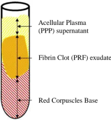
The centrifugation step is designed to separate the blood into three layers, red blood cells are found at the bottom, acellular plasma (PPP, Platelet Poor Plasma) is in the supernatant and a “buffy coat” layer appears in between, in which platelets are concentrated (Dohan, D. et al., 2008; Dohan, D. et al., 2010). During this process, a rapid activation of the coagulation cascade and synthesis of thrombin take place, which induces fibrin formation (Su, C., 2009; Choukroun, J. et al., 2017).
The mechanism followed here is that: fibrinogen which is initially concentrated in the high part of the tube, combines with the circulating thrombin due to centrifugation,
Acellular Plasma (PPP) supernatant Fibrin Clot (PRF) exudate
Red Corpuscles Base
12
to form fibrin (Saluja, H. et al., 2014). A fibrin clot is then obtained in the middle of the tube, just between the red corpuscles at the bottom and acellular plasma at top (Dohan, D. et al., 2006). The upper colored layer is then removed and middle fraction is collected, 2 mm below lower dividing line, which is the PRF (Saluja, H. et al., 2014). Thus, it is necessary to preserve a small red layer at the PRF clot end to collect as many platelets and leukocytes as possible (Nishimoto, S. et al., 2015; Bai, M. et al., 2017). This part of the procedure is done with scissors and remains operator-dependent (Dohan, D. et al., 2010). According to Gürbüzer and colleagues, in their cytologic evaluation study, the samples from the upper part of PRF were acellular and mainly composed of fibrin. Cellular elements usually appeared in lower parts of the samples taken from the middle section where PRF was connected to the red corpuscle beneath (Gürbüzer, B. et al., 2010). This clot is then removed from the tube and the attached red blood cells scraped off and discarded. The PRF clot is then placed on the grid in the PRF Box, and covered with the compressor and lid. This procedure produces an inexpensive autologous fibrin membrane in approximately 1 min. The PRF Box produces membranes of constant thickness that remain hydrated for several hours and recovers the serum exudate expressed from the fibrin clots. The exudate collected at the bottom of the box may be used to hydrate graft materials and to utilize in surgical sites. (Toffler, M et al., 2009).
PRF clot can also be placed into the cylinder in the PRF Box and slowly compressed with the piston which results in “plugs” or thick small discs of PRF measuring 1 cm in diameter. These are useful in protecting extraction sites, for example (Toffler, M et al., 2009; Kobayashi, M. et al., 2012).
However, another alternative describes in literature to obtain a PRF membrane is by pressing the clot between two gauzes thereby squeezing out the fluids of the fibrin clot (Kobayashi, M. et al., 2012; Mihaylova, Z. et al., 2016). This method gives to the membrane a tense-elastic consistency able to resist the pull of the suture (Giannini, S. et al., 2015).
Singh et al. and Mohanti et al., defend in their in vitro studies, that PRF membrane had acceptable elasticity and it could be stretched to some extent (1.52 mm) to cover the wound edges well. However, it is a fragile covering and needs careful manipulation to prevent tearing (Singh, S. et al., 2013; Mohanty, S. et al., 2014).
Other authors defended another device called PRF-compressor, proposed for obtaining PRF membrane, by which platelets keep their integrity. It has two spoon-like
13
surfaces. Between them PRF is pressed and 1 mm thick membrane is established (Mihaylova, Z. et al., 2016).
The serum exudate recovers from fibrin clots, rich in proteins like vitronectin and fibronectin, can be used for the hydration of graft materials. (Dohan, D. et al., 2006; Kumar, R. et al., 2012).
Fibronectin and vitronectin are two key cell adhesion and migration proteins. The fibronectin is present in great quantities in a blood circulating form and in the platelets (Rodriguez-Cuartero, A. et al., 1993), and is also a key component of the architecture of the fibrin clot (Jin, H. et al., 1991). The vitronectin was massively released from the membranes during the first 4 hours, and the release was then almost nil during the next 7 days. It means that if the surgeon need vitronectin on a surgical site, the membranes or the clot exudate should be used quite quickly after preparation (Dohan, D. et al., 2010).
These methods allow the production of a high quantity of PRF clots simultaneously using making it possible to produce even more clots for larger surgeries (Dohan, D. et al., 2008).
Moreover, by simply changing the settings of the centrifuge, it is possible to obtain a normal gelling if it is to be used as regenerative and stimulating material, or a more consistent substance to use as, for example, a filler in the split crest bone gap (Cortese, A. et al., 2016).
Besides that, according Miron et al., and Ghanaati et al., it may be hypothesized that the differences in spin protocols are suggested to have collected slightly different cell populations and/or total growth factors responsible for the variations in growth factor release over time (Miron, R. et al., 2017).
In conclusion, the PRF protocol makes possible to collect a fibrin clot charged with serum and platelets (Dohan, D. et al., 2008).
4.3. Role of PRF in Wound Healing
Although the application of PRF in the oral and maxillofacial surgery has been established by various clinical studies, their mechanisms of action on cellular level remain poorly understood. On the other hand, it is well known that these products contain a wide range of active molecules acting together in biological processes (Mihaylova, Z. et al., 2016).
Wound healing is a natural restorative response to tissue injury, that involves a cascade of complex, orderly, and elaborate events, driven by resident and circulating
14
cells, moving to the injury site, that release soluble mediators or signals generated from the extracellular matrix that are capable of influencing the transport of the circulating cells to damaged tissues (Guo, S. et al., 2010; Martínez, C. et al., 2015).
In adults, optimal wound healing involves four phases (Gosain, A. et al., 2004; Eming, S. et al., 2007):
1. Hemostasis;
2. Inflammatory phase (1-4 days);
3. Proliferation phase (Fibroblastic phase: 2-22 days);
4. Maturation (Remodeling phase: 6-12 months) (Agrawal, M et al., 2014); After tissue damage, the blood flows out from the vessels and occurs the platelet aggregation and activation of hemostasis cascade: they are the first cells recruited at the site of injury and involved to prevent the blood loss and to engage the healing cascade (Gillitzer, R. et al., 2001; Guo, S. et al., 2010).
In the existence of thrombin and Ca2+, the coagulation cascade leads to conversion
of soluble fibrinogen into a network of insoluble fibrin fibers, which stabilizes the platelet plug. In conjunction with fibronectin and vitronectin, fibrin provides a provisional matrix for migration of cells involved in wound healing (Clark, R., 2001).
Fibrin plays a crucial role in the recruitment of neutrophils, monocytes, endothelial cells, and fibroblasts to the wound site. In addition, the intrinsic characteristics of fibrin determine the cellular and humoral processes involved in epithelialization, granulation tissue formation and angiogenesis (Laurens, N. et al., 2006; Yoo, J. et al., 2008).
The second phase, inflammation, is defined by all reaction phenomena initiated in response to a specific aggression. The inflammatory process proceeds in 3 successive phases: vascular phase, cellular phase and cicatrization phase (Dohan, D. et al., 2006).
After this phase has been initiated, the wound healing response requires angiogenesis as a process that modulates the activation, proliferation, and migration of endothelial cells to establish new blood vessels from pre-existing vasculature. Platelets play a critical role in regulating angiogenesis. Nevertheless, their contribution to blood vessel repair in the course of wound healing is still poorly understood (Eming, S. et al., 2007; Klement, G. et al., 2013).
The initial vasoexudative phenomena allows leucocyte migration to the inflammatory site. All these cells secrete many cytokines and growth promoters. This inflammation mediators take part in the fibroblast recruitment, induce proliferation,
15
stimulate biosynthetic activity and leading to the secretion of proteases (Dohan, D. et al., 2006).
Although hemostasis is the major role of fibrin in wound repair, it is not the only one (Gaßling, Volker. et al., 2009).
During any phenomenon of hemostasis and healing, the fibrin clot traps the circulating stem cells brought to the injured site thanks to initial neovascularization (Choukroun, J. et al., 2006).
The bone graft healing is completed by the appearance of osteoblasts and new bone formation (third phase). Finally, the bone remodeling process is characterized by the interaction of osteoblasts and osteoclasts (Zipfel, G: et al., 2003).
The acceleration of the healing processes makes the treated site less sensitive to outside attacks (mechanical, bacterial and chemical) and crucially influences the aesthetic result on oral rehabilitations. Also, have an important role on the patient’s postoperative comfort, due to the anti‑inflammatory properties of PRF (El-Sharkawy, H. et al., 2007; Corso, M. et al., 2008).
4.3.1. Role of Platelets and Growth Factors
Even tough platelet and leukocyte cytokines play an important part in the biology of this biomaterial, the fibrin matrix supporting them certainly constitutes the determining element responsible for the real therapeutic potential of PRF (Gaultier, F. et al., 2004; Simonpieri, A. et al., 2004).
Actually, platelets are thought to contribute to the hemostatic process, where they adhere together to form a platelet plug in a severed vessel and actively extrude several initiators of the coagulation cascade (Arunachalam, M. et al., 2016).
Formed in bone marrow from megacaryocytes, platelets have discoidal and anuclear structures which circulate in blood for 8-10 days (Dohan, D. et al., 2006). Their cytoplasm contains many granules whose contents (growth factors, enzymes and other proteins) are secreted at the time of activation (Borie, E. et al., 2015).
The main function of platelets is known to be prevention of excessive bleeding and repair the blood vessels’ wall after injury (Amable, P. et al., 2013). Platelets are responsible for the first phase of blood clotting. It includes adhesion, activation and aggregation of the platelets. Platelets participate also in angiogenesis, inflammation, antibacterial safety and tissue regeneration (Broos, K. et al., 2012).
16
According to Dohan and coworkers, their activation is fundamental to initiate the interactions with coagulation mechanisms (Dohan, D. et al., 2006).
Degranulation of platelets implies their transformation into the activated state, along with the continuous release of growth factors after the fibrinolysis to promote the healing of hard and soft tissues (Zhang, J. et al., 2016). It is also responsible for the proliferation and differentiation of leukocytes and for playing an important role in immunology, specifically, in inflammation mechanism (Gupta, V. et al., 2011).
However, in the available literature, only a few reports can be found about their antimicrobial effects. At present, the components responsible for the antimicrobial activity of platelet concentrates remain poorly understood (Badade, P. et al., 2016).
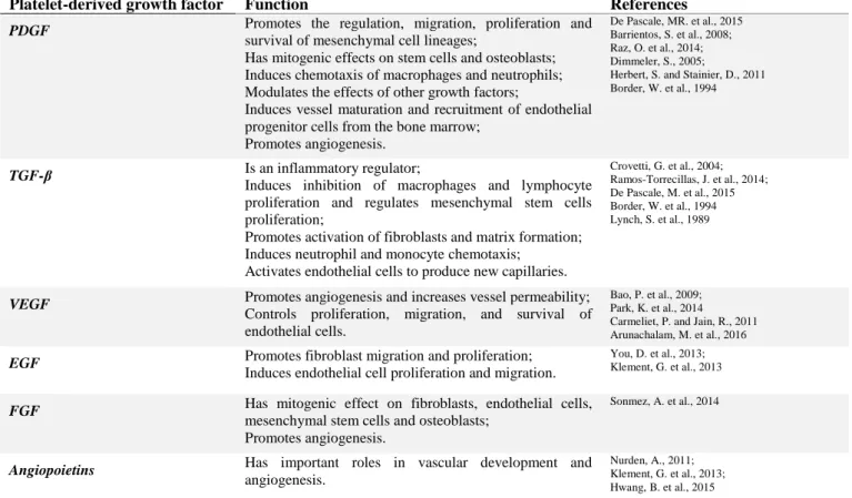
On the other hand, studies on PRF reveal that it is able to release growth factors like Platelet-Derived Growth Factor (PDGF), Transforming Growth Factor-β (TGF-β), Vascular Endothelial Growth Factor (VEGF), Epithelial Growth Factor (EGF), Insulin-like Growth Factor-1 (IGF-1), and Fibroblast Growth Factor (FGF) (Dohan, D. et al., 2006) and several blood proteins such as thrombospondin, fibronectin and vitronectin, during several days (Dohan, D. et al., 2009; Panda, S. et al., 2016). The following table summarizes the main functions of growth factors most frequently released by PRF.
Platelet-derived growth factor Function References
PDGF Promotes the regulation, migration, proliferation and
survival of mesenchymal cell lineages;
Has mitogenic effects on stem cells and osteoblasts; Induces chemotaxis of macrophages and neutrophils; Modulates the effects of other growth factors;
Induces vessel maturation and recruitment of endothelial progenitor cells from the bone marrow;
Promotes angiogenesis.
De Pascale, MR. et al., 2015 Barrientos, S. et al., 2008; Raz, O. et al., 2014; Dimmeler, S., 2005;
Herbert, S. and Stainier, D., 2011 Border, W. et al., 1994
TGF-β Is an inflammatory regulator;
Induces inhibition of macrophages and lymphocyte proliferation and regulates mesenchymal stem cells proliferation;
Promotes activation of fibroblasts and matrix formation; Induces neutrophil and monocyte chemotaxis;
Activates endothelial cells to produce new capillaries.
Crovetti, G. et al., 2004; Ramos-Torrecillas, J. et al., 2014; De Pascale, M. et al., 2015 Border, W. et al., 1994 Lynch, S. et al., 1989
VEGF Promotes angiogenesis and increases vessel permeability;
Controls proliferation, migration, and survival of endothelial cells.
Bao, P. et al., 2009; Park, K. et al., 2014 Carmeliet, P. and Jain, R., 2011 Arunachalam, M. et al., 2016
EGF Promotes fibroblast migration and proliferation;
Induces endothelial cell proliferation and migration.
You, D. et al., 2013; Klement, G. et al., 2013
FGF Has mitogenic effect on fibroblasts, endothelial cells,
mesenchymal stem cells and osteoblasts; Promotes angiogenesis.
Sonmez, A. et al., 2014
Angiopoietins Has important roles in vascular development and
angiogenesis.
Nurden, A., 2011; Klement, G. et al., 2013; Hwang, B. et al., 2015
Table 1. Summary of the main functions of the growth factors and blood proteins present in the PRF.
(PDGF: Platelet-Derived Growth Factor; TGF-β: Transforming Growth Factor-β; VEGF: Vascular Endothelial Growth Factor; EGF: Epidermal Growth Factor; FGF: Fibroblast Growth Factor)
17
According to literature, it has been demonstrated that the growth factors released from activated platelets in PRF are biologically active substances that are involved in tissue repair mechanisms, such as chemotaxis, cell proliferation, angiogenesis, extracellular matrix deposition and remodeling (Singh, S. et al., 2013; Dar, M. et al., 2016), and can work together or cooperate with other cytokines to promote the differentiation of osteoblast as well as inhibiting the function of osteoclast (Jang, E. et al., 2010).
Moreover, they can accelerate bone repair and promote fibroblast proliferation, increase tissue vascularity, raise the rate of collagen formation, promote mitosis of mesenchymal stem cells and endothelial cells and play key roles in the bone formation (Kumar, A. et al., 2009; Anilkumar, K. et al., 2009).
Although the growth factors and the mechanisms involved are still poorly understood, the ease of applying PRF in the dental clinic and its beneficial outcomes, including reduction of bleeding and rapid healing, and definitely a good promise for further procedures (Anilkumar, K. et al., 2009).
4.3.2. Role of Leukocytes
The leukocytes have a great impact on the intrinsic biology and the properties of the platelet concentrates, not only because of their immune and antibacterial potential but also because these cells have a crucial role in the wound healing process and in the biology of a complex bio-material like the PRF (Bielecki, T. et al., 2012).
During the production of PRF, apart from platelets, other cellular elements are activated, such as leukocytes. These are able to release three pro-inflammatory cytokines (IL-1β, IL-6 e TNFα), an anti-inflammatory cytokine (IL-4) and an angiogenesis promoter (VEGF) and, for that reason, have important roles such as anti-infectious action, immune regulation (Everts, P. et al., 2008), and capacity to release cytokines (Inchingolo, F. et al., 2010).
As demonstrated in in vitro studies by Dohan et al. and Choukroun et al., the number of leukocytes in PRF are as many as in 10 mL of blood, from which the secretion and slow release of cytokines make a very big difference to the immunoregulatory effect of the graft materials (Dohan, D. et al., 2006). Thus, leucocytes, when present in moderate quantities in platelet concentrates, are essential actors in wound healing (Martin, P. et al., 2005).
18
PRF is also able to regulate inflammation and stimulate the immune process (Inchingolo, F. et al., 2010) because fibrin mesh stimulates the migration of neutrophils, modulates phagocytosis and promote enzymatic degradation of the immune cells (Dohan, D. et al., 2006).
4.4. Advantages and Disadvantages of Using PRF
Some advantages are reported in the literature related to the use of PRF, such as the following:
Its preparation is a simplified and efficient technique, with centrifugation in a single step, free and openly accessible for all clinicians (Dohan, D. et al., 2006);
It is obtained by autologous blood sample (Fernández-Delgado, N. et al., 2012);
Minimized blood manipulation (Kawase, T. et al., 2015);
It does not require the addition of external thrombin because polymerization is a completely natural process, without any risk of suffering from an immunological reaction (Ross, R. et al., 1974);
It has a natural fibrin framework with growth factors within that may keep their activity for a relatively longer period and stimulate tissue regeneration effectively (Gupta, V. et al., 2011);
It can be used solely or in combination with bone grafts, depending on the purpose (Harrison, P., 2005);
Increases the healing rate of the grafted bone (Harrison, P., 2005);
It is an economical and quick option compared with recombinant growth factors when used in conjunction with bone grafts (Wu, C. et al., 2012);
When used as a membrane, it avoids a donor site surgical procedure and results in a reduction in patient discomfort during the early wound-healing period (Saluja, H. et al., 2011);
The studies of PRF seems to be more efficient and with less controversies on its final clinical results when compared to PRP (Simonpieri, A. et al., 2012).
On the other hand, PRF also may present some disadvantages as follows:
The final amount available is low because it is autologous blood (Choukroun, J. et al., 2006);
The success of the PRF protocol depends directly on the handling, mainly, related to blood collection time and its transference for the centrifuge (Dohan, D. et al.,
19 2006);
Need of using a glass-coated tube to achieve clot polymerization (Dohan, D. et al., 2007);
Possible refusal of the treatment by the puncture required for blood collection at surgery time (Simonpieri, A. et al., 2012);
This procedure needs a minimal experience of clinician for PRF manipulation (Gupta, V. et al., 2011; Simonpieri, A. et al., 2012).
Although not many disadvantages of PRF are known, further studies are needed to better understand its importance in oral surgery.
4.5. Current Applications of PRF in Dentistry
Platelet Rich Fibrin is a new biomaterial with many applications. Briefly, it is a user-friendly and economical procedure, and has huge potential to be used routinely to reduce postoperative discomfort. It may also be used to accelerate natural healing in immune-compromised patients, those taking drugs that interfere with natural healing, and those with a history of radiotherapy. As minimal cost is involved, it can be used for all patients independently of their economical possibilities (Kumar, Y. et al., 2015).
Therefore, the interest in such membrane, mainly to protect open wounds and accelerate healing, is evident (Pierce, G. et al., 1991; Bolander, M.,1992).
Recently, a lot of research has been done on PRF and numerous cases have been reported regarding the use of PRF membranes (Agrawal, M. et al., 2014).
In Oral and Maxillofacial Surgery:
PRF membrane can help in wound healing, protecting the surgical site (Del Corso, M. et al., 2010) and promoting soft and hard tissue repair. When mixed with bone graft, it may act as a “biological connector” which attracts stem cell, favors the migration of osteoprogenitor cells to the center of the graft and provides neo-angiogenesis (Toffler, M. et al., 2009).
The immunological properties of the PRF, resulting from its content in leukocytes, could be useful to prevent the surgical site infections, such as postextraction alveolitis for example (Marenzi, G. et al., 2015).
Studies show that PRF can also be used as a filling material in extraction sockets to expedite socket healing and reduce postoperative pain, dryness or purulent complications (Hoaglin, D. et al., 2013; Yelamali, T. et al., 2015).
20
According to the literature, PRF is a useful tool in post extraction hemostasis control (Dohan, D. et al., 2008), in prevention of hemorrhagic complications and in reducing postoperative hematoma (Matras, H., 1985; Sammartino, G. et al., 2011).
PRF membranes have strictly no contraindications, they can be used in all kinds of patients and can even be recommended in patients under anticoagulants or smokers (Corso, M. et al., 2012). Can also be used to improve wound healing in immunocompromised, diabetic patients and as an adjuvant in patients on anticoagulation therapy (Corso, M. et al., 2010).
The use of PRF in oral surgery has also been implicated in other procedures such as the repair of potentially malignant lesions (Pathak, H. et al., 2015), to reconstruct the defects following cyst enucleation and tumor excision and as an adjunct to palatal wound treatment (Femminella, B. et al., 2016) or alveolar cleft repair (Jain, V. et al., 2012; Kulkari, M. et al., 2014).
Some studies advocated that cavities filled with PRF showed two times faster healing as compared to physiologic healing (Saluja, H. et al., 2014). Choukroun and colleagues defend that the physiologic healing time of cystic cavity normally last between 6 months and 1 year, but filled with PRF will be totally healed in just 2 months (Choukroun, J. et al., 2006).
In cases of wide sockets and lesions where primary closure is difficult, PRF membrane can also be used as a covering and protective membrane that promotes re-epithelialization of the site and accelerates the merging of the wound margins. The elasticity and strength of PRF fibrin membrane makes it easy to suture (Eren, G. et al., 2014).
Another possible clinical application, that have been extensively used, is in sinus lift procedures. Several studies demonstrate the use of PRF as the sole filling material during sinus lift procedures (Agrawal, M. et al., 2014).
Therefore, some clinical studies (Mazor, Z. et al., 2009; Simonpieri, A. et al., 2011), that use PRF membrane as a sole grafting material to achieve maxillary sinus floor augmentation, presents promising results. Other authors, including Toffler et al., recommended the use of PRF membrane to seal undetected sinus membrane perforation during a lateral window osteotomy in a maxillary sinus lift procedure (Toffler, M. et al., 2009; Borie, E. et al., 2015).
PRF can also serve as a resorbable membrane that can be used in pre-prosthetic surgery as well as in implantology to cover bone augmentation sites (Choukroun, J. et al.,
21
2006; Öncü, E. et al., 2016). Choukroun et al., concluded that, with the aid of PRF, the healing time is significantly reduced and the implant can be placed only 4 months (120 days) after surgery (Tatullo, M. et al., 2012).
In Tissue Engineering: As a membrane for guided bone regeneration, the PRF dense matrix architecture covers, protects and stabilizes bone graft material and may facilitate better and faster bone regeneration due to the presence of growth factors (Rao, S. et al., 2012; Agrawal, M. et al., 2014). A clinical advantage of PRF as a graft material is related to avoidance of a donor site and a major decrease in patient discomfort after surgery (Eren, G. et al., 2014).
PRF is a potential tool in tissue engineering but clinical aspects of PRF in this field requires further investigation (Agrawal, M. et al., 2014).
In Periodontics: PRF membrane has exhibited favorable clinical results in the treatment of periodontal infrabony defects (Chang, Y. et al., 2011) and gingival recessions (Agrawal, M. et al., 2014).
In Endodontics: Studies have shown that PRF can be used as a scaffolding material in an infected necrotic immature tooth for pulpal regeneration and tooth revitalization (Shivashankar, V. et al., 2012). Also, it can induce faster periapical healing in cases with large periapical lesions (Geeta, I. et al., 2013). The use of this biomaterial in regenerative pulpotomy procedures have also been documented (Geeta, I. et al., 2013).
Other Clinical Applications: In other medical procedures, PRF had been additionally utilized for the successful management of hard-to-heal leg ulcers, including diabetic foot ulcers, venous leg ulcers, and chronic leg ulcers (O’Connell, S., 2008; Miron, R. et al., 2017).
Furthermore, hand ulcers, facial soft tissue defects, laparoscopic cholecystectomy, deep nasolabial folds, facial defects, superficial rhytids, acne scars, lipostructure surgical procedures, chronic rotator cuff tears, and acute traumatic ear drum perforations have also all been treated with PRF (Miron, R. et al., 2017).