R E S E A R C H
Open Access
Therapeutic angiogenesis induced by
human umbilical cord tissue-derived
mesenchymal stromal cells in a murine
model of hindlimb ischemia
Ana Rita S. Pereira
1†, Teresa F. Mendes
1†, Augusto Ministro
1,2, Mariana Teixeira
3, Mariana Filipe
3, Jorge M. Santos
3,
Rita N. Bárcia
3, J. Goyri-O
’Neill
4, Fausto Pinto
1,5, Pedro E. Cruz
3, Helder J. Cruz
3and Susana Constantino Rosa Santos
1,5*Abstract
Background: Mesenchymal stem cells derived from human umbilical cord tissue, termed UCX®, have the potential to promote a full range of events leading to tissue regeneration and homeostasis. The main goal of this work was to investigate UCX® action in experimentally induced hindlimb ischemia (HLI).
Methods: UCX®, obtained by using a proprietary technology developed by ECBio (Amadora, Portugal), were delivered via intramuscular injection to C57BL/6 females after unilateral HLI induction. Perfusion recovery, capillary and collateral density increase were evaluated by laser doppler, CD31 immunohistochemistry and diaphonisation, respectively. The activation state of endothelial cells (ECs) was analysed after EC isolation by laser capture microdissection microscopy followed by RNA extraction, cDNA synthesis and quantitative RT-PCR analysis. The UCX®-conditioned medium was analysed on Gallios flow cytometer. The capacity of UCX® in promoting
tubulogenesis and EC migration was assessed by matrigel tubule formation and wound-healing assay, respectively. Results: We demonstrated that UCX® enhance angiogenesis in vitro via a paracrine effect. Importantly, after HLI induction, UCX® improve blood perfusion by stimulating angiogenesis and arteriogenesis. This is achieved through a new mechanism in which durable and simultaneous upregulation of transforming growth factorβ2, angiopoietin 2, fibroblast growth factor 2, and hepatocyte growth factor, in endothelial cells is induced by UCX®.
Conclusions: In conclusion, our data demonstrate that UCX® improve the angiogenic potency of endothelial cells in the murine ischemic limb suggesting the potential of UCX® as a new therapeutic tool for critical limb ischemia. Keywords: UCX®, Mesenchymal stem cells, Angiogenesis, Arteriogenesis, Critical limb ischemia, Endothelial cells, Hindlimb ischemia
Background
Critical limb ischemia (CLI) is a severe form of periph-eral artery disease (PAD) in which patients with occlu-sive arterial disease of the legs experience chronic ischemic rest pain, ulcer, or gangrene [1]. This syndrome
is associated with severe prognosis, with 1-year mortality
exceeding 25 % and about 30–50 % major limb
amputa-tion at 1 year from diagnosis [1]. The limitaamputa-tions of surgical/endovascular revascularization, due the distribu-tion and diffuseness of arterial occlusions, are well recognized and amputation, despite its associated morbidity and mortality rates, is often recommended [2]. The goal of limb salvage has stimulated research into alternative methods, including therapeutic angiogenesis which can be achieved either by local administration of * Correspondence:sconstantino@medicina.ulisboa.pt
†Equal contributors 1
Centro Cardiovascular da Universidade de Lisboa, Av. Prof Egas Moniz 1649-028 Lisbon, Portugal
5Faculdade de Medicina da Universidade de Lisboa, Av. Prof Egas Moniz
1649-028 Lisbon, Portugal
Full list of author information is available at the end of the article
© 2016 The Author(s). Open Access This article is distributed under the terms of the Creative Commons Attribution 4.0 International License (http://creativecommons.org/licenses/by/4.0/), which permits unrestricted use, distribution, and reproduction in any medium, provided you give appropriate credit to the original author(s) and the source, provide a link to the Creative Commons license, and indicate if changes were made. The Creative Commons Public Domain Dedication waiver (http://creativecommons.org/publicdomain/zero/1.0/) applies to the data made available in this article, unless otherwise stated.
pro-angiogenic growth factors and gene- or cell-based therapies [3].
Mesenchymal stem cells (MSCs) hold great promise as a therapy for PAD, mainly due to their paracrine activity and immunosuppressive capacity [4]. MSCs are known to home specifically to hypoxic tissues following injury [5] where they potentiate vascular growth through the release of pro-angiogenic factors [6]. It was previously reported that autologous, allogeneic and xenogeneic MSC administration induced therapeutic angiogenesis in animal models of hindlimb ischemia (HLI) [7]. In a meta-analysis of cell therapy for PAD, it was found that autologous bone marrow MSCs delivery led to improved indices of ischemia and pain-free walking [8]. Although first harvested from the bone marrow, MSCs have since been identified in many other tissues, namely adipose tissue [9] and umbilical cord tissue [10]. UCX® in particular are MSCs obtained from the human umbilical cord Wharton’s jelly that are isolated, expanded and cryo-preserved according to a patented method (PCT/IB2008/ 054067; WO 2009044379) and produced according to advanced therapy medicinal product (ATMP) guide-lines [11]. UCX® fulfil the MSC criteria as defined by the International Society for Cellular Therapy (ISCT) [12]. UCX® cells are advantageous in comparison to other sources due to the absence of invasiveness in their collection process, their faster self-renewal, higher cell yield and being more potent modulators of the immune system than bone marrow MSCs, making them more attractive for allogeneic cellular therapies.
Recently, it was demonstrated that UCX® paracrine activity represses T cell activation and promotes the expansion of regulatory T cells (Tregs) better than bone marrow MSCs [13]. Furthermore, by using an acute arthritis in vivo model it was found that UCX® can re-duce paw edema more efficiently than bone marrow MSCs. The use of a chronic arthritis model showed that UCX® induce faster remission of local and systemic arth-ritic manifestations [13]. The UCX® tissue regeneration capacity suggested for rheumatoid arthritis was later cor-roborated for myocardial infarction. By using a mouse model of myocardial infarction, it was shown that UCX® preserve cardiac function and attenuate adverse tissue remodelling after intramyocardial transplantation [14]. Interestingly, it was demonstrated that this cardioprotec-tive effect was exerted through paracrine mechanisms involving angiogenesis promotion [14]. Furthermore, in vitro studies, performed with conditioned medium (CM) produced by UCX® grown in classical two-dimensional monolayer cultures, have demonstrated the potential to induce keratinocyte migration in the early stages of wound healing [15]. Moreover, UCX® were able to attract bone marrow MSCs in vivo with potential to promote
the formation of granulation tissue, contraction by myo-fibroblasts, angiogenesis, vasculogenesis and epitheliza-tion [15]. These results strongly suggest that UCX® have the potential to promote a full range of events leading to tissue regeneration and homeostasis. Interestingly, it was shown recently that the later proliferative and re-modelling stages of wound healing promoted by the CM of the two-dimensional monolayer cultures were improved by using three-dimensional culture-derived CM [16]. An enhanced secretion of healing-inducing paracrine factors by UCX® including the extracellular
cellular matrix metalloproteinase-2 (MMP-2) and
matrix metalloproteinase-9 (MMP-9), collagen I, fibro-nectin, laminin, collagen IV and angiogenic factors such as vascular endothelial growth factor A (VEGF-A), granulocyte-colony stimulating factor (G-CSF), transform-ing growth factorβ1 (TGF-β1), fibroblast growth factor 2 (FGF-2), hepatocyte growth factor (HGF) and interleukin 6 (IL-6) was observed in the CM of three-dimensional cul-tures based on self-assembled spheroids [16].
In this work, our data corroborate that UCX® enhance angiogenesis in vitro and in vivo, by modulating endo-thelial cells. In vitro, we found that UCX® promote tubule formation and endothelial cell migration. Import-antly, our results have demonstrated that UCX® in the setting of experimentally induced unilateral hindlimb ischemia (HLI) stimulate angiogenesis and collateral ves-sel development and thereby improve blood perfusion in the ischemic limb demonstrating the potential of UCX® as new therapeutic tool for CLI. Furthermore, and to the best of our knowledge, this is the first study in an ex-perimental model of hindlimb ischemia showing that, in vivo and in a sustained way human MSCs upregulate the endothelial gene expression of several pro-angiogenic
players (such as transforming growth factor β2 (Tgfβ2),
angiopoietin 2 (Ang-2), Fgf-2, Hgf ), shedding light on the potential mechanisms of action of this particular ATMP.
Methods
Ethics and regulations
All animal procedures were performed according to Direct-ive 2010/63/EU. The procedures were approved by the in-stitutional Animal Welfare Body and licensed by DGAV, the Portuguese competent authority for animal protection (license number 023861/2013).
Cell culture
UCX® were cultured in static monolayers in α-MEM
with 1 g/L glucose and 2 mM glutamine (Sigma-Aldrich, St. Louis, MO, USA), hereafter designated basal medium (BM), supplemented with 20 % foetal bovine serum (FBS; Gibco®, Madrid, Spain), in a humidified incubator at 37 °C and 7 % CO2.
Human umbilical vein endothelial cells (HUVECs) (Sciencell, Carlsbad, CA, USA) were cultured in M199 media (Sigma-Aldrich, St. Louis, MO, USA), hereafter designated endothelial basal medium (EBM) supple-mented with 10 % FBS (Gibco™, Thermo Fisher
Scien-tific, Waltham, MA, USA), 50 μg/ml endothelial cell
growth supplement (ECGS) (Sigma-Aldrich), 100 μg/ml
heparin (Sigma-Aldrich) and 1 % penicillin-streptomycin (10,000 U/mL, Sigma-Aldrich), hereafter designated endothelial growth medium (EGM). Cells were grown in flasks coated with 0.2 % gelatin (Sigma-Aldrich) until 70 % confluence and used up to passage 6.
Preparation of conditioned media
UCX® were seeded in BM supplemented with 5 % FBS and grown until 90 % confluence. After washing cells with phosphate-buffered saline (PBS), BM (without serum) was added for 24 hours. Thereafter, BM was re-placed and conditioned for 48 hours at 37 °C and 7 % CO2after which it was collected, centrifuged at 300 g for
10 minutes to remove cell debris, filtered (0.22μm pore size, Merck Millipore, Billerica, MA, USA), and concen-trated using 5 kDa cut-off spin concentrators (Agilent Technologies, Santa Clara, CA, USA) as per manufac-turer’s recommendations. Control conditioned medium samples underwent the same procedure as described above, but in the absence of cells.
Quantification of secreted factors
The quantification of HGF, VEGF-A, TGF-β1, IL-8,
platelet-derived growth factor-AA (PDGF-AA) and
FGF-2 in the conditioned medium was performed using a commercially available kit (FlowCytomix™; eBioscience, San Diego, CA, USA) according to the manufacturer’s instructions. Samples were acquired on a Gallios flow cytometer (Beckman Coulter, Brea, CA, USA) and the results obtained using FlowCytomix Pro 3.0 Software (eBioscience).
Tubule formation assay
The tubule formation assay was performed using the thick gel method of preparation. Briefly, after thawing over-night, the matrigel (Corning, Corning, NY, USA) was plated into a pre-cooled 48-well plate (Nunc™, Thermo Fisher Scientific) (180μl per well) using a chilled pipet tip. After polymerization of matrigel at 37 °C, 5 % CO2, for
45 minutes, HUVECs were inoculated at a density of 4.5 × 10⁴ cells/cm2 on top of the matrigel in 350 μl of EBM. Cells were then incubated for 1 hour at 37 °C, 5 %
CO2. 1 × 10⁶UCX® were resuspended in 350μl BM and
loaded on a 1-μm insert (Brand, Wertheim, Germany) and carefully placed in a 48-well plate and incubated for
16 hours at 37 °C, 5 % CO2. Controls with no UCX®
included HUVECs in (i) EBM, (ii) EGM and (iii) EBM
supplemented with FGF-2 (50 ng/ml). After incubation, inserts were removed and all wells were photographed (×10 amplification) using a Nikon Eclipse Ti-U inverted microscope with a conjugated Nikon DS-Qi1Mc camera (Nikon, Tokyo, Japan). Tubule formation was quantified on four random fields per replicate, using the Angiogen-esis analyser from Image J.
Wound-healing assay
HUVECs were plated to confluence in a 24-well plate (previously coated with gelatine 0.2 %) with EBM supplemented with 5 % FBS. After culturing overnight, wounds were created in the monolayer by scrapping the plate with a sterile pipette tip. After that, 1 × 10⁶ UCX®, resuspended in EBM supplemented with 5 % FBS and loaded on a 1-μm insert (Brand, Wertheim, Germany), were carefully placed in a 24-well plate and incubated for 9 hours at 37 °C, 5 % CO2. In controls, the same
pro-cedure was performed in the absence of UCX®. Photo-graphs were taken in Primovert (Carl Zeiss Microscopy, Jena, Germany) on a × 4 magnification and three inde-pendent wound areas were measured with ImageJ imme-diately after wounding and 9 hours later.
Mice
Twenty-two-week-old C57BL/6 female mice, purchased from Charles River Laboratories, Barcelona, Spain, were used in all experiments. The animals were anaesthetised with a ketamine-medetomidine cocktail (75 mg/kg BW and 1 mg/kg BW, respectively) intraperitoneally for the surgical procedure as well as for the other analysis procedures. The anaesthesia was partially reverted with atipamezole (5 mg/kg BW). Postoperatively, analgesia
was performed (buprenorphine 100 μl/15–30 g BW
every 8–12 h) and the animals were closely monitored. HLI model
A surgical procedure was performed to induce unilateral HLI in the mice. Briefly, an incision in the skin overlying the thigh of the right hindlimb of each mouse was made and the distal external iliac artery and the femoral artery and veins were ligated and excised. The vein was ligated both to increase the severity of the ischemia as well as to increase the technical reproducibility of the model, as isolation of the femoral artery alone often results in tear-ing of the vein resulttear-ing in haemorrhage.
UCX® administration
A dose of 2 × 10⁵ UCX® was injected intramuscularly,
5 hours post-ischemia induction on the gastrocnemius muscle (right hindlimb) in a volume of 50μl per animal, divided into two injections of 25μl. As a control, UCX® vehicle (PBS) was administered at the same conditions.
Laser doppler perfusion imaging
The laser doppler perfusion imager (MoorLDI V6.0, Moor Instruments Ltd, Axminster, UK) was used to as-sess limb perfusion. Hair was removed 1 day before laser doppler analysis using an electrical shaver followed by depilatory cream. Blood flow was measured both in the ischemic leg and the contralateral one, before HLI in-duction (PRE-HLI), immediately post-HLI (POST-HLI), and at day 7, 14 and 21 post-HLI (d7 POST-HLI, d14 POST-HLI and d21 POST-HLI, respectively). Colour-coded images of tissue perfusion were recorded and poor or no perfusion was displayed as dark blue, and the highest perfusion level was displayed as red. Mean flux values were calculated using the MoorLDI V6.0 image processing software. To account for variables such as temperature and ambient light, blood perfusion is expressed as the ratio of ischemic to non-ischemic limb. The mice were placed on a 37 °C heating pad to reduce heat loss during measurements.
Immunohistochemistry and capillary density analysis Mice were sacrificed at day 90 post-HLI (to assure capil-lary stabilization after the ischemic injury). The gastro-cnemius muscles of both legs were harvested, placed in transverse orientation on a small cork disc with the help of 10 % tragacanth, snap frozen in liquid nitrogen-cooled isopentane and stored at -80 °C until sectioned. Seven-micrometer sections were labeled with CD31 monoclonal antibody (Pharmingen, San Diego, CA, USA). After fixation in acetone for 10 minutes, hydrogen peroxidase (0.3 % diluted in methanol) was added for 30 minutes, at room temperature (RT) and followed by two washes in PBS for a total of 10 minutes. Blocking solution (5 % rabbit serum in PBS) was applied for 30 minutes, at RT, and the slides were then incubated for 1 hour at RT with rat monoclonal antibody against mouse CD31 at 1:500, diluted in 1 % bovine serum albu-min (BSA) in PBS. After three washes in PBS for a total of 30 minutes, a secondary biotinylated rabbit anti-rat IgG antibody was added at 1:200 in 1 % BSA in PBS and 5 % rabbit serum for 30 minutes, at RT. Washes were performed as before and labeled avidin-conjugated per-oxidase complex (Vectastain ABC kit; Vector
Laborator-ies, Burlingame, CA, USA) was used for color
development according to the manufacturer’s recom-mendations for 30 minutes, at RT. After rinsing in PBS (three times for 5 minutes), DAB peroxidase substrate kit (Dako, Glostrup, Denmark) was added for 5 minutes to localize the immune complexes. The sections were counterstained with haematoxylin (Merck, Kenilworth, NJ, USA) for 10 seconds and mounted with entelan (Merck). Omission of the first antibody was used as a
negative control. Analysis of tissue samples was
conducted using a Leica DM2500 upright brightfield
microscope (Leica Microsystems, Wetzlar, Germany). Capillary densities, i.e. number of capillaries per number of myocytes, were measured in two different sections of four distinct anatomic areas of each specimen using the ImageJ software.
Contrast agent perfusion and diaphonisation
Ninety days post-ischemia induction (to assure vascular stabilization after the ischemic injury), mice were deeply anaesthetised and the torso and limbs were shaved. A medial thoracotomy was performed to expose the heart and a needle (26 gauge), attached to an automatic in-jector, was introduced in the left ventricle. An incision was performed in the right atrium to allow venous drainage. Mice were primarily perfused with heparinized serum (3000 IU/L) until the blood was completely re-moved from circulation. A vasodilatation mixture of ad-enosine (1 mg/L) and papaverine (4 mg/L) was subsequently administered, right before the contrast agent, for 2 minutes. The contrast used was a mixture of barium sulfate (50 %) and gelatin (5 %). This solution was kept warm until the injection time to avoid thicken-ing. Contrast agent was perfused manually until the feet blanched. Right after the injection the mice were trans-ferred to a cold chamber, so that the contrast agent became solidified. All the solutions were injected with a perfusion rate of 0.7 mL/minute. Then, the skin of each mouse was removed from the lower body and diaphoni-sation was performed by using a modified version of the Spalteholz technique. Briefly, the mice were fixated, decalcified, whitened, washed, dehydrated by freeze substitution and placed into a vacuum pump with Spalteholz solution (benzyl benzoate and methyl salicyl-ate) until transparency was acquired.
Collateral vessels quantification
Mice were kept in Spalteholz solution during image acquisition, in order to achieve a homogenous density between the tissues and the media, with minimal absorp-tion or reflecabsorp-tion of light. Mice entire limbs were photo-graphed in a magnifier with a light source. After acquisition, images were aligned and stitched together using Adobe Photoshop CS6®, and entire limb photo-graphs were obtained. In order to exclude the femoral artery and all venous structures from this quantification, only the collateral vessels were manually segmented by highlighting them using Adobe Photoshop CS6®. We considered as collateral vessels all the vessels with a
diam-eter between 20 and 300 μm. For every mouse, an
ana-tomically determinable region comprising the ligation site was selected and defined as a region of interest (ROI). Collateral vessels density (CVD) was quantified in equiva-lent ROIs corresponding to 20 % of the total limb area.
The CVD was calculated as the ratio between the vascular and the ROI areas.
To exclude variations in the anatomy, perfusion and diaphonization procedures, the CVD value of the non-ischemic limb obtained for each mouse was assumed to correspond to 100 %. According to this assumption, the CVD percentage in the ischemic limb was calculated relatively to the non-ischemic one. The percentage of CVD increase was determined as the difference between the CVD percentage among the ischemic and non-ischemic limbs. Means of CVD percentages and stand-ard deviations were calculated for each experimental group. All density measurements were performed using ImageJ software.
Laser capture microdissection of capillaries
Mice were sacrificed at day 70 post-HLI. Twelve-micrometer sections of the gastrocnemius muscles were labeled with CD31 monoclonal antibody (Pharmingen).
The sections were stored at –80 °C until
microdissec-tion. The immunohistochemistry protocol described above was modified to improve RNA preservation by using high salt buffer, 2 M NaCl in PBS 1× (at 4 °C) in all incubation and washing steps [17]. Briefly, slides were placed in acetone, for 5 minutes, air-dried, rehydrated with 2 M NaCl/PBS (4 °C) and incubated, overnight, at 4 °C with rat monoclonal antibody against mouse CD31, at 1:500, in 2 M NaCl/PBS. After two washes in 2 M NaCl/PBS for a total of 6 minutes, a secondary biotinyl-ated rabbit anti-rat IgG antibody was added at 1:200 in 2 M NaCl/PBS and 5 % rabbit serum for 30 minutes, at 4 °C. Washes were performed as before and labeled avidin-conjugated peroxidase complex (Vectastain ABC kit; Vector Laboratories) was used for color development
according to the manufacturer’s recommendations for
30 minutes, at 4 °C. After rinsing, DAB peroxidase substrate kit (Vector Laboratories) was added for 5 mi-nutes to localize the immune complexes. Sections were dehydrated in ice-cold 90 % ethanol followed by 100 % ethanol and allowed to dry. Ten thousand capillaries were microdissected using a Zeiss PALM MicroBeam Laser Microdissection System (Carl Zeiss Microscopy) equipped with a pulsed solid-state 355 nm laser. Dissected capillaries were catapulted into a microfuge tube adhesive cap.
RNA extraction, cDNA synthesis, pre-amplification and RT-PCR Total RNA from the microdissected capillaries was isolated using an RNeasy Micro Kit (QIAGEN, Hilden,
Germany), including DNase treatment to remove
potential genomic DNA contamination. For synthesis
and preamplification of cDNA, RT2 PreAMP cDNA
Synthesis Kit (QIAGEN) was used with two rounds of pre-amplification using the following primers: Fgf-2_F
(5′- ACTCCAGTTGGTATGTGGCACTGA-3′); Fgf-2_ R (5′-AACAGTATGGCCTTCTGTCCAGGT-3′); Tgfb-2_F (5′-GCTTTGGATGCGGCCTATTGCTTT-3′); Tgf b-2_R (5′-CTCCAGCACAGAAGTTGGCATTGT-3′); Ang-2_F (5′-ATCCAACACCGAGAAGATGGCAGT-3′); Ang-2_R (5′-AACTCATTGCCCAGCCAGTACTCT-3′); Hgf_F (5′-GCATTCAAGGCCAAGGAGAAGGT T-3′); Hgf_R (5′-TCATGCTTGTGAGGGTACTGCG AA-3′); 18s_F (5′-GCCCTATCAACTTTCGATGGTA GT-3′); 18s_R (5′- CCGGAATCGAACCCTGATT-3′). RT-PCR was performed according to the manufac-turer’s protocol using Power SYBR® Green (Invitro-gen, Carlsbad, CA, USA) and an Applied Biosystems (Waltham, MA, USA) 7500 Fast Real-Time PCR for the same targets described above. The housekeeping gene used to normalize was 18S. The RT-PCR pro-gram consisted of an initial denaturation step, at 95 ° C, for 10 minutes followed by 50 cycles, at 95 °C, for 15 seconds and at 60 °C, for 1 minute. The relative quantification was performed according to the
com-parative method (2-ΔΔCt; Applied Biosystems User
Bulletin no. 2P/N 4303859), with the non-ischemic muscle as an internal calibrator. The formula used is 2-ΔΔCt =2-[ΔCt (sample) - ΔCt (calibrator)], where ΔCt
(sample) = Ct (sample) –Ct (reference gene). For the
internal calibrator ΔΔCt = 0 and 20= 1. For the
remaining samples the value of 2-ΔΔCt indicates the
fold change in gene expression relative to the
calibra-tor. The ΔCt value for each sample is the average of
triplicates.
Statistical analysis
For the wound-healing assay, the values assume normal distribution and unequal variances and therefore an inde-pendent two-tailed t test was performed. For doppler ana-lysis, an independent two-tailed t test was also performed at days 7, 14 and 21. The values assume normal distribu-tion. Only at day 7, we cannot assume equal variances. For capillary and collateral analysis, the values also assume normal distribution and equal variances and therefore an independent two-tailed t test was also performed. For the master junctions and segment length, one-way independ-ent analysis of variance (ANOVA) was developed as data followed a normal distribution, though equality of variances could not be assumed for the first. Therefore, a Games-Howell corrected post hoc test was used to iden-tify differences between groups. For the meshes area, as normality could not be assumed and so a Kruskal-Wallis non-parametric test was developed followed by its paired analysis to identify differences between groups. The effect size and power was determined by using the G-Power software in all type of analyses. p < 0.05 was interpreted to denote statistical significance.
Results
UCX® promote tubulogenesis and endothelial cell migration
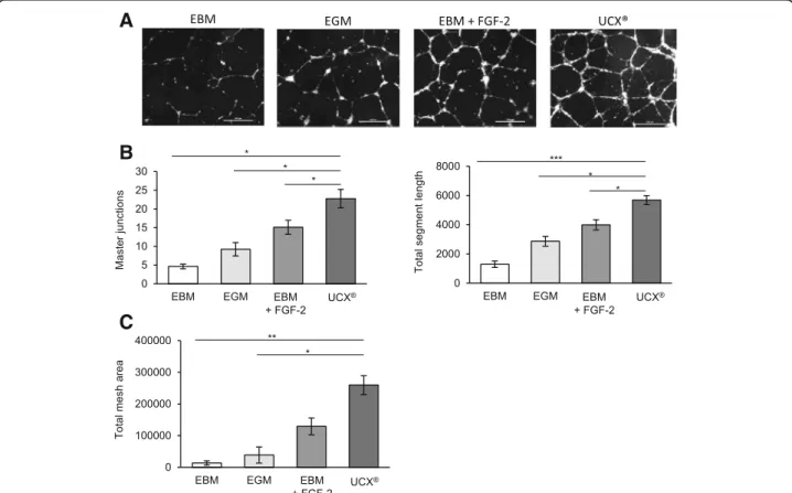
We first evaluated the effect of UCX® on the capillary structure formation. A matrigel tubule formation assay was used as an in vitro model. HUVECs were seeded onto matrigel in endothelial basal medium (EBM) and co-cultured with UCX®. As a control HUVECs were seeded in EBM, where we do not expect to visualize capillary-like structures formed by HUVECS. For that reason, two positive controls were added and HUVECs were seeded both in endothelial growth medium (EGM) that contains several pro-angiogenic factors such as VEGF and FGF-2 and in EBM supplemented with FGF-2 (EBM-FGF-2). As shown in Fig. 1a and quantified in Fig. 1b, UCX® induce tubule formation by HUVECs and these structures consistently showed a significant in-crease in the number of master junctions, total mesh
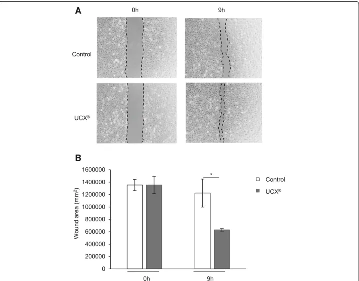
area and total segment length when compared to those from HUVECs cultured in EBM or EGM. With the ex-ception of the total mesh area, all the measured parame-ters were also significantly different between HUVECs cultured with UCX® in EBM and HUVECs cultured in EBM supplemented with FGF-2. Next, we assessed the migratory capacity of HUVECs seeded in EBM supple-mented with 5 % of serum co-cultured with UCX® with-out cell to cell contact. With this objective, a wound-healing assay was performed and as a control HUVECs were seeded in the same culture conditions but in the absence of UCX®. As shown in Fig. 2a and b, the wound area decreased more rapidly in the presence of UCX® than in the control condition, suggesting that UCX® promote the migration of HUVECs via a paracrine secretion. Taken together, these results clearly show that, in vitro, UCX® induce angiogenic processes in endothe-lial cells.
A
B
C
Fig. 1 UCX® promote tubulogenesis. Matrigel assay was performed by seeding human umbilical vein endothelial cells (HUVECs) in endothelial basal medium (EBM), endothelial growth medium (EGM), EBM supplemented with fibroblast growth factor 2 (FGF-2) (EBM + FGF-2) or EBM co-cultured with UCX®. (a) Representative images from the different experimental conditions are shown. (b) Quantitative evaluation demonstrated significantly enhanced master junctions, total mesh area and total segment length in capillary-like structures formed in HUVECs co-cultured with UCX® when compared to the other experimental conditions. For master junctions, following Games-Howell post hoc test the mean differences between UCX® and the other experimental conditions were: EBM (mean dif. =24.27; p = 0.01); EGM (mean dif. = 19.67; p = 0.03) and EBM + FGF-2 (mean dif. = 13.8; p = 0.034). The effect size to basal was 2.08 and power 0.98. For the meshes area, following pairwise comparisons, the equivalent to a post hoc Kruskal-Wallis test, significant differences between UCX® and EBM or UCX® and EGM were observed with KW = 22.66, p < 0.01 or KW = 18.77, p = 0.01, respectively. For segment length, following Games-Howell post hoc test, UCX® show significant difference when compared to the other groups: EBM (mean dif. = 4920; p < 0.001); EGM (mean dif. = 3346; p = 0.01) and EBM + FGF-2 (mean dif. = 2223; p = 0.016). Effect size to basal was 3.85 and power 1. Scale bar, 500μm
UCX® secrete pro-angiogenic factors
Since it was already demonstrated that the beneficial effects promoted by UCX® result from a paracrine secre-tion, we decided to evaluate the secretion of six repre-sentative trophic factors critical for angiogenesis in the conditioned medium of an UCX® monolayer seeded in basal medium (BM) for 48 hours. As a control, the same trophic factors were measured in BM that had not been in contact with cells. The expression of HGF, VEGF-A, TGF-β1, interleukin 8 (IL-8), platelet-derived growth factor-AA (PDGF-AA) and FGF-2 were analysed. Our results show a significant increase of HGF, TGF-β1, IL-8 and PDGF-AA in the conditioned medium of UCX® when compared to the control (Table 1), demonstrating that UCX® secrete pro-angiogenic factors to the extracel-lular medium.
UCX® increase perfusion recovery after HLI
In order to evaluate the therapeutic potential of UCX® in a HLI mouse model, the perfusion recovery was assessed over time after ischemia induction and UCX® adminis-tration. Five hours after surgical induction of unilateral HLI, UCX® or their vehicle (as a control) were adminis-tered in the ischemic muscle and perfusion was mea-sured over time. As shown in Fig. 3a and quantified in Fig. 3b, a dramatic reduction in blood flow was observed in the ischemic limb immediately after surgery, in com-parison to the contralateral limb, followed by a gradual normalization of ischemic blood flow over time. Import-antly, UCX® markedly improved blood flow recovery at 7, 14 or 21 days post-HLI, comparing with control mice, demonstrating that UCX® administration contributes to-ward the functional recovery of ischemic tissues.
A
B
Fig. 2 UCX® stimulate endothelial cell migration. Human umbilical vein endothelial cells (HUVECs) were seeded to confluence in endothelial basal medium (EBM) supplemented with 5 % FBS and cultured for 16 hours after wounding. Immediately after wound, HUVECs were co-cultured with UCX® or not (control) for 9 hours. (a) Representative images are shown. The left and right panels concern HUVEC monolayer immediately after or 9 hours post-wounding, respectively. (b) The wound area was measured immediately after and 9 hours post-wounding by using ImageJ software (three independent measurements were performed for each experimental group; t (2.03) = 4.66; *p < 0.05); effect size was 3.82 and power 0.93)
UCX® increase capillary and collateral vessel density after HLI
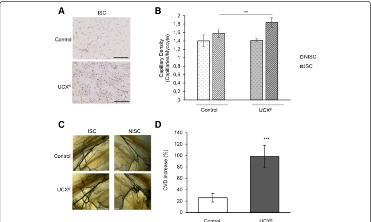
As blood flow recovery depends on both angiogenesis and arteriogenesis we examined whether UCX® would affect capillary and collateral vessel densities in hindlimb muscles. Five hours after surgical induction of unilateral HLI, UCX® or their vehicle (as a control) were adminis-tered in the ischemic muscle and mice were sacrificed 90 days post-HLI induction. Capillary density was assessed through quantification of CD31-positive capil-laries on histological sections of gastrocnemius muscle. As expected, the capillary density was greater in the
ischemic versus the non-ischemic hindlimb in both ex-perimental groups (Fig. 4b). Notably, the level of capil-lary density in the ischemic hindlimb treated with UCX® was significantly higher than the one observed in the control ischemic hindlimb, as shown in Fig. 4a and quantified in Fig. 4b. In order to evaluate the collateral vessel density (CVD), mice were diaphonised and an equivalent region of interest (ROI), corresponding to 20 % of the limb area, was selected for CVD quantifica-tion (Fig. 4c). A significantly higher CVD increase was observed in UCX®-treated mice when compared to the control (Fig. 4d). Taken together, our results show that
A
B
Fig. 3 UCX® increase perfusion recovery. UCX® or their vehicle (as a control) were administered in the ischemic gastrocnemius muscle 5 hours after HLI induction. (a) Representative laser doppler flow images before (PRE-HLI), immediately after (d0 POST-HLI) and at 7, 14 and 21 days post-HLI induction (d7 POST-HLI, d14 POST-HLI, d21 POST-HLI). (b) Quantitative evaluation of blood flow expressed as a ratio of ISC to NISC limb demonstrated significantly enhanced limb blood perfusion in UCX®-treated mice at 7, 14 and 21 days post-HLI. (n = 16 for each experimental group; D7: t (22.69) = 4.26; ***p < 0.001; effect size was 1.51 and power 0.98; D14: t (30) = 4.7; ***p < 0.001; effect size was 1.66 and power 0.99; D21: t (30) = 7.22; ***p < 0.001; effect size was 2.56 and power 0.99). HLI hindlimb ischemia, ISC ischemic, NISC non-ischemic
Table 1 Pro-angiogenic factors present in the conditioned medium from UCX® or basal medium UCX® CM (pg/ml) Mean±SEM Basal CM (pg/ml) Mean±SEM t-test HGF 2,64 ± 0,718 0,001 ± 0 0,0144 VEGF-A 2,025 ± 1,043 0,001 ± 0 0,1099 TGF-β1 0,983 ± 0,122 0,001 ± 0 0,0005 IL-8 0,765 ± 0,069 0,001 ± 0 0,0001 PDGF-AA 0,123 ± 0,045 0,001 ± 0 0,0412 FGF-2 0,0317 ± 0,009 0,02 ± 0 0,2874 CM conditioned medium
UCX® significantly augment capillary density and CVD increase after ischemic injury.
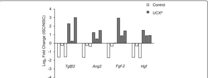
UCX® upregulate the endothelial Tgf-β2, Ang-2, Fgf-2 and Hgf expression in response to HLI
To elucidate the mechanism of action of UCX® in our HLI model we investigated if UCX® could change the endothelial gene expression. For this purpose, 5 hours after surgical induction of unilateral HLI, UCX® or their vehicle (as a control) were administered in the ischemic muscle and mice were sacrificed at day 70 post-HLI in-duction. The gastrocnemius muscle sections were stained for CD31 and visualized using a Laser Capture Microdissection microscope. CD31-positive cells were dissected, isolated and analysed by quantitative RT-PCR for the expression of several pro-angiogenic factors. Our results show that transcripts for Tgf-β2, Ang-2, Fgf-2 and Hgf were clearly upregulated in endothelial cells isolated from muscle of the ischemic limb, comparing with those found in endothelial cells from the contralateral limb.
This is observed exclusively in mice treated with UCX®. Control mice show the opposite trend, downregulating the expression of the angiogenic genes in endothelium from the ischemic limb, when compared with the contralateral limb (Fig. 5). These results suggest that the mechanism of action of UCX® in the HLI mouse model involves the upregulation of several angiogenic genes in the endothelial cells present in the ischemic muscle.
Discussion
CLI is a manifestation of peripheral arterial disease that describes patients with chronic ischemic rest pain, ulcers or gangrene [1]. Available therapies are limited and pa-tients may require amputation. The goal of limb salvage has stimulated research into alternative methods, includ-ing therapeutic angiogenesis. Stem cell therapy holds great potential for therapeutic angiogenesis, but its clin-ical translation has been slow due to (i) lengthy proce-dures performed ex vivo in which stem cells are manipulated and often lose viability, differentiate or
A
B
C
D
Fig. 4 UCX® increase capillary and collateral densities. UCX® or their vehicle (as a control) were administered in the ischemic gastrocnemius muscle 5 hours after HLI induction. (a) Representative sections from control and UCX®-treated ischemic gastrocnemius muscles at 90 days post-HLI. Capillaries and myocytes were identified by CD31 immunohistochemistry and haematoxylin, respectively. Scale bar, 125μm. (b) Quantitative analysis, at 90 days post-HLI, revealed increased capillary density (capillaries/myocyte) in UCX®-treated ischemic gastrocnemius muscles compared to control ischemic ones. (n = 3 and n = 7 for control and UCX®, respectively; t (8) = 4.44; **p = 0.0057; effect size was 3.064 and power 0.97). (c) Illustrative images of se-lected regions of interest (ROI) for control and UCX®-treated mice. ISC and NISC limbs are shown. Scale bar, 1 mm (d) Data are represented as the per-centage of collateral vessel density (CVD) increase of the ISC limb relatively to the NISC one. At 90 days post-HLI, UCX®-treated mice presented significantly higher CVD increase (%) when compared to control mice (n = 5 for each experimental group; t (8) = 7.63; ***P = 0.000062; effect size was 4.82 and power 0.99). ISC ischemic, NISC non-ischemic
change their characteristics, and (ii) the high costs in-volved. In addition, the clinical use of bone marrow MSCs has demonstrated some caveats, mainly due to low yields which further decrease with donor’s age and medical condition, leading to variable and/or limited cell doses.
Among the other possible sources for MSCs, there is the umbilical cord tissue [10], from which UCX® are de-rived. UCX® have already demonstrated the potential to lead to tissue regeneration and homeostasis, promoting faster remission of local and systemic manifestations of inflammatory arthritis [13]; preserving cardiac function after intramyocardial transplantation in a myocardial in-farction murine model [14]; and accelerating wound healing [15]. Those studies strongly suggest that UCX® act in different cell types through paracrine mechanisms. In addition, it was found that conditioned medium of UCX® cultures induces fibroblast and keratinocyte
mi-gration and UCX® are chemotactic to CD34-/CD45
-bone marrow MSCs [15]. Further, it was also shown that in vitro UCX®-conditioned medium induces angiogenesis by promoting the formation of capillary-like structures by HUVECs [13]. Herein, we corroborated those find-ings showing that UCX® promotes tubulogenesis and endothelial cell migration. We observed that UCX® when co-cultured with HUVECs induce tubule formation by HUVECs to a higher extent than the known pro-angiogenic factor FGF-2, and also promote migration. As already demonstrated for other cell types, our data strongly suggest that UCX® act in endothelial cells through paracrine mechanisms since a significant
increase of pro-angiogenic factors such as HGF, TGF-β1, IL-8 and PDGF-AA was observed in the conditioned medium of UCX® when compared to the control.
In order to investigate if UCX® could promote thera-peutic angiogenesis in a HLI context, we developed an experimental model of unilateral HLI and two important parameters were assessed: angiogenesis and arteriogen-esis. Our data indicate that UCX® administration signifi-cantly induces blood perfusion recovery in the limb after HLI induction. Accordingly, a significantly higher capil-lary density and CVD increase was found in the ische-mic muscles after UCX® treatment. These evaluations were performed at day 90 post-HLI suggesting that the effect induced by UCX® in angiogenesis and arteriogen-esis is maintained over time. Besides, the non-ischemic muscles are unaffected by UCX® since capillary density and CVD are similar in non-ischemic muscles both from UCX®-treated and control mice, indicating that UCX® might have a local action. These data suggest that UCX® could secrete cytokines/chemokines and paracrinally interfere with the ischemic microenvironment contribut-ing to a therapeutic angiogenesis. More importantly, a new mechanism of action of UCX® is demonstrated for the first time: UCX® modulate the expression of pro-angiogenic players in endothelial cells. We found that 70 days post-HLI and UCX® treatment, endothelial cells isolated from UCX®-treated microenvironment present
an upregulation in their expression levels for Tgfβ2,
Ang-2, Fgf-2 and Hgf when isolated from the ischemic gastrocnemius muscle in comparison with the contralat-eral one. This effect is UCX®-specific as the opposite is
Fig. 5 UCX® upregulate the expression of Tgf-β2, Ang-2, Fgf-2 and Hgf in endothelial cells isolated from ischemic gastrocnemius muscles. UCX® or their vehicle (as a control) were administered in the ischemic gastrocnemius muscle 5 hours after HLI induction. At 70 days post-HLI, the expression of pro-angiogenic factors and their receptors was evaluated by qRT‐PCR exclusively on endothelial cells isolated from gastrocnemius muscles. Each bar represents the relative gene expression in one animal. White and grey bars represent control and UCX®-treated mice, respectively. Values were normalized to 18S to obtain relative expression levels. Results expressed as log2 fold changes between ISC and NISC samples demonstrated relative abundance of the transcripts in UCX®-treated mice; in contrast, a downregulation is observed in control mice. ISC ischemic, NISC non-ischemic
verified in control mice. According to these findings, we show that UCX® simultaneously upregulate the expres-sion of several angiogenic factors in endothelial cells and by this mechanism we may hypothesize that UCX® im-prove their endogenous angiogenic potency in an ische-mic context. This could be very relevant when compared to other strategies where only a single angiogenic factor is administered, especially if we understand angiogenesis as a complex process that involves multiple cytokines. It is also very interesting to note that the same factors modulated by UCX® have been extensively studied in therapeutic angiogenesis. Specifically, in gene and pro-tein therapy, FGF-2 was used in a rabbit model of HLI [18], where it was shown to promote the development of collateral vessels. The efficacy of FGF-2 was also evalu-ated in clinical trials such as FIRST (FGF Initiating Re-vascularization Trial) [19] and TRAFFIC (Therapeutic Angiogenesis with Recombinant FGF-2 for Intermittent Claudication) [20]; however, it did not show a sustained success [21]. Concerning HGF, it was also used in clinical trials through gene transfer to treat CLI, where its safety was shown [22]. More rigorous controlled trials (randomised and placebo-controlled studies with larger numbers of patients) are currently ongoing. Given the fact that CLI is a common complication of diabetes mellitus, future studies should assess the UCX® efficacy in a diabetic experimental model since diabetes may adversely affect EPC function and thus limit therapeutic efficacy. Recent data have further shown that the secreted proangiogenic protein osteopontin (OPN), significantly downregulated in diabetic EPCs, could increase the secretion of angiogenic proteins from EPCs increasing their therapeutic efficacy [23]. Importantly, these findings show that the function of EPCs could be modulated and raise interesting questions for future investigation, such as, (a) which molecules could eventu-ally improve the therapeutic efficacy of UCX® in an ischemic microenvironment; (b) could different ischemic microenvironments modulate differently the function of UCX®; if so, by which mechanisms; and (c) should UCX® be modulated by the administration of selected molecules according to the pathologies associated with CLI.
Conclusions
In conclusion, here we propose a model of enhanced and sustained angiogenesis induction by using UCX® as a promising therapeutic approach for CLI. UCX® signifi-cantly induce blood perfusion, capillary density and collateral development. This could be at least in part achieved by a new mechanism since we show that UCX® upregulate the simultaneous expression of several pro-angiogenic factors in endothelial cells that could improve their angiogenic potency.
Abbreviations
ATMP:advanced therapy medicinal product; Ang-2: angiopoietin 2; BM: basal medium; CLI: critical limb ischemia; CM: conditioned medium; CVD: collateral vessels density; EBM: endothelial basal medium; EGM: endothelial growth medium; ECs: endothelial cells; FGF-2: fibroblast growth factor 2; G-CSF: granulocyte-colony stimulating factor; HGF: hepatocyte growth factor; HLI: hindlimb ischemia; HUVECs: human umbilical vein endothelial cells; IL-6: interleukin 6; IL-8: interleukin 8; 2: matrix metalloproteinase-2; MMP-9: matrix metalloproteinase-9; MSCs: mesenchymal stem cells;
PAD: peripheral artery disease; PDGF-AA: platelet-derived growth factor-AA; POST-HLI: post-HLI; PRE-HLI: before HLI; ROI: region of interest; RT: room temperature; TGF-β1: transforming growth factor β1; Tgfβ2: transforming growth factorβ2; VEGF-A: vascular endothelial growth factor A Funding
This study was partially supported by PORLisboa-FEDER (project QREN 2013/ 30196– ClinUCX).
Availability of data and materials
All data generated or analysed during this study are included in this published article.
Authors’ contributions
PC and HC devised the overall preclinical development strategy of UCX® for peripheral arterial disease and CLI and sourced for funding of the research. SCRS conceived and designed the experiments, in discussion with FP, MS, RB, PC and HC. SCRS wrote the manuscript and FP, MS, RB, PC and HC critically revised it. ARP performed laser doppler perfusion imaging,
immunohistochemistry and capillary density analysis, analysed the data and revised the manuscript; TM performed collateral vessel quantification and gene expression analysis, analysed the data and revised the manuscript; ARP and TM performed endothelial culture and wound-healing assays and analysed the data. AM and JON performed diaphonisation. AM performed the surgical HLI and UCX® administration in vivo, performed contrast agent perfusion before diaphonisation and participated in data analysis, interpretation and critically revised the manuscript. JMS and RB designed the experiments on tubule formation and preparation of conditioned medium. MT and MF performed UCX® culture and tubule formation assays, prepared conditioned medium and performed the quantification of secreted factor and revised the manuscript. All the authors read and approved the final version of the manuscript.
Competing interests
HC and PC are shareholders of ECBio; JMS, MF, MT are employees of ECBio. RB was an employee of ECBio when this work was performed. The other authors declare that they have no competing interests.
Consent for publication Not applicable.
Ethics approval and consent to participate
Umbilical cord donations, with written informed consents, as well as umbilical cord procurement, were made according to Directive 2004/23/EC of the European Parliament and of the Council of 31 March 2004 on setting standards of quality and safety for the donation, procurements, testing, processing,
preservation, storage and distribution of human tissues and cells. Human umbilical cord tissue-derived mesenchymal stem cells (UCX®) were isolated according to a patented proprietary technology developed by ECBio (Amadora, Portugal). All animal procedures were carried out with the permission of the local animal ethics committee in accordance with the Directive 2010/63/EU. The procedures were approved by the institutional Animal Welfare Body and licensed by DGAV, the Portuguese competent authority for animal protection (license number 023861/2013).
Author details
1Centro Cardiovascular da Universidade de Lisboa, Av. Prof Egas Moniz
1649-028 Lisbon, Portugal.2Centro Hospitalar Lisboa Norte, Av. Prof. Egas
Moniz 1649-035 Lisbon, Portugal.3ECBio, Investigação e Desenvolvimento
em Biotecnologia S.A., R. Henrique Paiva Couceiro, 27, 2700-4511 Amadora, Portugal.4Nova Medical School/Faculdade de Ciências Médicas, Universidade
Nova de Lisboa, 1169-056 Lisbon, Portugal.5Faculdade de Medicina da
Received: 19 July 2016 Accepted: 9 September 2016
References
1. Norgren L, Hiatt WR, Dormandy JA, Nehler MR, Harris KA, Fowkes FG, TASC II Working Group. Inter-society consensus for the management of peripheral arterial disease (TASC II). J Vasc Surg. 2007;45(Suppl S):S5–S67.
2. Ouma GO, Zafrir B, Mohler 3rd ER, Flugelman MY. Therapeutic angiogenesis in critical limb ischemia. Angiology. 2013;64:466–80.
3. Shimamura M, Nakagami H, Koriyama H, Morishita R. Gene therapy and cell-based therapies for therapeutic angiogenesis in peripheral artery disease. Biomed Res Int. 2013;2013:186215.
4. Yan J, Tie G, Xu TY, Cecchini K, Messina LM. Mesenchymal stem cells as a treatment for peripheral arterial disease: current status and potential impact of type II diabetes on their therapeutic efficacy. Stem Cell Rev. 2013;9:360–72. 5. Nekanti U, Dastidar S, Venugopal P, Totey S, Ta M. Increased proliferation and
analysis of differential gene expression in human Wharton’s jelly-derived mesenchymal stromal cells under hypoxia. Int J Biol Sci. 2010;6:499–512. 6. Tao H, Han Z, Han ZC, Li Z. Proangiogenic features of mesenchymal stem
cells and their therapeutic applications. Stem Cells Int. 2016;2016:1314709. 7. Liew A, O’Brien T. Therapeutic potential for mesenchymal stem cell
transplantation in critical limb ischemia. Stem Cell Res Ther. 2012;3:28. 8. Fadini GP, Agostini C, Avogaro A. Autologous stem cell therapy for
peripheral arterial disease meta-analysis and systematic review of the literature. Atherosclerosis. 2010;209:10–7.
9. Zuk PA, Zhu M, Mizuno H, Huang J, Futrell JW, Katz AJ, Benhaim P, Lorenz HP, Hedrick MH. Multilineage cells from human adipose tissue: implications for cell-based therapies. Tissue Eng. 2001;7:211–28.
10. Erices A, Conget P, Minguell JJ. Mesenchymal progenitor cells in human umbilical cord blood. Br J Haematol. 2000;109:235–42.
11. Martins JP, Santos JM, de Almeida JM, Filipe MA, de Almeida MV, Almeida SC, Agua-Doce A, Varela A, Gilljam M, Stellan B, et al. Towards an advanced therapy medicinal product based on mesenchymal stromal cells isolated from the umbilical cord tissue: quality and safety data. Stem Cell Res Ther. 2014;5:9. 12. Dominici M, Le Blanc K, Mueller I, Slaper-Cortenbach I, Marini F, Krause D,
Deans R, Keating A, Prockop D, Horwitz E. Minimal criteria for defining multipotent mesenchymal stromal cells. The International Society for Cellular Therapy position statement. Cytotherapy. 2006;8:315–7. 13. Santos JM, Barcia RN, Simoes SI, Gaspar MM, Calado S, Agua-Doce A,
Almeida SC, Almeida J, Filipe M, Teixeira M, et al. The role of human umbilical cord tissue-derived mesenchymal stromal cells (UCX®) in the treatment of inflammatory arthritis. J Transl Med. 2013;11:18.
14. Santos Nascimento D, Mosqueira D, Sousa LM, Teixeira M, Filipe M, Resende TP, Araujo AF, Valente M, Almeida J, Martins JP, et al. Human umbilical cord tissue-derived mesenchymal stromal cells attenuate remodeling after myocardial infarction by proangiogenic, antiapoptotic, and endogenous cell-activation mechanisms. Stem Cell Res Ther. 2014;5:5.
15. Miranda JP, Filipe E, Fernandes AS, Almeida JM, Martins JP, De la Fuente A, Abal M, Barcia RN, Cruz P, Cruz H, et al. The Human umbilical cord tissue-derived MSC population UCX((R)) promotes early motogenic effects on keratinocytes and fibroblasts and G-CSF-mediated mobilization of BM-MSCs when transplanted in vivo. Cell Transplant. 2015;24:865–77.
16. Santos JM, Camoes SP, Filipe E, Cipriano M, Barcia RN, Filipe M, Teixeira M, Simoes S, Gaspar M, Mosqueira D, et al. Three-dimensional spheroid cell culture of umbilical cord tissue-derived mesenchymal stromal cells leads to enhanced paracrine induction of wound healing. Stem Cell Res Ther. 2015;6:90. 17. Brown AL, Smith DW. Improved RNA preservation for immunolabeling and
laser microdissection. RNA. 2009;15:2364–74.
18. Li J, Wei Y, Liu K, Yuan C, Tang Y, Quan Q, Chen P, Wang W, Hu H, Yang L. Synergistic effects of FGF-2 and PDGF-BB on angiogenesis and muscle regeneration in rabbit hindlimb ischemia model. Microvasc Res. 2010;80:10–7. 19. Simons M, Annex BH, Laham RJ, Kleiman N, Henry T, Dauerman H, Udelson JE,
Gervino EV, Pike M, Whitehouse MJ, et al. Pharmacological treatment of coronary artery disease with recombinant fibroblast growth factor-2: double-blind, randomized, controlled clinical trial. Circulation. 2002;105:788–93. 20. Lederman RJ, Mendelsohn FO, Anderson RD, Saucedo JF, Tenaglia AN,
Hermiller JB, Hillegass WB, Rocha-Singh K, Moon TE, Whitehouse MJ, et al. Therapeutic angiogenesis with recombinant fibroblast growth factor-2 for intermittent claudication (the TRAFFIC study): a randomised trial. Lancet. 2002;359:2053–8.
21. Ellis LM, Hicklin DJ. Pathways mediating resistance to vascular endothelial growth factor-targeted therapy. Clin Cancer Res. 2008;14:6371–5. 22. Morishita R, Makino H, Aoki M, Hashiya N, Yamasaki K, Azuma J, Taniyama Y,
Sawa Y, Kaneda Y, Ogihara T. Phase I/IIa clinical trial of therapeutic angiogenesis using hepatocyte growth factor gene transfer to treat critical limb ischemia. Arterioscler Thromb Vasc Biol. 2011;31:713–20.
23. Vaughan EE, Liew A, Mashayekhi K, Dockery P, McDermott J, Kealy B, Flynn A, Duffy A, Coleman C, O’Regan A, et al. Pretreatment of endothelial progenitor cells with osteopontin enhances cell therapy for peripheral vascular disease. Cell Transplant. 2012;21:1095–107.
• We accept pre-submission inquiries
• Our selector tool helps you to find the most relevant journal • We provide round the clock customer support
• Convenient online submission • Thorough peer review
• Inclusion in PubMed and all major indexing services • Maximum visibility for your research
Submit your manuscript at www.biomedcentral.com/submit