Bone marrow mesenchymal stem cells
overexpressing human basic fibroblast
growth factor increase vasculogenesis
in ischemic rats
J.C. Zhang
1, G.F. Zheng
2, L. Wu
1, L.Y. Ou Yang
1and W.X. Li
11Department of Vascular Surgery, The First Affiliated Hospital of Fujian Medical University, Fuzhou, China 2Department of Vascular Surgery, The People’s Hospital of Ganzhou, Ganzhou, China
Abstract
Administration or expression of growth factors, as well as implantation of autologous bone marrow cells, promotein vivo
angiogenesis. This study investigated the angiogenic potential of combining both approaches through the allogenic transplantation of bone marrow-derived mesenchymal stem cells (MSCs) expressing human basic fibroblast growth factor (hbFGF). After establishing a hind limb ischemia model in Sprague Dawley rats, the animals were randomly divided into four treatment groups: MSCs expressing green fluorescent protein (GFP-MSC), MSCs expressing hbFGF (hbFGF-MSC), MSC controls, and phosphate-buffered saline (PBS) controls. After 2 weeks, MSC survival and differentiation, hbFGF and vascular endothelial growth factor (VEGF) expression, and microvessel density of ischemic muscles were determined. Stable hbFGF expression was observed in the hbFGF-MSC group after 2 weeks. More hbFGF-MSCs than GFP-MSCs survived and differentiated into vascular endothelial cells (P,0.001); however, their differentiation rates were similar. Moreover, allogenic transplantation of hbFGF-MSCs increased VEGF expression (P=0.008) and microvessel density (P,0.001). Transplantation of hbFGF-expressing MSCs promoted angiogenesis in anin vivo hind limb ischemia model by increasing the survival of transplanted cells that subsequently differentiated into vascular endothelial cells. This study showed the therapeutic potential of combining cell-based therapy with gene therapy to treat ischemic disease.
Key words: Angiogenesis; Basic fibroblast growth factor; Bone marrow mesenchymal stem cells; Gene transfection; Ischemia
Introduction
Increasingin vivoangiogenesis is a therapeutic strategy for treating ischemic diseases, including peripheral artery disease, heart disease, myocardial infarction and stroke. Administration of angiogenic factors, such as vascular endothelial growth factor (VEGF), human basic fibroblast growth factor (hbFGF), and hepatocyte growth factor (HGF) (1-3), or gene transfer of these growth factors (4) have been reported. Intramuscular transfer of both theVEGF165and
HGFgenes increased perfusion and decreased necrosis in ischemic mouse hind limbs via neovascularization (5). Similar results were obtained with AGGF1 (angiogenic factor with G patch and FHA domains 1), a relatively newly isolated angiogenic factor that significantly reduced the ambulatory impairment associated with limb ischemia (6).
In addition to direct or systemic administration of growth factors to promote angiogenesis, implantation of autologous
bone marrow cells promoted angiogenesis in a rat ischemic hind limb model (7,8). Specifically, injection of purified bone marrow cells (BMCs) that have the potential to differentiate into endothelial cells (9) and produce angiogenic growth factors (10) increased blood flow in ischemic hind limbs 4 weeks after transplantation (7). Furthermore, increased exercise capacity was observed after implantation of autologous BMCs in a rat ischemic hind limb model (8). Similar functional recovery has been observed with allogenic transfer of mesenchymal stem cells (MSCs) derived from bone marrow or adipose tissue in an in vivo model of ischemic stroke (11). Although stem cell transplantation has been used to stimulate vasculogenesis with favorable efficacy, the reparative capacity of MSCs decline with age, which may be enhanced by lentiviral-mediated expression of myocardin and telomerase reverse transcriptase (12) or
Correspondence: J.C. Zhang, Department of Vascular Surgery, Institute of Vascular and Endovascular Surgery, The First Affiliated Hospital of Fujian Medical University, No. 20, ChaZhong Road, TaiJiang District, Fuzhou 35005, China. Fax: ++86-591-8335-6180. E-mail: [email protected]
culture expansion in media supplemented with growth factors prior to transplantation (13). Moreover, stem cell survival after transplantation remains low, negatively impacting their therapeutic efficacy.
Because bFGF has the potential to promote angiogen-esis (14,15) and also increase the in vivo survival and proliferation of stem cells (16), this study aimed to test the hypothesis that bone marrow-derived MSCs overexpressing hbFGF would increase angiogenesis to a greater extent than MSCs alone. To test this hypothesis, rats with hind limb ischemia were treated with MSCs expressing green fluorescent protein (GFP-MSC group), MSCs expressing hbFGF (hbFGF-MSC group), MSCs alone (MSC group), or phosphate-buffered saline (PBS control group). After 2 weeks, MSC survival was determined by GFP fluorescence, and differentiation was determined by performing immuno-fluorescence assays of CD31 expression. hbFGF and VEGF expression in gastrocnemius muscle was determined by Western blot analyses. In addition, microvessel density (MVD) was determined by immunohistochemical analysis of von Willebrand factor (vWF) expression. Our results may form the basis for further clinical studies assessing the therapeutic efficacy of hbFGF overexpression in MSCs prior to autologous transplantation.
Material and Methods
Animals
Thirty healthy 6-week-old male Sprague Dawley rats were purchased from the Shanghai SLAC Laboratory Animal Co., Ltd. (China). Animals were acclimated in the Fujian Medical University Animal Center for 1 week with free access to food and water. The animals were fasted for 12 h prior to the beginning of the experiments. The study was approved by the Institutional Review Board of The First Affiliated Hospital of Fujian Medical University.
MSC isolation
The isolation and identification of MSCs were performed as we previously described (17). Briefly, 6 Sprague Dawley rats were euthanized by cervical dislocation, and bilateral femurs and tibias were collected. After the metaphysis was removed, the exposed bone marrow cavity was flushed with Dulbecco’s modified Eagle medium (DMEM; Hyclone, USA) containing 10% fetal bovine serum (FBS, Hyclone). The fluid was harvested and centrifuged at 180gfor 10 min. After the supernatant was removed, the cells were resuspended in culture medium and lymphocyte separation medium at a ratio of 2:1 (v:v). The cell suspension was added to 1.077 g/ L lymphocyte separation medium (d=1.077 g/mL, H&Y Bio, China) followed by centrifugation at 360gfor 15 min. The white, cloudy cell layer was collected, and the cells were washed twice with L-DMEM. Single-cell suspensions were prepared with L-DMEM containing 10% FBS. Cells at 16106 cells/cm2 were seeded into 25-cm flasks and maintained in an incubator at 376C in a humidified
environment with 5% CO2.
MSC surface antigen expression assayed by flow cytometry
MSCs were observed with an inverted microscope (Olympus, Japan). Cells of the third passage (P3) were used for all subsequent experiments as previously described (18). Cells were harvested at 80-90% confluency by digestion with 0.125% trypsin. Cell suspensions contain-ing 26106cells were transferred into 10 1.5-mL Eppendorf tubes and then incubated with 10mL of the following mouse anti-rat monoclonal antibodies: CD90-R-phycoerythrin (PE), CD34-fluorescein isothiocyanate (FITC), CD44-PE, CD11b/c-FITC or isotype controls (all from Beckman Coulter, USA) for 15 min in the dark at 376C. Cells were washed twice with 1 mL PBS followed by centrifugation at 360gfor 5 min. After resuspension at 0.56106cells/mL in PBS, surface antigens were detected by flow cytometry (Beckman Coulter); the System II software (Beckman Coulter) was used for data acquisition and analysis. MSCs were characterized as negative for CD11b and CD34 hematopoietic cell marker expression (19) and positive for CD44 and CD90 MSC marker expression (20).
Preparation of hbFGF-expressing lentivirus
Briefly, human cDNA was amplified by polymerase chain reaction (PCR) using the followinghbFGF-specific primers (the AgeI restriction site is underlined) from Shanghai Genechem Co., Ltd. (China): sense: 59-CATGGGCTGGAC GAGGAATGGCAGCCGGGAGCATC-39and antisense: 59 -TCACCATGGTGGCGACCGGTACGTAGCTCTTAGCAG ACATTGGAAG-39.
The PCR conditions were as follows: denaturation at 946C for 30 s; 30 cycles of 946C for 30 s, 556C for 30 s, 726C for 30 s, and a final extension at 726C for 10 min. After the PCR product was subjected to 1.1% agarose gel electrophoresis and purified using a plasmid extraction kit (Qiagen, The Netherlands), it was inserted into the pGC FU vector (Shanghai Genechem Co., Ltd.) at the AgeI site. pGC FU-hbFGF vectors were identified by PCR followed by DNA sequencing to confirm the insertion of the target gene.
The pGC FU-hbFGF vector, pHelper 1.0 vector, and pHelper 2.0 vector (all from Shanghai Genechem Co., Ltd.) were transfected into 293T cells (Shanghai Genechem Co., Ltd.) in the presence of Lipofectamine 2000 (Invitrogen, USA). After 2 days, the cell culture supernatant was collected and repeatedly centrifuged at 4000 g for 10 min. After the lentivirus was concentrated by filtration, it was stored at ––806C. Real-time quantitative PCR was performed to determine the lentivirus titer.
Lentiviral-mediated MSC transduction
1 mL fresh medium was added. When the cell confluency reached 30%, cells were infected with GFP- or hbFGF-expressing recombinant virus at a multiplicity of infection of 50 as previously described (21). The hbFGF-expres-sing lentiviruses also expressed GFP. After 10 h, the medium was removed, and cells were washed with PBS followed by addition of fresh medium. Cells were passaged using routine methods and observed daily by fluorescence microscopy to confirm continued GFP expression.
Establishment of anin vivoischemia model and MSC transplantation
Rats were fasted for 12 h before surgery but were given
ad libitumaccess to water. Animals were intraperitoneally anesthetized with 10% chloral hydrate (3 mL/kg). Under aseptic conditions, a longitudinal incision was made in the hind limb 2-3 cm from the femoral artery. After the femoral artery was ligated at the level of the inguinal ligament, it and its branches were removed while protecting the femoral nerve.
At 24 h post-induction of hind limb ischemia, the rats were randomly divided into four groups (n=6/group): control, MSC, GFP-MSC, and hbFGF-MSC. Animals in the MSC, GFP-MSC, and hbFGF-MSC groups received injections of 56106cells in 0.5 mL PBS into the adductors, tibialis anterior, and gastrocnemius muscles of the ischemic limb. Animals in the control group received an injection of 0.5 mL PBS into the ischemic limb.
No difference in tissue necrosis was observed between the hbFGF-MSC and control groups. Specifically, after induction of hind limb ischemia, ischemic necrosis was found in one or two toes of 3 animals in the PBS group; no necrosis was observed in the other groups. In addition, wound infection and delayed wound healing were found in 2 rats in the PBS control group. However, all animals survived. Apparent ambulatory impairment was observed within 3-5 days after surgery; however, normal activity was observed 1 week after surgery.
At 2 weeks post-transplantation, which was considered a chronic condition, blood was collected from the abdom-inal aorta, after which the animals were sacrificed and the adductor muscle and gastrocnemius were collected. A fraction of skeletal muscle was used for histological examination, and the remaining tissues were stored at –806C for later use.
Immunohistochemistry and immunocytochemical analyses
Muscle cross-sections (8mm) were prepared for immu-nohistochemistry using the SP-9000 two-step method (Beijing Zhongshan Biotech, China). Primary antibodies specific for vWF (Cat No. sc-53466; Santa Cruz Biotech-nology, USA) and CD31 (Cat No. sc-13537, Santa Cruz Biotechnology), goat anti-rabbit IgG secondary antibodies (Beijing Zhongshan Biotech) and a diaminobenzidine (DAB)
substrate kit (Beijing Zhongshan Biotech) were used. vWF-positive cells appeared brownish-yellow, and the number of positive cells (i.e., microvessel number) was counted at a high magnification of 2006(excluding the tubes formed by more than three positive cells). CD31-positive cells were identified by their red fluorescence using a confocal laser-scanning microscope (ZEISS LSM510, Germany).
Western blot analysis
Muscles were cut into pieces and homogenized mechanically for 10 min (170 rpm). After determination of protein concentration, 100mg protein was separated by sodium dodecyl sulfate polyacrylamide gel electrophore-sis (SDS-PAGE), and the proteins were transferred onto polyvinylidene difluoride (PVDF) membranes. After the membranes were washed with PBS four times, they were incubated in blocking solution consisting of 5% skimmed milk powder in Tris-buffered saline with Tween 20 (TBS-T) for 60 min. The membranes were then incubated with rabbit anti-human bFGF (diluted 1:3000, Santa Cruz Biotech-nology), rabbit anti-rat VEGF (diluted 1:1000; Santa Cruz Biotechnology), or rabbit anti-ratb-actin antibodies (diluted 1:500; Sigma, USA) for 60 min. After washing with PBS 4 times, the membranes were incubated with horseradish peroxidase-conjugated goat anti-rabbit secondary antibo-dies (diluted 1:1000; Santa Cruz Biotechnology) at room temperature for 60 min. After washing in PBS, visualization was performed with alkaline phosphatase (Santa Cruz Biotechnology). The ImageJ analysis software (National Institutes of Health, USA) was used to detect the absorb-ance of VEGF bands, which were normalized to values obtained for actin to determine relative VEGF expression.
Statistical analysis
Continuous variables are reported as means±SD. Independent-sample t-tests were performed to compare GFP+and GFP+CD31+cell counts and the ratios in the GFP-MSC and hbFGF-MSC groups. ANOVA and Bonferronipost hoctests were performed to compare the differences among the four treatment groups in VEGFvs
actin and capillaries vs fibers (as ratios) and capillary counts. Two-sided P values less than 0.05 were consid-ered to be significant. The SPSS 17.0 statistics software (SPSS Inc., USA) was used for the statistical analyses.
Results
MSC surface marker expression
morphology, confirmed the identity of isolated cells as MSCs. The corresponding negative controls were also included (Figure 1A-D).
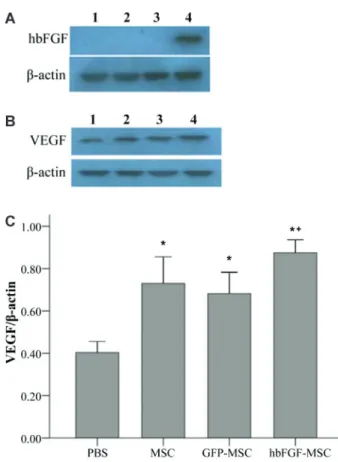
Effects of hbFGF-MSCs on VEGF expression in ischemic muscle
As shown in Figure 2A, hbFGF expression was only observed in animals in the hbFGF-MSC group. In addition, although VEGF expression was observed in the skeletal muscle of animals of all groups, significant differences in relative VEGF expression were observed among the groups (Figure 2B). Specifically, significantly increased VEGF expression was observed in all MSC-treated animals compared to the PBS control group (P,0.001). Furthermore, the highest VEGF expression was observed in the hbFGF-MSC group, which was significantly higher than in the GFP-MSC group (0.87vs0.68; P=0.008).
Survival and differentiation of transplanted MSCs
To assess the differentiation of MSCs that did not express CD31 or Factor VIII (data not shown) into vascular endothelial cells, the coexpression of CD31 and GFP in the gastrocnemius was determined by immunofluorescence analysis 2 weeks after cell transplantation. As shown in Figure 3A, low CD31 expression was observed in the MSC group; however, CD31 expression was increased in both the GFP-MSC and hbFGF-MSC groups. In addition, a fraction of cells was both GFP++ and CD31++, suggesting that differe-ntiation of MSCs into vascular endothelial cells had occurred in these groups. Analysis of the immunofluorescence data
revealed that the number of GFP++ cells in the hbFGF-MSC group was significantly higher than in the GFP-MSC group (26.83 vs 15.50, P,0.001; Figure 3B). In addition, the number of GFP++CD31++ cells was also significantly higher in the hbFGF-MSC group compared to the GFP-MSC group (14.33vs6.83, P,0.001; Figure 3C). However, as shown in Figure 3D, no significant difference was observed between the GFP-MSC and hbFGF-MSC groups in the ratio of GFP++CD31++ to GFP++ cells, indicating that the differ-entiation rate did not differ between these two treatment groups.
Effects of hbFGF-MSCs on vasculogenesis
The in vivo angiogenic potential of hbFGF-MSCs was then determined. The number of microvessels was determined immunohistochemically by analysis of the endothelial cell marker vWF. Representative images of immunohistochemical vWF staining in each of the 4 groups Figure 1.Surface marker expression of isolated rat MSCs (P3).
The expression of CD11b/c (A), CD34 (B), CD44 (C), and CD90 (D) was determined using flow cytometry (red lines). Their respective negative controls were also included (black lines).
Figure 2.Overexpression of human basic fibroblast growth factor (hbFGF) by mesenchymal stem cells (MSCs) in a rat hind limb ischemia model.A, hbFGF andB, vascular endothelial growth factor (VEGF) protein expression was determined by Western blot analysis.1, PBS group (n=6);2, MSC group (n=6);3, green fluorescent protein (GFP)-MSC group (n=6); 4, hbFGF-MSC group (n=6).C, Relative VEGF expression was determined after normalization to b-actin. *P,0.05, compared to PBS group; +P
are shown in Figure 4A-D. Compared to the PBS control group, the ratio of capillary to fiber counts was significantly higher in the MSC, GFP-MSC, and hbFGF-MSC groups (P,0.001). However, the highest capillary to fiber ratio was observed in the hbFGF-MSC group (0.70vs 0.41 in the MSC group and 0.42 in the GFP-MSC group, both P,0.001; Figure 4E). Finally, as shown in Figure 4F, capillary counts (microvessel density, MVD) were signifi-cantly higher in the GFP-MSC and hbFGF-MSC groups compared to the PBS control group (29.67 and 52.83vs
8.5, both P,0.001). The capillary counts were significantly higher in the hbFGF-MSC group than in the other groups (P,0.001).
Discussion
Inducing angiogenesis is a relatively new therapeutic strategy for the treatment of ischemia. Given the role of hbFGF in inducing angiogenesis, as well as stem cell
survival and proliferation (16,22), the effects of bone marrow-derived MSCs overexpressing hbFGF on angio-genesis were analyzed in anin vivoischemia model. In all of the treatment groups containing MSCs, significantly greater capillary-to-muscle fiber ratios were observed, which is similar to previous reports on the therapeutic potential of MSCs in this model (7,8). Compared to GFP-MSCs, a greater number of hbFGF-MSCs survived and differentiated into vascular endothelial cells; however, the differentiation rate was the same. In addition, hbFGF-MSCs induced the greatest VEGF expression and MVD.
2 weeks post-transplantation. Further long-term studies will be undertaken to confirm the continued skeletal-muscle expression of hbFGF. In addition to increased hbFGF, hbFGF-MSCs also induced in vivo VEGF expression. However, the exact mechanism by which overexpression of hbFGF in MSCs also increased VEGF expression remains to be determined. It may be an indirect effect of an increased number of MSCs differentiating into endothelial cells. Alternatively, hbFGF expression may directly induce VEGF expression, as observed in human aortic smooth muscle cells (24). Further studies will determine the mechanism responsible for increased skeletal muscle VEGF expression in hbFGF-MSC transplantation.
Although stem cell transplantation has been used to stimulate vasculogenesis with favorable efficacy, the survi-val rate of stem cells remains low, which negatively impacts their therapeutic efficacy. In the present study, increased MSC survival was observed in those overexpressing hbFGF, which is consistent with previous studies reporting that hbFGF increased survival rate of transplanted stem cells (16), as well as proliferation of stem cells (22). In addition, hbFGF overexpression by MSCs increased the number of cells that differentiated into endothelial cells, but not the relative differentiation rate, suggesting the increased number of GFP++CD31++ cells was due to increased survival and/or proliferation but not increased differentiation capacity. However, the mechanism by which hbFGF
influences survival was not determined. bFGF induced both vascular endothelial cell proliferation (25) and endothelial cell migration by inducing the secretion of cathepsin L by skeletal muscle cells (26). In addition, bFGF can simulta-neously promote the growth of capillaries and small arteries (27). Furthermore, bFGF can upregulate the expression of the platelet-derived growth factor (PDGF) receptor; there-fore, bFGF and PDGF may exert synergistic effects to promote vasculogenesis (28). Further studies will determine the mechanism by which hbFGF expression induces MSC-mediated endothelial cell differentiation. Furthermore, addi-tional studies will analyze the effect of coexpression of hbFGF and its coreceptor, syndecan-4, in MSCs, as this combination alone can stimulate neovascularization and increase perfusion in rats with hind limb ischemia (29).
In the present study, both GFP- and hbFGF-expres-sing MSCs differentiated into endothelial cellsin vivo. This differentiation capacity is thought to be the mechanism by which cell therapy restores cardiac function (30), improves blood flow, and reduces necrosis in ischemia (13,31). Specifically, MSCs have been shown to differ-entiate into cardiomyocytes, vascular smooth muscle cells, and endothelial cells in ischemic cardiomyopathy (30). Furthermore, in anin vivostroke model, transplanta-tion of brain-derived neurotrophic factor-differentiated, MSCs improved motor function to a greater degree than transplantation of undifferentiated MSCs (32). However, Figure 4.Overexpressing human basic fibroblast growth factor (hbFGF) in mesenchymal stem cells (MSCs) stimulates angiogenesis in a rat hind limb ischemia model.A-D, Representative images of sections of ischemic gastrocnemius muscles from the phosphate-buffered saline (PBS) control (A), MSC (B), green fluorescent protein (GFP)-MSC (C), and hbFGF-MSC (D) groups immunostained with anti-von Willebrand factor (vWF) antibodies. After counting the vWF-positive cells (microvessels) and muscle fibers, the ratio of microvessels to muscle fibers (E) and microvessel density (F) were determined. *P,0.05, compared to the PBS group; +P,0.05, compared to the MSC group; #P
the improved blood perfusion in ischemic hind limbs after transplantation of in vitro-differentiated endothelial cells suggests that this differentiation pathway may be neces-sary, and possibly sufficient, for angiogenic therapy in ischemia (31).
Induction of a microenvironment conducive to stem-cell differentiation and engraftment likely depends on paracrine effects (33) that promote vascularization (e.g., interleukin [IL]-4, IL-13, angiogenin, and VEGF), extracellular matrix production (e.g., PDGF-BB), and tissue remodeling (e.g., matrix metalloproteinases) (13). In addition, differential paracrine factor expression is thought to be responsible for the varying restorative capacity of different stem cells in ischemic diseases (33). That is consistent with the results of the present study, in which greater VEGF production was observed with hbFGF-MSCs than with GFP-MSCs. Further studies will assess the mechanism by which hbFGF-MSCs differentiate into endothelial cells, as well as the engraft-ment capacity of these cells.
In addition to inducing endothelial differentiation, hbFGF-expressing MSCs significantly increased the MVD by nearly 2-fold compared to the GFP-MSC group, which is similar to the results of previous studies (14,15). Injection of bFGF-expressing adenovirus into the cerebral ventricle (14) or injection of fibroblasts infected with bFGF-expres-sing adenovirus into ischemic muscles (15) has been shown to increase the local expression of bFGF, as well as the MVD and number of lateral branches. Furthermore, in a cranial defect model, Chen et al. (23) reported enhanced angiogenesis after treatment with poly-DL-lactide/hydrox-yapatite in combination with bFGF-expressing MSCs.
In addition to the direct effect of cytokines, stem cell transplantation may also directly promote vasculogenesis (34). Resident stem cells synthesize VEGF, which then activates vascular endothelial cells via paracrine signaling (35). However, because the extent of VEGF upregulation was not greatly increased in hbFGF-MSCs relative to controls, upregulation of other molecules that affect
angiogenesis is likely necessary. Stem cells may also synthesize HGF, tumor necrosis factor, and proteases, which promote vascular endothelial cell proliferation and migration (36). Moreover, stem cells have the potential to differentiate into vascular endothelial cells (37), and there-fore have a direct role in capillary network formation (38).
The present study is limited in that we did not determine the effects of the various treatment groups on blood flow. In addition, functional experiments to assess recovery after treatment were not carried out. Further studies will evaluate the effects of hbFGF-MSCs on the physiological status of the ischemic limb, including limb rescue, necrosis, and loss, as well as post-treatment exercise capacity. These studies will also help determine the extent of angiogenesis required to achieve clinical benefits. The small proportion of CD11b++ cells is indicative of myeloid cell contamina-tion, which may have been eliminated with further pas-saging. Although Singh et al. (18) also used P3 MSCs, additional studies will determine if this possible contamina-tion affected the findings. Moreover, although previous studies have successfully used hbFGF in rats (39,40), use of rat bFGF might have resulted in greater effectiveness.
Taken together, the data in this study suggest that transplantation of hbFGF-expressing MSCs promotes angiogenesis by increasing the survival of transplanted cells, which subsequently differentiate into vascular endothelial cells. Therefore, patients with ischemic dis-ease may benefit from therapeutic angiogenesis mediated by a combination of gene therapy (i.e., hbFGF over-expression) with allogenic MSC transplantation.
Acknowledgments
This research was supported by grants from the Provincial University Program of Science and Technology, Department of Fujian Province (#2008F5022) and the Natural Science Foundation of Fujian Province (#C0410023).
References
1. Schumacher B, Pecher P, von Specht BU, Stegmann T. Induction of neoangiogenesis in ischemic myocardium by human growth factors: first clinical results of a new treatment of coronary heart disease.Circulation1998; 97: 645-650, doi: 10.1161/01.CIR.97.7.645.
2. Sellke FW, Laham RJ, Edelman ER, Pearlman JD, Simons M. Therapeutic angiogenesis with basic fibroblast growth factor: technique and early results.Ann Thorac Surg1998; 65: 1540-1544, doi: 10.1016/S0003-4975(98)00340-3. 3. Aoki M, Morishita R, Taniyama Y, Kida I, Moriguchi A,
Matsumoto K, et al. Angiogenesis induced by hepatocyte growth factor in non-infarcted myocardium and infarcted myocardium: up-regulation of essential transcription factor for angiogenesis, ets.Gene Ther2000; 7: 417-427, doi: 10.1038/ sj.gt.3301104.
4. Losordo DW, Vale PR, Symes JF, Dunnington CH, Esakof
DD, Maysky M, et al. Gene therapy for myocardial angiogen-esis: initial clinical results with direct myocardial injection of phVEGF165 as sole therapy for myocardial ischemia. Circulation1998; 98: 2800-2804, doi: 10.1161/01.CIR.98.25. 2800.
5. Makarevich P, Tsokolaeva Z, Shevelev A, Rybalkin I, Shevchenko E, Beloglazova I, et al. Combined transfer of human VEGF165 and HGF genes renders potent angio-genic effect in ischemic skeletal muscle.PLoS One2012; 7: e38776, doi: 10.1371/journal.pone.0038776.
6. Lu Q, Yao Y, Yao Y, Liu S, Huang Y, Lu S, et al. Angiogenic factor AGGF1 promotes therapeutic angiogenesis in a mouse limb ischemia model.PLoS One2012; 7: e46998, doi: 10.1371/journal.pone.0046998.
implantation of autologous bone marrow cells: a novel and simple therapeutic method.Surgery2001; 130: 44-54, doi: 10.1067/msy.2001.114762.
8. Ikenaga S, Hamano K, Nishida M, Kobayashi T, Li TS, Kobayashi S, et al. Autologous bone marrow implantation induced angiogenesis and improved deteriorated exercise capacity in a rat ischemic hindlimb model.J Surg Res2001; 96: 277-283, doi: 10.1006/jsre.2000.6080.
9. Asahara T, Murohara T, Sullivan A, Silver M, van der Zee R, Li T, et al. Isolation of putative progenitor endothelial cells for angiogenesis.Science1997; 275: 964-967, doi: 10.1126/ science.275.5302.964.
10. Noishiki Y, Tomizawa Y, Yamane Y, Matsumoto A. Autocrine angiogenic vascular prosthesis with bone marrow transplan-tation.Nat Med1996; 2: 90-93, doi: 10.1038/nm0196-90. 11. Gutierrez-Fernandez M, Rodriguez-Frutos B,
Ramos-Cejudo J, Teresa Vallejo-Cremades M, Fuentes B, Cerdan S, et al. Effects of intravenous administration of allogenic bone marrow- and adipose tissue-derived mesenchymal stem cells on functional recovery and brain repair markers in experimental ischemic stroke.Stem Cell Res Ther2013; 4: 11, doi: 10.1186/scrt159.
12. Madonna R, Taylor DA, Geng YJ, De Caterina R, Shelat H, Perin EC, et al. Transplantation of mesenchymal cells rejuvenated by the overexpression of telomerase and myocardin promotes revascularization and tissue repair in a murine model of hindlimb ischemia.Circ Res2013; 113: 902-914, doi: 10.1161/CIRCRESAHA.113.301690. 13. Whiteley J, Bielecki R, Li M, Chua S, Ward MR, Yamanaka N,
et al. An expanded population of CD34++ cells from frozen banked umbilical cord blood demonstrate tissue repair mechanisms of mesenchymal stromal cells and circulating angiogenic cells in an ischemic hind limb model.Stem Cell Rev2014; 10: 338-350, doi: 10.1007/s12015-014-9496-1. 14. Yukawa H, Takahashi JC, Miyatake SI, Saiki M, Matsuoka
N, Akimoto M, et al. Adenoviral gene transfer of basic fibroblast growth factor promotes angiogenesis in rat brain. Gene Ther2000; 7: 942-949, doi: 10.1038/sj.gt.3301182. 15. Ohara N, Koyama H, Miyata T, Hamada H, Miyatake SI,
Akimoto M, et al. Adenovirus-mediated ex vivo gene transfer of basic fibroblast growth factor promotes collateral development in a rabbit model of hind limb ischemia.Gene Ther2001; 8: 837-845, doi: 10.1038/sj.gt.3301475. 16. Bhang SH, Lee TJ, La WG, Kim DI, Kim BS. Delivery of
fibroblast growth factor 2 enhances the viability of cord blood-derived mesenchymal stem cells transplanted to ischemic limbs.J Biosci Bioeng 2011; 111: 584-589, doi: 10.1016/j.jbiosc.2011.01.003.
17. Zhang JC, Wu MX, Wang YL, Guo PF, Liu XQ. Transfection and expression of basic fibroblast growth factor gene mediated by lentivirus vector into rat mesenchymal stem cells.Chin J Exp Surg2010; 27: 1436-1438.
18. Singh SP, Tripathy NK, Nityanand S. Comparison of phenotypic markers and neural differentiation potential of multipotent adult progenitor cells and mesenchymal stem cells.World J Stem Cells2013; 5: 53-60, doi: 10.4252/wjsc. v5.i2.53.
19. Soulas C, Arrighi JF, Saeland S, Chapuis B, Kindler V. Human CD34++ CD11b- cord blood stem cells generate in vitro a a CD34- CD11b++ subset that is enriched in langerin++Langerhans dendritic cell precursors.Exp Hematol
2006; 34: 1471-1479, doi: 10.1016/j.exphem.2006.06.011. 20. Tsai MJ, Tsai SK, Hu BR, Liou DY, Huang SL, Huang MC,
et al. Recovery of neurological function of ischemic stroke by application of conditioned medium of bone marrow mesenchymal stem cells derived from normal and cerebral ischemia rats.J Biomed Sci2014; 21: 5, doi: 10.1186/1423-0127-21-5.
21. Zhang JC, Wang YL, Wu MX, Guo PF. Construction and identification of the recombinant lentiviral vector expressing human bFGF gene.Chin J Exp Surg2010; 27: 580-582. 22. Takagi M, Nakamura T, Matsuda C, Hattori T, Wakitani S,
Yoshida T. In vitro proliferation of human bone marrow mesenchymal stem cells employing donor serum and basic fibroblast growth factor.Cytotechnology 2003; 43: 89-96, doi: 10.1023/B:CYTO.0000039911.46200.61.
23. Chen M, Song K, Rao N, Huang M, Huang Z, Cao Y. Effect of bone marrow mesenchymal stem cells transfected with rAAV2-bFGF on early angiogenesis of calvarial defects in rats.J Huazhong Univ Sci Technolog Med Sci2010; 30: 519-524, doi: 10.1007/s11596-010-0561-1.
24. Belgore F, Lip GY, Blann AD. Basic fibrobrast growth factor induces the secretion of vascular endothelial growth factor by human aortic smooth muscle cells but not by endothelial cells. Eur J Clin Invest2003; 33: 833-839, doi: 10.1046/ j.1365-2362.2003.01223.x.
25. Watanabe S, Morisaki N, Tezuka M, Fukuda K, Ueda S, Koyama N, et al. Cultured retinal pericytes stimulatein vitro angiogenesis of endothelial cells through secretion of a fibroblast growth factor-like molecule.Atherosclerosis1997; 130: 101-107, doi: 10.1016/S0021-9150(96)06050-9. 26. Chung JH, Im EK, Jin TW, Lee SM, Kim SH, Choi EY, et al.
Cathepsin L derived from skeletal muscle cells transfected with bFGF promotes endothelial cell migration. Exp Mol Med2011; 43: 179-188, doi: 10.3858/emm.2011.43.4.022. 27. Hao X, Mansson-Broberg A, Gustafsson T, Grinnemo KH,
Blomberg P, Siddiqui AJ, et al. Angiogenic effects of dual gene transfer of bFGF and PDGF-BB after myocardial infarction.Biochem Biophys Res Commun2004; 315: 1058-1063, doi: 10.1016/j.bbrc.2004.01.165.
28. Cao R, Brakenhielm E, Pawliuk R, Wariaro D, Post MJ, Wahlberg E, et al. Angiogenic synergism, vascular stability and improvement of hind-limb ischemia by a combination of PDGF-BB and FGF-2.Nat Med2003; 9: 604-613, doi: 10.1038/nm848.
29. Jang E, Albadawi H, Watkins MT, Edelman ER, Baker AB. Syndecan-4 proteoliposomes enhance fibroblast growth factor-2 (FGF-2)-induced proliferation, migration, and neo-vascularization of ischemic muscle.Proc Natl Acad Sci U S A2012; 109: 1679-1684, doi: 10.1073/pnas.1117885109. 30. Quevedo HC, Hatzistergos KE, Oskouei BN, Feigenbaum
GS, Rodriguez JE, Valdes D, et al. Allogeneic mesenchymal stem cells restore cardiac function in chronic ischemic cardiomyopathy via trilineage differentiating capacity.Proc Natl Acad Sci U S A2009; 106: 14022-14027, doi: 10.1073/ pnas.0903201106.
31. Lai WH, Ho JC, Chan YC, Ng JH, Au KW, Wong LY, et al. Attenuation of hind-limb ischemia in mice with endothelial-like cells derived from different sources of human stem cells.PLoS One2013; 8: e57876, doi: 10.1371/journal.pone.0057876. 32. Huang W, Mo X, Qin C, Zheng J, Liang Z, Zhang C.
promotes motor functional recovery in rats with stroke. Neurol Res2013; 35: 320-328, doi: 10.1179/1743132812Y. 0000000151.
33. Hsieh JY, Wang HW, Chang SJ, Liao KH, Lee IH, Lin WS, et al. Mesenchymal stem cells from human umbilical cord express preferentially secreted factors related to neuropro-tection, neurogenesis, and angiogenesis.PLoS One2013; 8: e72604, doi: 10.1371/journal.pone.0072604.
34. Shintani S, Murohara T, Ikeda H, Ueno T, Sasaki K, Duan J, et al. Augmentation of postnatal neovascularization with autologous bone marrow transplantation.Circulation2001; 103: 897-903, doi: 10.1161/01.CIR.103.6.897.
35. Salvolini E, Orciani M, Vignini A, Mattioli-Belmonte M, Mazzanti L, Di Primio R. Skin-derived mesenchymal stem cells (S-MSCs) induce endothelial cell activation by para-crine mechanisms.Exp Dermatol 2010; 19: 848-850, doi: 10.1111/j.1600-0625.2010.01104.x.
36. Kachgal S, Putnam AJ. Mesenchymal stem cells from adipose and bone marrow promote angiogenesis via distinct cytokine and protease expression mechanisms. Angiogenesis2011;
14: 47-59, doi: 10.1007/s10456-010-9194-9.
37. Tang J, Xie Q, Pan G, Wang J, Wang M. Mesenchymal stem cells participate in angiogenesis and improve heart function in rat model of myocardial ischemia with reperfusion.Eur J Cardiothorac Surg2006; 30: 353-361, doi: 10.1016/j.ejcts. 2006.02.070.
38. Li TS, Hamano K, Nishida M, Hayashi M, Ito H, Mikamo A, et al. CD117++ stem cells play a key role in thera-peutic angiogenesis induced by bone marrow cell im-plantation. Am J Physiol Heart Circ Physiol 2003; 285: H931-H937.
39. Nakae M, Kamiya H, Naruse K, Horio N, Ito Y, Mizubayashi R, et al. Effects of basic fibroblast growth factor on experimental diabetic neuropathy in rats.Diabetes 2006; 55: 1470-1477, doi: 10.2337/db05-1160.