Universidade de Lisboa
Faculdade de Farmácia
The links between membrane actin assembly by
mycobacterial phagosomes and phagosome
maturation, macrophage activation and pathogen
killing
Maria Luísa Forte Marques Jordão
Doutoramento em Farmácia ( Biologia e Genética Molecular)
Universidade de Lisboa
Faculdade de Farmácia
The links between membrane actin assembly by
mycobacterial phagosomes and phagosome
maturation, macrophage activation and pathogen
killing
Maria Luísa Forte Marques Jordão
Doutoramento em Farmácia ( Biologia e Genética Molecular)
Tese orientada pela Professora Doutora Elsa Anes e co-orientada pelo Doutor Gareth Griffiths
Dissertação de candidatura ao Grau de Doutor em Farmácia, apresentada à Faculdade de Farmácia, Universidade de Lisboa.
A presente dissertação foi realizada na Unidade de Retrovírus e Infecções Associadas da Faculdade de Farmácia de Lisboa (Portugal) e no Laboratório Europeu de Biologia Molecular (EMBL) em Heidelberg (Alemanha), sob a orientação da Professora Doutora Elsa Anes e co-orientação do Professor Doutor Gareth Griffiths.
O financiamento foi suportado pela Fundação para a Ciência e Tecnologia através das verbas atribuídas a dois projectos (FCT/FEDER POCTI/BCI/38983/2001 e POCI/BIA-BCM/55327/2004) e da bolsa de doutoramento com a referência SFRH / BD / 14284 / 2003.
Preface
The paradigm of mycobacteria infection is the fact that, in most cases, the host response is capable of controlling infection effectively: only a minority of infected individuals develops the active disease. Nevertheless tuberculosis remains one of the major infectious disease killers in the world.
Since the seminal studies of Armstrong and Hart, more than 30 years ago, is accepted in the field that pathogenic mycobacteria block phagosome maturation persisting in an immature phagosome. Although is not yet clear how this inhibition is achieved there is growing evidence that lipids and proteins secreted by the pathogens into the phagosomal membrane play a role. Phagosomes are complex organelles capable of a range of intricate functions after isolation even when they enclose an inert particle as a latex bead. Among its functions we can point acidification, nucleation of actin, binding to F-actin, binding and bi-directional motility along microtubules and fusion with early and late endosomes.
Taking advantage of the previous work done by Griffiths’ group on latex bead phagosomes with the proposal of the actin track model for actin assembly and the knowledge of Anes group in the mycobacteria field a profitable collaboration started in the beginning of the present century. In the early years was possible to establish a relation between mycobacteria killing, phagosome maturation and actin assembly by the phagosome membrane. It was also shown that lipids are important modulators of the system. The present work is a follow up of these interesting results in which we try to contribute to the elucidation of the molecular mechanisms involved in mycobacteria killing and /or survival.
In the Introduction a general overview of the key aspects of mycobacteria/ macrophage interaction is given to the reader. The main goals of the work are also presented.
The second chapter presents a systematic study of J774 macrophages interaction with the non-pathogenic mycobacteria: M. smegmatis. This is an interesting model to identify the macrophage killer factors since M. smegmatis is cleared intracellularly within 48 h.
The third chapter is a follow up of the studies started with M. smegmatis now using different host macrophages and slow growing mycobacteria with different virulence profiles. The avirulent M. bovis BCG and the virulent M. bovis were used.
After the identification of some of the mechanisms involved in mycobacteria killing we modulate the system using lipids with different inflammatory properties. Since MAP kinase p38 plays an important role in pro-inflammatory response to bacterial infection its modulation by lipids was followed up in detail. A preliminary study of a lipid diet in animal models of intracellular pathogens infection was also conducted. All the results are presented in chapter 4.
In the final chapter the main conclusions are presented.
Part of the results obtained so far was presented in scientific meetings, published or submitted to publication.
A. Publications
1. Elsa Anes, Pascale Peyron, Leila Staali, Luisa Jordao, Maximiliano G. Gutierrez, Monica Hagedorn, Isabelle Maridonneau-Parini, Mhairi A. Skinner, Alan G. Wildeman, Stefanos A. Kalamidas, Mark Kuehnel and Gareth Griffiths, 2006, Dynamic life and death interactions between Mycobacterium smegmatis and macrophages, Cell
Microbiol., 8:939-960
2. Luisa Jordao, Gareth Griffithsand Elsa Anes, 2006, Understanding Host-pathogen interactions during macrophage infection with Mycobacterium bovis. Int J Infect Dis 10(S1):S313(66018)
3. Luisa Jordao, Christopher K. E. Bleck, Luis Mayorga, Gareth Griffiths and Elsa Anes, On the killing of mycobacteria by macrophages, Accepted for publication in Cell
Microbiol..
4. Luisa Jordao, Carlos Guzman, Pablo D. Becker, Andreas Lengeling, Yann Bordat, Frederic Boudou, Brigitte Gicquel, Olivier Neyrolles, Gareth Griffiths and Elsa Anes, Lipid regulation of inflammatory states during macrophage infection with
Mycobacterium, Submitted.
5. Maximiliano Gabriel Gutierrez, Bibhuti B. Mishra, Luisa Jordao, Edith Elliot, Elsa Anes and Gareth Griffiths, NF-κB activation is required for the late endosome/lysosome
B. Communications in scientific meetings
1. Luisa Jordao, Pascale Peyron, Leila Staali, Maximiliano Gutierrez, Monica Hagedon, Mark Kuehnel, Gareth Griffiths and Elsa Anes. 2004. A role for p38 MAP kinase in the dynamic life and death interactions of Mycobacterium smegmatis in macrophages. Euresco conference Frontiers of Cellular Microbiology and Cell Biology: Spacial and temporal dynamics of the endomembrane system, San Féliu de Guixols, 2004. (Poster and Invited Conference by Gareth Griffiths).
2. Gareth Griffiths, Mark Kuehnel, Pascale Peyron, Leila Staali, Maximiliano Gutierrez, Luisa Jordao and Elsa Anes. Phagosome Biology : from a Latex Bead model to mycobacteria. EMBO/ Novartis Foundation Workshop on Innate Immunity in the Lung, Cape Town, South Africa, 2005. (Plenary Conference by Gareth Griffiths)
3. Luisa Jordao, Gareth Griffiths and Elsa Anes. Cell Biology of Mycobacterium bovis interaction with macrophages. Sixth International Conference on the Pathogenesis of Mycobacterial Infections, Saltsjobaden, Sweeden, 2005 (Poster)
4. Luisa Jordao, Gareth Griffiths and Elsa Anes. Cell Biology of Mycobacterium bovis interaction with macrophages. Workshop Cryo Methods in Analytical and Immuno-Electron Microscopy. Sociedade Portuguesa de Microscopia Immuno-Electronica e Biologia Celular, Lisbon, Portugal, 2005 (Oral communication)
5. Luisa Jordao, Gareth Griffiths, and Elsa Anes. Cell Biology of Mycobacterium bovis interaction with macrophages The EMBO Practical Course on Electron Microscopy and Stereology in Cell Biology. Ceske Budejovice, Czech Republic, 2005 (Oral communication)
6. Luísa Jordão, Gareth Griffiths, and Elsa Anes. Cell Biology of Mycobacterium bovis interaction with macrophages. EMBO/ University of Cape Town Practical Course Functional Microscopy of host pathogen interactions. Cape Town, South Africa, 2005 (Oral communication)
7. Elsa Anes, Mark Kuehnel, Luisa Jordao, Pascale Peyron, Leila Staali, Maximiliano Gutierrez, Mónica Hagedorn and Gareth Griffiths. Lipids and P38 MAP kinase signalling for actin assembly on phagosomal membranes: implications on phagosome maturation and mycobacterial killing in macrophages. “Tuberculosis: Integrating Host and Pathogen Biology” Keystone Symposia, Whistler, British Columbia, 2005 (Poster) 8. Luisa Jordao, Gareth Griffiths and Elsa Anes. Mycobacterium bovis interaction with macrophages. 2nd Workshop on host-pathogen interactions and cell signalling. Faculty
9. Luisa Jordao, Pascale Peyron, Leila Staali, Maximilliano Gutierrez, Mark Kuehnel, Gareth Griffiths and Elsa Anes, A role for p38 MAP kinase in the dynamic life and death interactions of Mycobacterium smegmatis in J774 macrophages. FEBS 15th Protein Kinase Meeting- Spatial and Temporal Regulation of Signalling. Oslo, Norway, 2006 (Poster)
10. Luisa Jordao, Gareth Griffithsand Elsa Anes, 2006, Understanding Host-pathogen interactions during macrophage infection with Mycobacterium bovis. 12th International Congress on Infectious Diseases, Lisbon, Portugal, 2006 (Poster)
11. Luisa Jordao, Gareth Griffiths and Elsa Anes, Dietary lipids and host response to tuberculosis. “Tuberculosis: From Lab Research to Field Trials” Keystone Symposia, Vancouver, British Columbia, 2007 (Poster)
12. Luisa Jordao, Gareth Griffiths and Elsa Anes. Role of sphingolipids in MAP kinase signalling during Mycobacterium spp infections. Lipid signalling pathways: from cell biology to novel drug targets, FEBS Advanced Course, Ortona, Itália, 2007 (Poster and selected oral communication)
Acknowledgments
I would like to thank Elsa Anes and Gareth Griffiths for having supervised my research in the last years. I’m also grateful to URIA-CPM (Faculty of Pharmacy, University of Lisbon) and EMBL (Heidelberg) for the use of their facilities to develop the work presented in this thesis.
I would like to acknowledge Prof Aida Duarte and her group for the open access to her laboratory equipment in FFUL; Prof Cecilia Rodrigues and her group for technical support and use of their facility in FFUL; Prof Isabel Portugal for molecular characterization of Mycobacterium strains; Drª Rosário Cidadão for providing access to and help in collecting blood from bovine cattle; Prof Carmo Fonseca for the use of the confocal facility at IMM and her team for technical advise and support; Prof António Pedro for the use of the electron microscopy facility at Hospital Curry Cabral and the laboratory at the Faculty of Dental Medicine as well as the technical support provided by Mena; everybody in the FFUL that contributed with blood samples and reagents for this work. I’m also grateful to everyone mentioned in the acknowledgment sections of the different chapters of this thesis. Finally I thank the people that formerly shared the laboratory with me (Zita and Ana), my colleagues from the neighbouring laboratories specially Alexandra Silva, Ana Rodolfo and Filipa Grosso, my present laboratory colleague Marta Simões and all the people working in URIA-CPM and the Animal and Vegetal Biology sub-group for their support.
I’m also grateful to the members of Griffiths’ group, ALMF and EM facility for technical support during my stay in EMBL. I would like to particularly acknowledge Birgit, Daniela, Leila, Sabrina, Sonja and Uta not only for their technical advices but also for their friendship that help me to overcome a lot of difficult situations. To Leila and Sabrina I would also like to thank for their availability to analyse my data and for sharing with me their comments and ideas.
I also thank all the people that showed interest for the data I presented in several meetings and contribute for the present work with their comments and suggestions. I thank my parents, my brother and my friends specially Sandra Lopes and Cláudia Valente for all the support. I’m sure that without the support of all these people I would have never finished this work.
Resumo
A infecção pelo Mycobacterium tuberculosis e por outra micobactérias pertencentes ao complexo Mycobacterium tuberculosis (MTC) é eficazmente controlada pelo sistema imunitário do hospedeiro em 90% dos casos. Contudo, embora os mediadores do sistema imunitário envolvidos neste processo estejam devidamente identificados e se saiba que o γ-IFN desempenha um papel central é dificíl prever o desfecho de uma infecção por micobactérias. Por esta razão é premente estudar a interacção micobactéria-hospedeiro ao nível molecular de forma a identificar potenciais alvos terapêuticos para o controlo da tuberculose.
As micobactérias são microrganismos intracelulares facultativos que sobrevivem nos macrófagos alveolares ao nível dos fagossomas. Os macrófagos são células fagocíticas profissionais com capacidade para eliminar invasores através da resposta imune inata. Esta inclui mecanismos como a produção de espécies radicalares de oxigénio e azoto, a maturação do fagossoma em fagolisossoma, produção de quimiocinas e a apresentação de antigénios micobacterianos ao sistema imunitário que resulta no desenvolvimento de uma imunidade adaptativa. As micobactérias patogénicas não só forçam a sua entrada nos macrófagos como são capazes de persistir intracelularmente resistindo aos vários mecanismos bactericidas dos macrófagos. A inibição da maturação do fagossoma em fagolisossoma pelas micobactérias patogénicas é apontada, à mais de três décadas, como a principal razão para o sucesso destes patogenes.
Uma vez que a capacidade de subverter os mecanismos bactericidas dos macrófagos é característica das micobactérias patogénicas iniciamos este estudo com um parente distante do M. tuberculosis, o M. smegmatis. Nesta parte do trabalho pretendemos identificar os mecanismos bactericidas do macrófago activados por uma micobactéria não patogénica no decorrer da infecção que resultam na eliminação da mesma. Utilizando macrófagos de ratinho (J774 e BMM) verificamos que o M. smegmatis apresenta um perfil de sobrevivência intermédio entre o de uma micobactérias de crescimento lento como o M. tuberculosis e o apresentado por bactérias de crescimento rápido como a E. coli ou o Streptococcus. Estas últimas são eliminadas pelos macrófagos em 4 h, enquanto que o M. smegmatis sobrevive intracelularmente até 48 h apresentando mesmo ciclos de multiplicação intracelular.
Para verificar se este perfil de sobrevivência se deve apenas à micobactéria ou se o hospedeiro também desempenha um papel importante no desfecho da infecção usámos macrófagos de vários hospedeiros . Em macrófagos de ratinho Raw e em macrófagos isolados do sangue periférico de voluntários saudáveis (HMDM) verificamos que o M.
smegmatis era eliminado de uma forma contínua ao longo de 48 h.
Assim, tomando partido da capacidade de replicação de M. smegmatis em J774 e das vantagens técnicas proporcionadas por uma linha celular de rato iniciámos uma abordagem sitemática dos mecanismos envolvidos na eliminação desta micobactéria não patogénica de crescimento rápido pelos macrófagos ao longo de 24 h.
O sistema estudado revelou-se extremamente dinâmico envolvendo um período inicial de morte (1-4 h), seguido de um período de replicação (4-8 h) e dois períodos de morte (8-12 h e 12-24 h). A síntese de óxido nítrico (NO) constitui o primeiro mecanismo de defesa dos macrófagos mas a sua acção está circunscrita à primeira fase de morte. A organização de uma rede de actina e a fusão com organelos endocíticos tardios coincidem com a primeira e última fase de morte. A reciclagem de marcadores de fase fluida e membranar do fagossoma coincidem com a fase de replicação micobacteriana (4-8 h). A acidificação e a aquisição de sub-unidades da bomba protónica (V-ATPase) pelo fagossoma apresentam um perfil diferente e coincidente com as fases tardias de morte. Além do mais, a colocalização da V-ATPase não é coincidente com a dos marcadores característicos de endossomas tardios e lisosomas. A MAP cinase p38 é crucial para a regulação de todos os processos investigados, excepto a produção de NO, funcionando alternadamente a favor do hospedeiro ou do patogene. Um modelo matemático suporta a hipótese de que períodos alternados de actividade bactericida da célula são mais eficazes para a elimição total de parasitas intracelulares do que uma actividade contínua.
Umas vez definidos os perfis de sobrevivência/ morte intracelular de uma micobactéria não patogénica de crescimento rápido resolvemos estender a mesma abordagem a micobactérias de crescimento lento avirulentas (Mycobacterium bovis BCG) ou virulentas (M. bovis). Neste estudo foram utilizados macrófagos de ratinho (J774, Raw e BMM) e de dois hospedeiros naturais de M. bovis. Os macrófagos humanos utilizados foram isolados a partir do sangue periférico ou da linha celular THP-1. Os macrófagos de bovino foram isolados a partir do sangue periférico de animais saudáveis (BMDM).
contínua em todos os macrófagos excepto nos HMDM onde após um ciclo de replicação atinge um estádio estacionário ao fim de 3 dias de infecção. Os perfis de sobrevivência observados para o M. bovis são distintos dos do BCG. A micobactéria virulenta permanece num estado de latência ou consegue replicar-se activamente em todos os macrófagos estudados. Em nenhum dos casos se observou a eliminação desta micobactéria pelos macrófagos.
Os macrófagos de ratinho J774 e os HMDM foram escolhidos para a realização de estudos mais detalhados. A nossa escolha recaiu nestas células porque apresentam perfis de sobrevivência distintos para as duas micobactérias utilizadas. O facto de serem de hospedeiros distintos (rato e Homem) permitiu avaliar a importância do hospedeiro no desfecho da infecção.
A produção de NO pelos macrófagos de ratinho desempenha um papel importante no controlo das infecções por estas micobactérias. À semelhança do anteriormente observado com M. smegmatis, a acção do NO e dos radicais reactivos de azoto (RNI) está limitada aos estádios precoces da infecção (3h-3dias) embora seja possível detectar a produção de NO até ao sétimo dia de infecção. Em nenhum momento da infecção foi possível detectar uma associação entre a membrana do fagossoma micobacteriano e a sintetase inductível do óxido nítrico (iNOS).
Nas células humanas não foi possível detectar a produção de NO ao longo da infecção o que pode, pelo menos parcialmente, explicar a diferença observada nos perfis de sobrevivência.
As micobactérias patogénicas são conhecidas por bloquearem a fusão do fagossoma com o lisossoma evitando desta forma a exposição a alguns dos agentes bactericidas mais potentes do macrófago. Por esta razão, a acidificação do fagossoma contendo
M.bovis spp e a sua fusão com vesículas endocíticas tardias e/ou lisossomas foi seguida
ao longo de 7 dias. Os resultados obtidos demonstram que as duas micobactérias são capazes de interferir com a maturação do fagossoma. O fagossoma micobacteriano não adquire marcadores de maturação de fase tardia como a V-ATPase ou o ácido liso-bis-fosfatídico (LBPA), não acidifica o suficiente para acumular o corante acidotrópico lysotracker ou fundir com endossomas tardios e/ou lisossomas. Contudo, o mais surpreendente nestes resultados é o facto de duas micobactérias com perfis de sobrevivência distintos apresentarem perfis de aquisição de marcadores de maturação
efectores como os RNI e o hidrogenião podem explicar estes resultados. In vitro a estirpe virulenta de M. bovis revelou ser mais resistente à acção do pH ácido e do NO do que o BCG. A realização de estudos em culturas de macrófagos com inibidores da bomba protónica e da iNOS confirmaram os resultados obtidos in vitro.
A última parte do trabalho consistiu na modulação da resposta inflamatória dos macrófagos e no estudo do seu impacto nas infeções por micobactérias. A modulação do sistema foi efectuada através da adição de lípidos. A nossa escolha recaiu sobre estas substâncias porque (i) existem estudos epidemiológicos que suportam a associação entre uma dieta rica em lípidos omega-3 e uma elevada incidência de tuberculose, (ii) estudos anteriores em culturas celulares infectadas com o bacilo da tuberculose demonstraram que o tratamento com estes lípidos provocam efeitos semelhantes, (iii) os lípidos são importantes para a regulação da nucleação da actina e consequentemente para a maturação do fagossoma em fagolisossoma.
O tratamento das culturas de macrófagos com ácido eicosopentaenoico (EPA), ácido araquidónico (AA) e ceramida (Cer) produziram efeitos distintos. O lípido omega-3 (EPA) estimula a sobrevivência do bacilo da tuberculose enquanto que o omega-6 (AA) e a ceramida exercem o efeito contrário. A ceramida foi incluída neste estudo por estar intimamente ligada com as vias biosintéticas do AA.
A MAP cinase p38 é importante na sinalização pró-inflamatária durante as infecções bacterianas. Neste contexto resolvemos estudar o efeito dos diferentes lípidos na actividade desta cinase. Uma vez mais o EPA apresentou efeitos distintos do AA e da ceramida. O primeiro lípido não estimula a activação da p38 ao contrário do AA e da ceramida. A infecção das culturas de macrófagos com o bacilo da tuberculose altera radicalmente a sinalização para a p38. O bacilo é capaz de bloquear a activação desta cinase nos estádios precoces da infecção. A sinalização para a p38 é modificada pelo tratamento com os lípidos e condiciona a sobrevivência do M.smegmatis nesta linha celular.
O conjunto dos nossos resultados demonstra que no decorrer das infecções por micobactéria o hospedeiro utiliza de uma forma faseada os vários mecanismos de defesa de que dispõe. Numa fase inicial a produção de NO e de RNIs desempenha um papel primordial. A fusão do fagosoma micobacteriano com o fagolisossoma constitui
micobactéria pode ocorrer num fagosoma imaturo. A eliminação das micobactérias no compartimento precoce é mais lenta. Finalmente, a modulação do sistema com lípidos afecta dramaticamente o desfecho da infecção, contudo, devido à complexidade dos mecanismos activados é difícil extrapolar os resultados obtidos em linhas celulares para animais de experiência e para o Homem.
Palavras chave: Mycobacterium spp, macrófago, interacção patogéne-hospedeiro,
Abstract
Non pathogenic mycobacteria are cleared from macrophages within 24 - 48 h while pathogenic mycobacteria can persist in the same cells for long periods of time. Using the non-pathogenic fast growing Mycobacterium smegmatis and two slow growing mycobacteria with distinct virulence (M. bovis BCG Pasteur and M. bovis) we start a systematic study to elucidate the role of different killing mechanisms during mycobacteria infection. Although all mycobacteria reside in phagosomes the acquisition of maturation markers were distinct for M. smegmatis and M. bovis spp. Thus different mycobacteria live and are killed in distinct compartments. Nevertheless, macrophage control of mycobacteria infections seems to follow a well established program where different killing mechanisms play a role at different times of infection. NO production is the first killing mechanism involved then phagossome maturation resulting in acidification of phagosome content and acquisition of lysosomal enzymes are also involved. Since inhibition of phagosome maturation plays a central role in mycobacteria pathogenesis we try to identify key regulators of this process. Previous studies shown that actin nucleation plays an important role in this process. In the present study we provide evidence that MAP kinase p38, a central player in the pro-inflamatory response to bacteria infection, regulates actin assembly by non-pathogenic mycobacteria containing phagosomes at early stages of infection. This kinase is a key regulator of all killing mechanisms except NO production.
In the last part of the work we studied the modulation of the host defense mechanism by lipid treatment. This approach is supported by (i) the well established link between particular diets and tuberculosis incidence and (ii) recent results by Anes group showing that omega-3 and omega-6 lipids had opposite effects in M. tuberculosis survival in macrophages mediated probably by their ability to regulate actin nucleation and phagosome maturation. At the macrophage level, in general, the effects of Cer and/or AA were opposite to those of EPA, but in many infection stages these pro- and anti-inflammatory lipids were capable of reversing the signalling states between killing and survival. The effects of Cer and EPA were in part regulated by p38 MAP Kinase. These results argue against the idea of considering a simple recommended lipid-based diet against mycobacteria.
Abbreviations
AA Arachidonic acid
AhpC Alkyl hydroperoxide redutase subunit C
AIDS Acquired immune deficiency syndrome
ARP Actin related proteins
BCG Calmette-Guérin bacillus
BMDM Bovine monocyte derived macrophages
BMM Bone marrow macrophages
BSA Bovine serum albumin
CDV Cardiovascular
CEMOVIS Cryo electron microscopy of vitrified sections
Cer Ceramide
CFU Colony forming unit
COPASI Complex pathway simulator
COX Ciclooxygenase
EEA1 Early endosome antigen 1
EM Electron microscopy
EPA Eicosapentaenoic acid
ERM Ezrin, radixin, moesin
ERK Extracellular signal-reculated kinases
HIV Human immunodeficiency virus
HMDM Human monocyte derived macrophages
IFN Interferon
IL Interleukin
iNOS Inducible nitric oxide synthase
JAK/STAT Janus kinase/ signal transducer and activator of transcription
JNK c-Jun-N-terminal kinase
LAM Lipoarabinomannan
LAMP-1 Lysosomal associated membrane protein 1
LBPA Lysobisphosphatidic acid
LB Latex bead
LBP Latex beads phagosomes
LDL Low density lipoproteins
LPS Lipopolisaccharides
LT Leukotrienes
LYAAT Lysosomal amino acid transporter
MAP Mitogen activated protein
MOI Multiplicity of infection
NF-κB Nuclear factor κB
NFP Nucleation-promoting factor
NO Nitric oxide
OD Optical density
PA Phosphatidate
PBS Phosphate buffer saline
PC Phosphatidylcholine
PG Prostaglandins
PI Post-infection
PIP2 Phospatidylinositol biphosphate PIP-3P Phosphatidylinositol 3-phosphate
PPAR Peroxisome proliferator activactor receptor
PPR Pattern recognition receptor
PUFAs Polyunsaturated fatty acids
RNI Reactive nitrogen intermediates
Sph Sphingosine
S1P Shingosine-1-phosphate
ROI Reactive oxygen intermediates
TB Tuberculosis
TfRs Transferrin receptors
TLR Toll like receptor
WASP Wiskott –Aldrich syndrome protein
WAVE WASP family verpolin homologus protein
Figures Index
Chapter 1
Figure 1. Phagolysosome biogenesis……….. 7
Figure 2. Effect of lipids in actin nucleation by LBP………. 11
Figure 3. Synthesis of eicosanoids from AA and EPA……….. 13
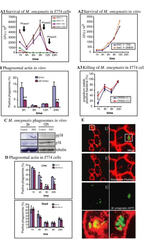
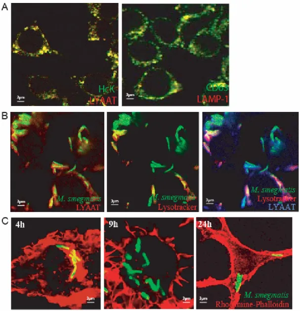
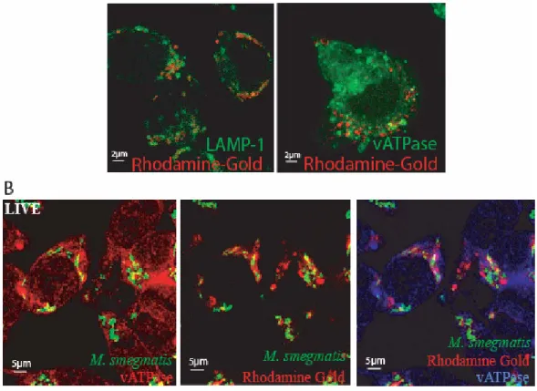
Chapter 2 Figure 1. Intracellular fate of M. smegmatis and phagosome assembly of actin………... 34
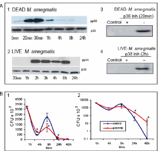
Figure 2. p38 activity in infected cells………... 38
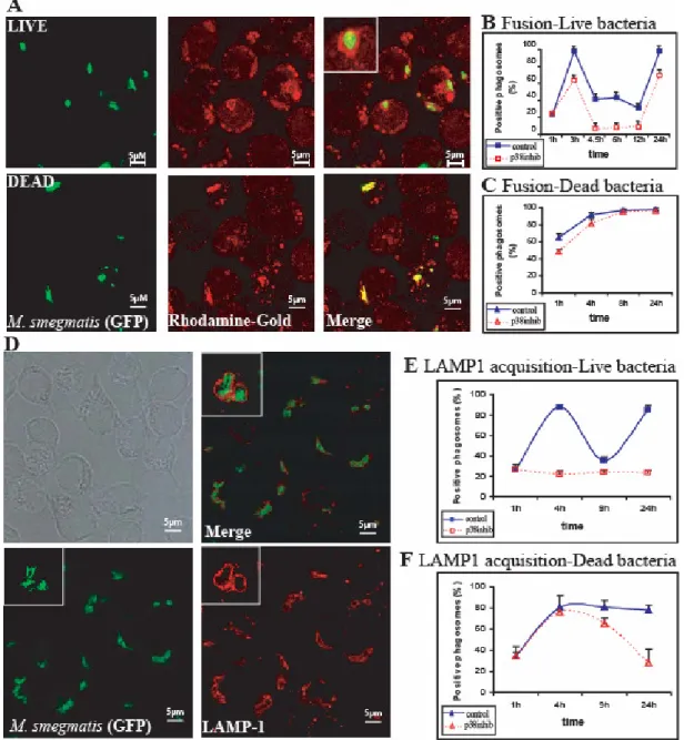
Figure 3. Phagosome fusion with late endosomes and lysosomes………. 42
Figure 4. Acquisition of late endocytic markers by M. smegmatis phagosomes………... 45
Figure 5. Nitric oxide release by infected J774 cells……….. 49
Figure 6. Mathematical modelling of infection……….. 52
Figure 7. Summary models………. 55
Figure S1. Co-localization of late endocytic markers in J774 cells………... 35
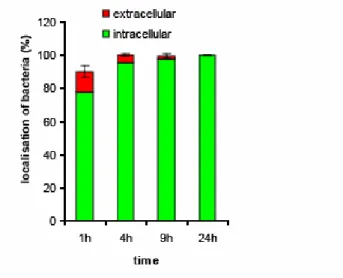
Figure S2. Quantitation of extracellular versus intracellular M. smegmatis……….. 36
Figure S3. Co-localization of markers with phagosomes………... 41
Chapter 3 Figure 1. Intracellular fate of M. smegmatis in different macrophages……….. 79
Figure 2. Intracellular fate of BCG (GFP) or M. bovis in different host macrophages….. 80
Figure 3. Role of nitric oxide in M. bovis spp survival……….. 83
Figure 4. Localization of iNOS in J774 macrophages………... 86
Figure 5. Phagosomes acidification and fusion with late endosome and lysosomes……. 88
Figure 6. Mycobacterium spp reside in a phagosome compartment at all infection times 92 Figure 7. Evaluation of mycobacteria growth/ survival and killing by different methods and the COPASI models for BCG……….. 96
Figure S1. Comparison of CFU of M. tuberculosis in J774 macrophages up to 7 days infection with and without γ-IFN treatment………... 85
Chapter 4 Figure 1A. Persistence of S. enterica serovar Thyfimurium in organs from mice receiving diets enriched in specific fatty acids………... 119
Figure 1B. Persistence of M. tuberculosis in organs from mice receiving diets enriched in specific fatty acids……….. 120 Figure 2. Effects of EPA or AA during the course of M. tuberculosis H37Rv infection in J774 macrophages……….. 121 Figure 3. Effects of ceramide during the course of M. tuberculosis H37Rv infection in J774 macrophages……….. 122 Figure 4. Effects of lipids on TNF-α pro-inflammatory response in J774 cell cultures infected with M. tuberculosis H37Rv………. 123 Figure 5. Lipid- induced phosphorylation of p38 in J774 macrophages during M.
tuberculosis infection………. 125
Figure 6A. Persistence of M. smegmatis in J774 macrophages in cultures supplemented with specific fatty acids during distinct phases of intracellular killing cycles (24 h post-infection)………. 126 Figure 6B-D. Persistence of M. smegmatis in J774 macrophages in cultures supplemented with specific fatty acids during distinct phases of intracellular killing cycles (4 h contact with the lipids)………. 128 Figure 7A. Effect of EPA treatment of macrophages prior to infection on the intracellular survival of M. smegmatis………... 129 Figure 7B-C. Effect of EPA treatment of macrophages prior to infection on the intracellular survival of M. smegmatis………... 130 Figure 8. Ceramide-induced phosphorylation of p38 in J774 macrophages……….. 131 Figure 9. Differential lipid induced phosphorylation of p38 in J774 macrophages during M. smegmatis infection………... 132
Tables Index
Chapter 2
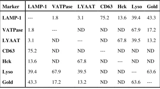
Table 1. Co-localization of different late endocytic markers by immunofluorescence
microscopy in uninfected J774 cells………... 40
Chapter 3
Table 1. Nitric oxide release by infected J774 macrophages………. 84 Table 2. Effects of pH on BCG and M. bovis in vitro……… 89
Table of contents
Preface... i Acknowledgments... v Resumo... vi Abstract... xi Abbreviations... xiii Figures Index... xv Tables index... xviiChapter 1 Introdution
Tuberculosis: The new old disease………... 1 The causal agent………... 2 The pathogenesis of tuberculosis………... 2 Entry of mycobacteria into host cells………... 3 Mycobacteria persistence and host defence mechanisms……… 5 NO and reactive nitrogen radicals (RNI) synthesis………... 5 Phagolysosome biogenesis………... 6 Role of actin cytoskeleton in host-pathogen interaction………... 9 Dietary fatty acids and immune response………... 12 Interplay between nutrition and tuberculosis……….... 14 Thesis goals……….. 15 Reference list………... 18
Chapter 2
Dynamic life and death interactions between Mycobacterium smegmatis and J774 macrophages
Abstract………... 30 Introduction………... 31 Results………... 33 Kinetics of survival of M. smegmatis in J774 macrophages……… 33
Phagosomal actin in infected macrophages………... 37 Macrophage activation of p38 MAP kinase………. 37 Role of p38 in intracellular survival of M. smegmatis………... 39 Use of markers for late endocytic organelles………... 39 Phagosome – late endocytic organelle fusion……….. 41 Content mixing……… 41 LAMP-1……….. 43 The amino acid transporter LYAAT………... 44 Phagosomal pH………... 46 Vacuolar ATPase localisation………... 46 Effect of bafilomycin on M. smegmatis survival………... 47 Role of lysosomal enzymes in bacterial survival………... 48 Role of inducible NO synthase and NO release………... 48 Effects of NO on survival of M. smegmatis in macrophages……….. 50 Mathematical modelling of intracellular bacterial growth………... 51 Discussion………... 54 Killing phase 1………... 54 Intracellular growth of M. smegmatis……….. 56 Killing phase 2………... 56 Killing phase 3………... 57 Role of p38 MAP kinase……….. 58 Material and methods………... 61 Acknowledgments………... 66 Reference list……… 68
Chapter 3
On the killing of mycobacteria by macrophages
Abstract………... 75 Introduction………... 76 Results………... 79 Kinetics of M. smegmatis survival in different host macrophages……….. 79 Kinetics of M. bovis spp survival in different host macrophages……… 79 Role of inducible NO synthase and nitric oxide release……….. 82 Phagosome-lysosome fusion and pH………... 86
All mycobacteria are exclusively in phagosomes……… 90 Killing of mycobacteria occurs in mature and non-mature phagosomes………. 93 Modeling intracellular transport and killing of BCG………... 95 Discussion………. 99 Acknowledgements………... 102 Material and methods………... 104 Reference list……… 109
Chapter 4
PUFAs and ceramide regulation of inflammatory states during macrophage infection with intracellular pathogens
Abstract………... 115 Introduction………... 116 Results………... 119
Effects of omega-3 and omega-6 enriched diets on the infection of mice with
Salmonella and M. tuberculosis……….. 119
Effect of EPA, Cer and AA on M. tuberculosis growth in J774
macrophages………... 121
Pro and anti-inflammatory lipids regulate TNF-α secretion………... 123 The links between lipids (Cer and EPA) and p38 MAP kinase activity during
M. tuberculosis infection……… 124
Effect of EPA, Cer and AA on M. smegmatis growth in J774 macrophages….. 125 Pre-treatment of cells with EPA……….. 128 The links between lipids (Cer and EPA) and p38 MAP kinase activity during
M. smegmatis infection……… 131
Discussion……… 134 Acknowledgments……… 138 Material and methods………... 139 Reference list……… 142
Chapter 5
Chapter 1
TUBERCULOSIS: THE NEW OLD DISEASE
Tuberculosis (TB) is one of the oldest infectious diseases affecting the human kind. Bone TB was identified in skeletons, from Europe and Middle East, as the cause of death showing that 4000 years ago this disease was a widespread health problem. In recorded history, Hippocrates writes of patients with consumption (the old definition of TB), i e, wasting away associated with chest pain and coughing, frequently with blood in sputum. The frequency of descriptions of patients with these symptoms indicated that the disease was already well entrenched.
The explosion of European population and the growth of large urban centres made this continent the epicentre of many TB epidemics during the 16th and 17th centuries. Although, during the first half of the 19th century the incidence of TB peaked causing death to approximately one quarter of European population, in the second half of this century TB mortality decreased due to improving sanitation and housing. The 20th century brought a steadily drop of morbidity and mortality due to TB, in developed world, due to better public health practices, massive vaccination with Calmette-Guérin bacillus (BCG) vaccine and the advent of antibiotics. This downward trend ended in the mid-1980s triggered by emergence of acquired immunodeficiency syndrome (AIDS) and an increase in homeless and poverty in the developed word. This fact pointed the important role played by the immune system in this disease and also the importance of socio-economical factors. More recently the identification of multi-drug resistant (MDR) and extensively drug resistant (XDR) strains of mycobacteria turned this disease even more frightening 1,2.
Actually more than one-third of the world’s population is infected with Mycobacterium
tuberculosis (M. tuberculosis). One in every ten of these individuals will develop TB at
some point in their lifetime and around 2 million people die of this disease every year 3. New drugs, better vaccines and new diagnostic methods are desperately needed to change and revert this situation. Despite the big effort made in order to develop new tools to fight this plague the successful candidate has not been found. In the present work we favour the hypothesis of boosting the host natural defence mechanisms to contribute to solve the problem. The first step towards this goal will be a better
THE CAUSAL AGENT
The Mycobacterium tuberculosis complex includes strains from five species- M.
tuberculosis, M. canettii, M. africanum, M. microti and M. bovis and two subspecies – M. caprae and M. pinnipedii 4. These mycobacteria are characterized by 99,9%
similarity at nucleotide level and virtually identical 16S rRNA sequences 5-8 but differ widely in terms of host tropisms, phenotypes and pathogenicity 4,9,10.
The most notable member of the complex is Mycobacterium tuberculosis the causative agent of human tuberculosis which has an exclusive tropism for this host. In contrast,
M. bovis, the etiologic agent of bovine tuberculosis, cause only 5 -10% of human
tuberculosis cases and has a wide host spectrum. The impact of M. bovis in human health declined sharply after the advent of pasteurization but there are records of new cases among immunocompromised individuals and re-activation in elderly individuals. Although in some countries eradication of bovine tuberculosis was possible due to programs based on a test and slaughter strategy it still constitutes an economic burden in developed countries as New Zealand and England 11,12. The persistence of M. bovis among wildlife that can serve as a reservoir for domestic animals is the aim of the problem. The disease in wild and domestic animals presents a wide spectrum of manifestations that mimic the progression of tuberculosis in humans. These animals when experimentally infected can serve as useful models of human disease since they are outbred, natural host of disease, allow experimental challenge in vaccine trials and is possible to monitor the immune response through the progression of the disease13,14. The major limitation of the model is the use of M. bovis instead of M. tuberculosis and the relative high cost.
THEPATHOGENESISOFTUBERCULOSIS
The infectious bacilli are inhaled as droplets from the atmosphere. In the lung, the bacteria are phagocytosed by the alveolar macrophages. The interaction of mycobacteria components with macrophage receptors as the Toll-like receptors (TLR) results in chemokines and cytokines production 15 that serve as signals of infection. The bacteria
The signals induced results in migration of monocyte derived macrophages and dendritic cells from the blood stream to the site of infection in the lung. The dendritic cells that engulf bacteria mature and migrate to the lymph nodes 16-18. Once there, CD4 and CD8 T cells are primed against mycobacterial antigens. Primed T cells expand and migrate back to the focus of infection in the lungs, probably in response to mediators produced by infected cells. This phenomenon of cell migration towards the infection focus culminates in formation of a granuloma the wall mark of tuberculosis. The granuloma is formed by T cells, macrophages, B cells, dendritic cells, endothelial and epithelial cells among others in a proportion that varies with the age of the structure. The granuloma walls the bacilli resident within macrophages preventing spreading and generating an immune microenvironment which facilitates the interaction between cytokines secreted by macrophages and T cells. However, the granuloma also provides housing for M. tuberculosis during a long period of time. The latent bacilli could be released if the cytokine balance is broken and originate a reactivation of the disease.
ENTRYOFMYCOBACTERIA INTOHOSTCELLS
M. tuberculosis entry into macrophages is mediated by receptor binding and
phagocytosis. Although the majority of studies indicate that the bacilli favour interaction with complement and mannose receptors, which are benign because they trigger minimal superoxide production, experimental data suggest that the receptor type has little impact on the intracellular survival of the bacteria 19,20. Selective receptor blockade during phagocytosis does not alter the survival and growth in human macrophages 20 although differential use of receptors may affect the invasion of different phagocytes populations.
For instance the macrophage mannose receptors are expressed on mature macrophages and allow uptake of virulent M. tuberculosis H37Rv but not the avirulent H37Ra. The interaction between the receptor and the mycobacteria is mediated at least by terminal residues present in mycobacteria lipoarabinomannam (LAM) 21,22 that are also involved in CD14 interaction. Since the expression of these receptors is downregulated by γ-IFN their role in mycobacteria ingestion is circumvented to early stages in infection and in
A scanveger receptors 20 are involved in mycobacteria uptake associated with a low pro-inflammatory response.
Another group of receptors, the pattern recognition receptors (PPR), are likely to be responsible for the immune recognition of pathogens and thus for the pro-inflammatory cell signalling rather than bacterial uptake. This group includes Fc receptors and Toll like receptors (TLR). Pathogenic mycobacteria avoid binding to this family of receptors being preferentially uptake by mannose receptors preventing a strong pro-inflammatory response at early stages of infection. If the host immune system secretes high levels of γ-IFN at this stage the mannose receptors are downregulated as stated before forcing M.
tuberculis uptake mediated by PRRs and TLR.
Mamalian TLR proteins derive their name from the Drosophila Toll protein, to which they have sequence similarity. TLRs were first identifies in Drosophila, where they are important for resistance to microbial antigens. Subsequently a large number of TLRs were identified in mammals and two of them, TLR2 and TLR4, have been implicated in the activation macrophages by mycobacteria involving MAP kinases (ERK 1 /2, p38 and JNK), Janus kinase/ signal transducer and activator of transcription (JAK/STAT) and NF-κB pathways.
The activation of host-cell signalling cascades, such as MAP kinases or JAK/STAT pathways results in the production of pro-inflammatory cytokines (such as IL-1, TNF-α and interferons) and chemokines. Pathogenic but not non-pathogenic mycobacteria have evolved mechanisms to suppress these signal transduction cascades and thereby attenuate the cytokine-induced immune response.
Extracellular signal-regulated kinases (ERK 1 /2) and p38 are members of MAP kinase family that became activated through the phosphorylation of tyrosine and threonine residues. Pathogenic mycobacteria such as M. avium and M. tuberculosis modulate MAP kinase activity. This leads to a decrease in pro-inflammatory response exemplified by a decrease in cytokine secretion such as TNF-α and their downstream effector nitric oxide (NO). Since TNF-α receptors are true death receptors the decrease production of this cytokine induced by blockade of NF-κB and MAP kinase activation results in apoptosis inhibition. The classical programmed cell death pathway is believed to constitute a effective mechanism of intracellular mycobacteria killing 25. However, many bacterial pathogens alter host apoptotic pathways 26. For example, infection of macrophages with virulent strains of M. tuberculosis induces much lower levels of
MYCOBACTERIA PERSISTENCE AND HOST DEFENCE MECHANISMS
Macrophages play a unique role in host response to mycobacteria infections. These cells represent both the primary effector cell for killing the pathogen and the habitat in whish mycobacteria reside. In order to survive pathogenic mycobacteria developed strategies to avoid important anti-mycobacteria mechanisms such as NO production and phagosome lysosome fusion.
NO and reactive nitrogen radicals (RNI) synthesis
The tolerance of mycobacteria in vitro to RNI is strain-, dose- and time-dependent, with pathogens being inherently more resistant than non-pathogens 28-31. This suggests that pathogenic mycobacteria express genes that counteract the bactericidal or bacteriostatic effects of RNI. Different experimental approaches lead to the identification of noxR1 and noxR3 which are able to confer RNI, and also reactive oxygen intermediates (ROI), resistance by a still unknown mechanism 32,33. Another gene involved in protection from oxidative stress is ahpC 34. The product of ahpC, the alkyl hydroperoxide reductase subunit C (AhpC), can metabolise peroxynitrite anion into nitrite, thereby contributing to detoxifying this highly reactive species 35. Peroxynitrite is a powerful oxidant produced by activated macrophages that can exert its toxic effects through protein modification 36. In vitro studies have shown that M. tuberculosis is resistant to this oxidant specie but M. smegmatis and BCG are susceptible 37.
NO produced by the inducible nitric oxide synthase (iNOS) and its derivatives are produced by macrophages in response to mycobacteria infection. Pro-inflammatory cytokines (e.g. γ-IFN and TNF-α) and bacterial lipopolisaccharides (LPS) enhance NO synthesis 38-40. The anti-mycobacterial effects of these intermediates were shown experimentally in macrophage cultures infected with Mycobacterium 41,42. Other studies in the murine model of infection involving iNOS inhibitors or mice with disruption in
nos2 gene highlighted the crucial role played by RNI in host defence against Mycobacterium infection 43-46. In contrast the importance of NO and RNI in human
defence against M. tuberculosis is a matter of controversy 47,48.
Although there is experimental evidence that NO made by iNOS or NOS2 (in humans) is required for mycobacteria killing it is unlikely that an effective killing would be
that NO and RNI derived from NOS2 mediated killing is insufficient to clear mycobacteria in the absence of a GTPase: LRG47. Experiments with infected macrophages have shown that LGR47 favour maturation of M. tuberculosis containing phagosomes through acquisition of vacuolar ATPase (V-ATPase) and ultimately enhance NO killing effects 49. More recently it was shown that LRG47 mediated lysosomal fusion is involved in autophagy rather than in phagosome maturation 50.
Phagolysosome biogenesis
Phagocytosis of pathogenic microorganisms by macrophages is the first step towards their eventual degradation. The phagosome eventually matures into a phagolysosome an organelle with acid pH, high content of hydrolases, defensins and the ability to generate toxic oxidative compounds, responsible for routine elimination of microorganisms 51,52. In order to avoid their degradation intracellular pathogens developed several strategies including escaping from the maturing phagosome to the cytoplasm (e.g. Listeria,
Shigella, Ricketsia and Trypanossoma cruzi)53,54, escaping from the endosomal pathway
to a endoplasmic reticulum derived vacuole (e.g. Chlamydia, Legionella, Brucella)55-58, modification of the phagosome or adaptation to the environment of the phagolysosome (e.g. Coxiella, Leishmania)59,60 and arrest phagosome maturation at late phase (e.g.
Salmonella, Histoplasma) 61,62 or at an early phage (e.g. Mycobacterium, Nocardia, Rhodococcus) 63-65. The notorious success of M. tuberculosis, a facultative intracellular
pathogen, rests upon the ability to arrest the biogenesis of the phagolysome. The ability of this pathogen to enter host macrophages and persist in friendly phagosomes, which do not mature into phagolysosomes 63,66-70, is crucial to tuberculosis infection, latency, disease activation, spread and suppression of immunological detection by the host 52,71-73. The maturation of a phagosome is a complex and ordered process involving interaction of the nascent phagosome with vesicles from the endocytic pathway (Figure 1).
When an inert particle as a latex bead (LB) is uptaked by macrophages the formed phagosome triggers a sequential series of fission and fusion events described by the kiss and run model of phagosome maturation 74. Shortly after their formation phagosomes bind to microtubules and start to fuse with early endosomes in a rab 5 dependent fashion. The early endosome is characterized by the presence of rab 5, transferin
proteases and a mildly acid pH around 6. As a consequence early endosomes contain, not only components characteristic of the plasma membrane but also of the early endosome.
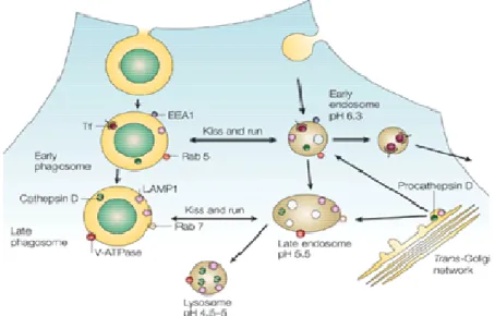
Figure 1. Phagolysosome biogenesis
Diagram showing the parallels between endosome progression to lysosome and maturation of a latex bead containing phagosome. The fusion of phagosomes with compartments of the endosomal pathway follows the kiss and run model proposed by M. Desjardins75. After internalization, the phagosome shows transient access to the rapid recycling pathway illustrated by the presence of transferin (Tf) its classical marker. During this early stage the phagosome has many of the markers found on early endosomes, such as EEA1 and rab5. However phagosomes rapidly acquire proteins associated with late endosomes, such as LAMP 1, rab7 and V-ATPase. Proforms of lysosomal hydrolases such as procathepsin D also appears in early phagosomes. Nevertheless, the processing of cathepsin D to its mature form seems to coincide with V-ATPase acquisition and pH drop. Figure from 76
These early markers are then recycled from the phagosomal membrane as maturation proceeds 77,78. Through fission and fusion events (kiss and run) phagosomes acquire new molecules and recycle others. Therefore the acquisition of rab 7 and the lost of rab 5 enable subsequent fusion of the phagossome with older organelles as late endosomes and lysosomes 74,75. Although the kinetics of maturation differ greatly depending both on the particle phagocytised and the cell, phagosomes begin to fuse with late endosomes and becoming refractory to early endosomes about 15-30 minutes after formation 78-80.
molecules best exemplified by rab 7, lysobisphosphatidic acid (LBPA) and the mannose-6-phosphate receptor cation independent 79,81. Nevertheless its presence is also transitory since the late phagosome evolved to a phagolysosome characterized by the presence of mature forms of lysosomal enzymes such as cathepsin D, lysosome associated membrane protein 1 (LAMP 1) and a luminal acidic pH 80.
Following phagocytosis, the bacteria continue to reside within a membrane-bound vacuole of host origin. The seminal studies of D’Arcy Hart in the early 1970s described how the absence of fusion correlated with viability of the infecting bacteria 66,82,83. The capacity of M. tuberculosis to regulate the fusogenicity of their phagosomes is shared with other pathogenic mycobacteria such as M. avium and M. bovis.
In 1986, Frehel and colleagues observed transient delivery of lysosomal tracers to phagosomes containing M. avium and suggest that these phagosomes had access to early endosomal compartment 84. Then in 1991 Crowle et al. 85 reported that phagosomes containing M. avium and M. tuberculosis were less acidic than neighbouring lysosomes. Sturgill-Kozycki 86 reported that pH of mycobacteria containing phagosomes is around 6.2-6.3 and the deficient recruitment of V-ATPases responsible for the acidification of phagosomes.
Later was shown that mycobacteria containing phagosomes are highly dynamic compartments. They had a paucity of V-ATPase complexes and a profile of endosomal constituents consistent with the arrest of phagosome maturation at a point that retained fusion with early endosomes 87. Consequently, markers of the recycling endosomal system, namely transferin receptor could be shown to traffic through the mycobacteria containing phagosome 87,88. Although certain “lysosomal” markers, such as cathepsin D, could be detected in this compartment, careful analysis revealed that it is an immature form of the enzyme 87,89. Most researchers in this field now work under the assumption that the phagosome containing pathogenic Mycobacterium spp are blocked at an early stage of their maturation process 52,70,76,90-92.
The exact point where mycobacteria stop the phagosome maturation process is not yet identified. Nevertheless the retention of rab5 in mycobacteria containing phagosomes indicates that the process is arrested at an early phase between the steps controlled by rab5 and rab7 (Figure 1) 81,93.
The regulation of membrane fusion in endosome biogenesis at this stage of the differentiation process is still debated although the major players have been identified 94,95
generates phosphatidylinosithol 3-phosphate (PI 3-P) on the cytosolic face of the phagosome. PI 3-P acts has an acceptor for the EEA1 which also complexes with rab5. EEA1 binds calmodulin in a Ca2+/calmodulin dependent process 96,97. Acquisition and accumulation of EEA1 are necessary for accumulation of rab7, the small GTPase that is enriched in lysosomes.
Malik et al. reported that M. tuberculosis blocked the normal calmodulin dependent signal transduction observed in maturation of phagosomes containing latex beads (LB) or dead bacteria 98,99. These authors also shown that blockage in Ca2+ mediated signalling by live bacteria was due to an inhibition in sphingosine kinase activity 100. In another study by Anes et al. it was demonstrated that sphingosine 1-phosphate content of the phagosome membrane influenced actin polymerization on isolated phagosomes and that M. tuberculosis containing phagosomes were deficient in actin nucleation 101. Fratti et al. showed that while M. bovis BCG containing phagosomes acquired rab5 and accumulated Vps34, they failed to bind the PI 3-P-binding protein EEA1 79. According to Vergne et al this would be due to the absence of PIP-3-P on the cytosolic face of the bacterium containing phagosome resulting of the interference with the kinase activity of Vps34 through a Ca2+/ calmodulin mediated process 102. For Corvera and collegues calmodulin did not affect Vps34 kinase activity but instead blocked EEA1 binding to PI 3-P 96.
Despite the apparent controversy between the data generated by different groups it is clear that the point of arrest in phagosome maturation is the accumulation of EEA1. This process is modulated by molecules that affect Ca2+/ calmodulin and class III PI 3-kinase activity.
ROLE OF ACTIN CYTOSKELETON IN HOST-PATHOGEN INTERACTION
The actin cytoskeleton is involved in several stages of host-pathogen interaction. For a long time was accepted that actin was involved in the first step of phagocytosis 103,104. More recently was suggested that organelle trafficking and spatial distribution was not only dependent on microtubules, but also involve actin cytoskeleton 105,106.
phagosome formation but most of F-actin was lost soon after this step. Many pathogens developed mechanisms to subvert the cellular actin cytoskeleton to trigger their internalization into normally non phagocytic host cells in order to escape the humoral immune defence. The invasive strategies used by pathogens to enter non-phagocytic cells are known as induced phagocytosis via the zipper and trigger mechanisms. In both cases actin assembly is stimulated by activation of the Rho-family GTPases Rac1 and Cdc42, both capable of eliciting Arp2/3 complex activation via proteins of the Wiskott-Aldrich syndrome protein (WASP) and WASP family verpolin homologus (WAVE) proteins families 107. In the trigger mechanism, adopted by Salmonella and Shigella, the pathogen delivers to the host cell virulence factors that induce actin polymerization via type III secretion systems. The first evidence for a role of actin in intracellular trafficking events came from studies on the interaction of Listeria and vaccinia virus 53,108. Both pathogens once within the cytosol are able to recruit G-actin and form actin tail comets thus providing the driving force within the infected cell and also to spread infection to the neighbouring cells. Since vaccinia virus and small endocytic vacuoles are similar in size, researchers wonder when formation of actin tail comets would be an ordinary event during small vesicles trafficking.
Pathogens that show actin based motility can be separated into two groups depending on whether they mimic a cellular nucleation-promoting factor (NFP) or they require the recruitment of a cellular NPF to the bacteria surface to promote Arp 2/3 mediated motility. The members of the Wiskott- Aldrich syndrome protein (WASP) family are an example of NFP recruited by pathogens as Shigella. Other pathogens such as Listeria and Ricketsia produce proteins that mimic WASP 109,110. Although the mechanism of tail formation induced by Mycobacterium marinum is largely unknown probably it is NFP (WASP) and actin related proteins complex 2/3 (Arp 2/3) dependent 111,112.
In recent years Griffiths provide evidence for the involvement of the actin cytoskeleton at late phases of phagocytosis/ endocytosis of latex beads by a mechanism independent of ARP 2/3 and alternative to actin tail comets113. In the actin track model the actin nucleation machinery is dependent of the ezrin, radixin and moesin (ERM) family of proteins and postulates that if a membrane phagosome is able to filament actin this will provide tracks for other organelles to bind and move towards the actin nucleation source. In the context of phagolysosome biogenesis this will aid phagosome maturation increasing the number of fission and fusion events. Moreover the actin cytoskeleton
cytochalasin D and latrunculin A can inhibit this process in 50 % 114,115. In addition actin was found associated with endosomes and lysosomes 116,117 as well as several actin binding proteins as myosins 114 and moesin 118. Indeed in vitro studies showed that actin filaments are able to mediate fusion between late but not early endocytic organelles and phagosomes 119. In the context of mycobacterial infections the role of the actin cytoskeleton was still in the infancy when our group started to work in this field in the beginning of the 3rd millennium. By that time Chantal de Chastellier group reported that pathogenic mycobacteria were able to disrupt the actin cytoskeleton within infected cells 120.
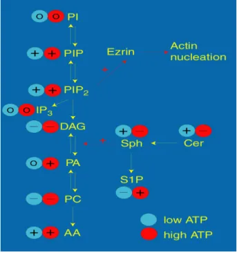
Using an in vitro assay that monitors membrane-dependent assembly of actin was possible to show that this process is regulated by lipids. The effect of different lipids at physiologic (1-5 mM: High ATP) and ischaemic (0.2 mM: Low ATP) concentrations of ATP are summarized in figure 2. In physiological conditions this class of lipids integrates the phagosome membrane in different amounts depending on the degree of vesicle maturation. While early phagosomes are enriched in phosphatidylcholine (a negative efector), late phagosomes are enriched in sphingomyelin the precursor of ceramide (both positive efectors). Thus by changing lipid composition we were able to switch on or block actin assembly/ fusion ability in vitro.
concentrations are indicated. Abbreviations: AA, arachidonic acid; Cer, ceramide; PC, phosphatidylcholine; PA, phosphatidate; Sph, sphingosine; S1P, sphingosine-1-phosphate. Figure from 101.
When instead of LBPs were used phagosomes enclosing the non pathogenic mycobacteria M. smegmatis we observed that these phagosomes were able to nucleate actin in vitro similarly to LBPs. However when phagosomes containing pathogenic mycobacteria, such as M. avium or M. tuberculosis, were used the actin nucleation was effectively blocked. This enable us to established a link between the ability of mycobacteria to assemble actin, phagosome maturation and intracellular pathogen killing 101. The most striking result arose from the fact that actin assembly by mycobacteria containing phagosomes could be controlled using signalling lipids such as arachidonic acid (AA), phosphatidylinositol biphosphate (PIP2) and ceramide (Cer). These lipids are potent inducers of actin assembly even in M. tuberculosis containing phagosomes allowing the overcome of phagosome maturation block and thus inducing an increase in mycobacterial killing within macrophages.
This study highlighted the opposite role played by two classes of dietary lipids in host defence to mycobacteria infections. The polyunsaturated fatty acids (PUFA) omega n-3 inhibited in vitro and in vivo actin nucleation stimulating mycobacteria survival within macrophages whereas the omega n-6 (AA) had the opposite effects. The positive effect on actin nucleation by phagosomes was observed not only by adding AA but also by addition of its metabolites such as prostaglandin F2 (PGF2) (Gordon Shore, University Sterling, Scotland, personal communication). The finding that another metabolite of AA, PGE2, when added to the system blocked actin assembly make hard to draw straigth prediction for the effect of AA in animal models of infection.
DIETARY FATTY ACIDS AND IMMUNE RESPONSE
In western diets omega-6-fatty acids (PUFA n-6) account for the majority of PUFA in the food supply. When diets are supplemented with omega-3-fatty acids, the latter partially replace the omega-6 fatty acid in the membrane of practically all cells 121,122. This change has profound effects on both inflammatory and immune response mediated by different mechanisms.
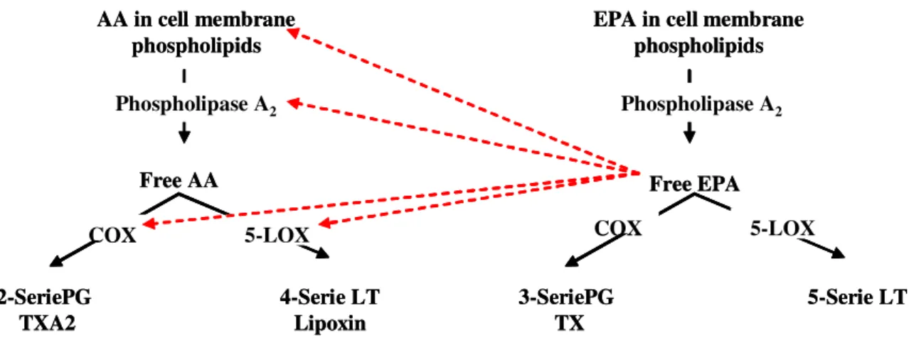
PUFA n-3 competes with AA as substrates for different enzymes (Fig 3). Competition at the level of ciclooxygenase (COX) and 5-lipoxygenase (5-LOX) 123,124 is of particular important since they are involved in the synthesis of key inflammatory mediators as prostaglandin, leukotriene (LT) and lipoxins. Thus higher levels of PUFA n-3 in cell membranes reduce the production, in the COX mediated pathway, of pro-inflammatory eicosanoids (PGE2, LTB4) from n-6 PUFA and increase production of eicosanoids from n-3 PUFA (PG3, LTB5) which have weaker or opposite activity. However, the situation is complicated, because eicosanoids derived from one precursor did not share all the effects. While LT4 tends to enhance NKcells activity and Th-1 cytokine production (IL-1, IL-6, TNF-α, IFN-γ) PGE2 tends to suppress this functions. The role played by products of 5-LOX pathway in host resistance to M. tuberculosis is less documented. Nevertheless, there is experimental evidence in mice of the importance of lipoxin A4 in infection control 125,126.
Figure 3: Synthesis of eicosanoids from AA and EPA
AA and EPA compete for prostaglandin (PG) and leukotriene (LT) synthesis at cyclooxigenase (COX) and lipoxygenase (LOX) level. The PG and LT derived from AA have an overall pro-inflammatory effect while the ones derived from EPA have anti-pro-inflammatory effect or a weaker potent pro-inflammatory effect. In this figure TXA2 means Tromboxane A2
Changes in membrane lipid composition can also interfere with signal transduction. The most obvious way would be synthesis of different signalling molecules or the same signalling molecule with different biological activity due to changes in fatty acid
AA in cell membrane phospholipids
EPA in cell membrane phospholipids
Free AA Free EPA
2-SeriePG TXA2 4-Serie LT Lipoxin 3-SeriePG TX 5-Serie LT Phospholipase A2 COX 5-LOX Phospholipase A2 COX 5-LOX AA in cell membrane phospholipids
EPA in cell membrane phospholipids
Free AA Free EPA
2-SeriePG TXA2 4-Serie LT Lipoxin 3-SeriePG TX 5-Serie LT Phospholipase A2 COX 5-LOX Phospholipase A2 COX 5-LOX
conducts to the synthesis of a less potent DAG 129 leading to a less potent anti-inflammatory response. Another mechanism that can account for a different inflammatory response is difference in binding of this and other molecules, as cytokines 130,131, to their receptors in cell membrane.
Omega-3 fatty acids can also modulate gene expression of important immune genes. Among these are cytokines (TNF-α, IL-1β and IL-6) 132-134, i-NOS135 and transcription factors as NF-κB 136. Changes in gene expression can result from direct or indirect effects of omega-3 fatty acids. The mechanisms are not fully understood may differ from cell types to cell type. For cytokine production, bind of EPA to a class of transcription factors known as peroxisome proliferator activactor receptor (PPAR) can account for inhibition of macrophage activation and production of TNF-α, IL-1β and IL-6 128. And at least, for TNF-α, another indirect mechanism mediated by inhibition of NF-κB activation can account for inhibition of TNF-α production. Collectively, we can conclude that omega-3 fatty acids can affect the balance between Th-1 and Th-2 cytokines that are of capital importance in mycobacteria infections 137.
INTERPLAY BETWEEN NUTRITION AND TUBERCULOSIS
Under or over nutrition and diet related chronic diseases are unbalanced nutrition status that constitutes a critical risk factor to infections. As stated before nutrients in the diet play a crucial role in maintaining an optimal immune response, such that deficient or excessive intakes can have negative consequences on susceptibility to a variety of pathogens. Under nutrition states and metabolic diseases as diabetes favour the establishment of infectious diseases including malaria, AIDS and TB while over nutrition states favours opportunistic and fungal infections 138,139.
The link between malnutrition and active TB has been recognized for many years. This notion is largely based on historical reports of the first and second world wars and also on more recent studies in animals 140,141. In fact food intake increases the levels of γ-IFN but not IL-4 secretion whereas starvation enhances IL-4 response but not γ-IFN levels. The severe malnutrition observed in tuberculosis patients is the major argument for the provision of nutritional supplementation of such individuals.
In the classical era the Greeks designated TB as “phthisis” meaning waste a way and