UNIVERSIDADE DE LISBOA
FACULDADE DE CIÊNCIAS
DEPARTAMENTO DE BIOLOGIA VEGETAL
SOURCES AND ROUTES OF TRANSMISSION OF Q FEVER: DETECTION,
IDENTIFICATION AND MOLECULAR TYPING OF COXIELLA BURNETII IN
DOMESTIC AND WILD ANIMALS
Aminata Cumbassá
DISSERTAÇÃO
MESTRADO EM MICROBIOLOGIA APLICADA
UNIVERSIDADE DE LISBOA
FACULDADE DE CIÊNCIAS
DEPARTAMENTO DE BIOLOGIA VEGETAL
SOURCES AND ROUTES OF TRANSMISSION OF Q FEVER: DETECTION,
IDENTIFICATION AND MOLECULAR TYPING OF COXIELLA BURNETII IN
DOMESTIC AND WILD ANIMALS
Aminata Cumbassá
Dissertação
Mestrado em Microbiologia Aplicada
Orientadores:
Dra. Ana Botelho (INIAV)
Prof. Dra. Lélia Chambel (FCUL)
SOURCES AND ROUTES OF TRANSMISSION OF Q FEVER: DETECTION,
IDENTIFICATION AND MOLECULAR TYPING OF COXIELLA BURNETII IN
DOMESTIC AND WILD ANIMALS
Aminata Cumbassá
MASTER THESIS
2013
This thesis was fully carried out at the Laboratory of Bacteriology of the National Institute of Agricultural and Veterinarian Research (INIAV, IP), under the direct supervision of Doutora Ana
Botelho, and was funded by the National Fundação para a Ciência e Tecnologia (FCT) Project PTDC/SAU-SAP/115266/2009 “Q fever – from diagnosis to eco-epidemiological investigation of
Coxiella burnetii in the context of human infection”.
Prof. Lélia Chambel was the internal designated supervisor in the scope of the Master in Applied Microbiology of the Faculty of Sciences of the University of Lisbon.
Instituto Nacional de Investigação Agrária e Veterinária, I.P.
ACKNOWLEDGEMENTS
At the end of this paper, I would like to express my deepest gratitude to all those who, in any way, contributed to its achievement.
I thank Prof. Rogério Tenreiro, for being the essential intermediate in the beginning of this study. I appreciate his demonstrated availability and the constant support.
Many thanks to Dr. Nuno Canada, Director of LNIV, for having accepted my internship at the Institute.
My warmest thanks to Dr. Ana Botelho, who at all times was able to bring together the rare qualities of clarity, objectivity and accuracy, the availability and unreserved solidarity, patience, encouragement and friendship throughout the internship, unique qualities that helped to bring this work to fulfillment. Nevertheless, I would like to express my appreciation for the material conditions made available, without which this work would not have been possible.
My expressed gratitude to Dr. Lélia Chambel (FCUL), for the attention and availability demonstrated whenever needed and at any moment.
I thank Dr. Ana Sofia Santos, from Centro de Estudos de vectores e Doenças Infecciosas, Instituto Nacional de saúde Dr. Ricardo Jorge (CEVDI/INSA), for providing us DNA sample of the reference strain of Coxiella burnetii Nine Mile ATCC 616-VR, and for her support and excellent work when sequencing the samples.
Many thanks to Dr. Maria José Barahona and Dr. Ana Canto, from INIAV, for supporting me whenever needed while performing several tasks.
To Dr. Mónica Cunha (INIAV), for providing us DNA samples of wild carnivores.
I thank Eng. Diogo Mendonça and Dra. Fernanda Matos from INIAV, for their support and excellent work when using the sequencer for the capillary electrophoresis.
Many thanks to Eng. Ana Cristina Ferreira, for her unconditional help during the interpretation of the MLVA typing results.
Thanks to Inês Guinote, Ana Sofia Ferreira, Célia Leão, and Pedro Costa, for being such good people, for all the help you have given me, for the friendship and constant teaching and, also for the moments shared at INIAV.
ABSTRACT
Query fever (Q fever) is a worldwide zoonosis, affecting many animal species, including Men. This zoonotic disease is caused by Coxiella burnetii, an obligate intracellular, Gram-negative filter-passing (0.3 μm) bacterium. Ticks are considered the natural primary reservoir of Coxiella burnetii, responsible for the spread of the infection in wild and domestic animals. Domestic ruminants contaminate the environment by shedding Coxiella in milk, feces, urine, saliva, vaginal secretions, placenta and amniotic fluids, being the main source of human infection. In animals, C. burnetii can cause abortion, premature birth, dead or delivery of weak offspring. Detection of sources of infection and routes of transmission, are essential for the control of C. burnetii spread among animals and transmission from animals to humans. The aim of this work is the molecular detection and characterization of Coxiella burnetii in animal samples, to elucidate the population structure of this agent in Portugal, for surveillance and epidemiological purposes.
A nested-touchdown PCR assay (Trans-PCR), targeting the repetitive transposon-like element of C. burnetii insertion sequence IS1111, was performed on 229 DNA tissues and cloacal swab samples, from domestic and wild animals. 19 of them tested positive for C. burnetii (14 in domestic and five in wild animals), revealing a prevalence of infection of 8.3% within this sample panel. All 19 cloacal swabs from vultures tested negative.
Multiple locus variable-number of tandem-repeat analysis (MLVA) was used to genotype the 19 C. burnetii positive samples, using a set of six VNTR loci (Ms23, Ms24, Ms27, Ms28, Ms33 and Ms34). Seven different completed profiles (M1 to M7) and nine partial profiles were observed. The calculated discriminatory power of MLVA was 0.94 for our sample setting, and the diversity indexes (D) of the individual markers ranged between 0.73 and 0.95, being loci Ms33 the most discriminatory one.
UPGMA clustering of the MLVA data grouped the C. burnetii samples into eight different clusters, being cluster IV the one that included more MLVA types: M1 and M4.
Clustering of the MLVA genotypes using the minimum spanning tree method (MST) grouped cattle and goat samples from this study in one branch, while two samples from the same sheep were grouped completely apart in another branch. Using only completed profiles, this analysis corroborate the hierarchical UPGMA data, confirming cluster IV as the most representative of our sample setting.
None of our samples clustered with animal or human data reported previously in Portugal, or with reference strains, showing a high diversity among the panel sample.
RESUMO
A febre Q é uma zoonose bacteriana com incidência a nível mundial, que afeta muitas espécies animais, incluindo o Homem. Foi descrita pela primeira vez em 1935, em Queensland, Austrália, devido a um surto de doença febril de origem desconhecida, entre os trabalhadores de um matadouro. Como não havia sido descrita até então, foi denominada “Query fever” que significa “febre desconhecida”.
Esta zoonose é causada por Coxiella burnetii, uma bactéria intracelular obrigatória, Gram-negativa. A espécie foi isolada pela primeira vez de uma carraça em 1938, em Nine Mile Creek, EUA. As carraças são consideradas o reservatório primário natural da bactéria e são os responsáveis pela sua transmissão aos animais domésticos (bovinos, ovinos, caprinos, suínos, cães, gatos) e selvagens (pássaros e outros), através das suas fezes e/ou mordidas.
Os animais domésticos, por sua vez, são os principais responsáveis pela transmissão da C. burnetii aos humanos, através do leite, fezes, urina, saliva, secreções vaginais (especialmente após o parto), placenta e fluidos amnióticos. As infeções por C. burnetii podem resultar em abortos, partos prematuros e morte, causando graves transtornos económicos a nível de produção animal.
A deteção das fontes de infeção e vias de transmissão da bactéria são essenciais para o controlo da sua disseminação entre os animais e da sua transmissão dos animais para os humanos.
Este estudo tem como objetivo a avaliação de métodos de deteção molecular e a caracterização de C. burnetii em amostras de tecidos animais (domésticos e selvagens), com o propósito de esclarecer qual a estrutura populacional deste microrganismo em Portugal, para fins epidemiológicos e de vigilância.
Analisamos um total de 229 amostras de DNA, extraídos de diferentes tecidos de animais domésticos e selvagens, através da técnica de Trans-PCR, que utiliza dois pares de iniciadores (Trans1/Trans2 e Trans3/Trans4), desenhados para amplificar a região repetitiva tipo-transposónica na sequência de inserção IS1111 existente no genoma de C. burnetii. Trata-se de um PCR “nested” altamente específico e sensível, em que o primeiro par de iniciadores (Trans1/Trans2) amplifica uma região de 687 pb, sendo o produto dessa primeira amplificação utilizado como molde na reação com o segundo par de iniciadores (Trans3/Trans4), que amplificam uma região de 243 pb.
Dezanove amostras (14 animais domésticos e 5 sacarrabos selvagens) foram positivas para C. burnetii, revelando uma prevalência de 8,3% no painel de amostras testadas. Todas as 19 zaragatoas cloacais de abutres selvagens analisadas foram negativas para o agente.
As amostras positivas foram posteriormente testadas por qRT-PCR, para quantificar o DNA alvo de C. burnetii existente em cada uma, através de valores de cycle threshold (Ct). Esses
valores são inversamente proporcionais à quantidade de DNA alvo existente, uma vez que Ct é
definido com o sendo o número de ciclos necessários para o sinal fluorescente exceder o limiar de interferências que possam existir na amostra. Quanto mais baixos os valores de Ct, maior a
quantidade de DNA na amostra.
Esta medida permitiu-nos ter uma indicação de quais as amostras que melhores resultados dariam na tipificação por “Multiple locus variable-number of tandem-repeats analysis” (MLVA) pois, de acordo com artigos publicados, amostras com valores de Ct >35 são dificilmente
tipificáveis. A tipificação molecular por MLVA foi feita com o objectivo de estudar as principais fontes de infeção e vias de transmissão de Coxiella. É uma técnica que se baseia nas variações que ocorrem naturalmente no número de sequências de DNA repetidas em “tandem”, no genoma das bactérias. Neste estudo, incluímos todas as 19 amostras, independentemente do Ct obtido, tendo conseguido tipificar algumas de Ct mais alto, apesar de outras de baixo Ct
não serem tipificáveis, usando um conjunto de seis loci VNTR com unidades repetitivas de seis (Ms27, Ms28 e Ms34) ou 7 pares de bases (Ms23, Ms24 e Ms33).
Obtivemos sete perfis completos diferentes, designados de M1 a M7, e nove perfis parciais, revelando esta técnica uma capacidade discriminatória (HGDI) de 0,93. Para cada marcador individual os índices discriminatórios situaram-se entre 0,73 e 0,95, sendo o locus Ms33 o mais discriminatório. Os dados obtidos por MLVA foram agrupados pelo método hierárquico com o coeficiente e método de aglomeração UPGMA [BioNumerics 6.6 (Applied Maths, Bélgica)] em sete grupos diferentes, sendo o grupo IV aquele com maior número de tipos MLVA (M1 e M4).
Utilizando apenas perfis completos para todos os loci, verificamos com o método do “Minimum spanning tree” (MST) que as amostras de DNA de C. burnetii de bovino, ovino e caprino se agrupavam num ramo, enquanto que duas amostras de ovino do mesmo animal ficavam noutro ramo. Esta análise veio corroborar os resultados da análise hierárquica, confirmando o grupo IV como sendo o mais representativo do nosso conjunto de amostras.
Nenhuma das amostras agrupou com outros perfis, de animais e de humanos, previamente publicados em Portugal, ou com estirpes de referência de C. burnetii sequenciadas, mostrando uma grande diversidade e exclusividade das amostras por nós analisadas.
Palavras chave: Febre Q, Coxiella burnetii, zoonose infeciosa, vias de infeção, abortos,
i
TABLE OF CONTENTS
1. INTRODUCTION ...1
1.1. Q fever ...1
1.2. Sources of infection ...2
1.3. Q fever in domestic and wild animals ...3
1.4. Q fever in humans ...3
1.5. Characteristics of Coxiella burnetii ...4
1.6. Coxiella burnetii pathogenesis ...5
1.7. Isolation and culture of Coxiella burnetii ...6
1.8. Diagnostic of Q fever: serological methods ...6
1.9. Diagnostic of Q fever: molecular methods based on PCR ...7
1.10. Molecular typing ...8
2. OBJECTIVES...11
3. MATERIAL AND METHODS...12
3.1. Bacterial strains ...12
3.2. Clinical samples ...12
3.3. DNA extraction...13
3.4. Trans-PCR assay ...13
3.5. Agarose gel electrophoresis ...14
3.6. Amplification of the constitutive gene of β-actin ...14
3.7. Purification and sequencing of the PCR amplicons ...15
3.8. Quantitative Real-time PCR (qRT-PCR) ...16
3.9. Plasmid-specific PCR to identify plasmids QpH1 and QpRS ...17
3.10. Multiple locus variable-number tandem-repeat analysis (MLVA) typing ...18
3.10.1. Capillary analysis of MLVA PCR products ...20
3.10.2. Agarose gel eletrophoresis analysis of MLVA PCR products ...20
3.10.3. Assessing the number of tandem repeats ...20
3.10.4. Analysis and clustering of MLVA data ...21
3.10.5. Determination of the discriminatory power (D) of the MLVA typing ...21
ii
4.1. Determination of the detection limit of the Trans-PCR assay ...22
4.2. Detection of C. burnetii DNA in clinical samples using Trans-PCR assay ...22
4.3. Amplification of the β-actin constitutive gene ...24
4.4. Amplicon sequencing ...25
4.5. Quantitative Real-time PCR (qRT-PCR) ...25
4.6. Plasmid-specific PCR to identify plasmids QpH1 and QpRS ...26
4.7. MLVA typing ...27
5. CONCLUSIONS...35
6. REFERENCES ...37
7. CYBERGRAPHIC REFERENCES ...42
iii
FIGURES INDEX
Figure 1 - Transmission cycle of Coxiella burnetii. ...2 Figure 2 - A Tandem repeat locus with 8 base pair long tandem repeat units. . ...9 Figure 3 – Example of variation in the number of tandem repeats in multiple VNTR loci in two different strains. ...10
Figure 4 – DNA purification procedure scheme. ...15 Figure 5 - Sensitivity of Trans-PCR for the detection of C. burnetii Nine Mile strain ATCC
616-VR DNA. ...22
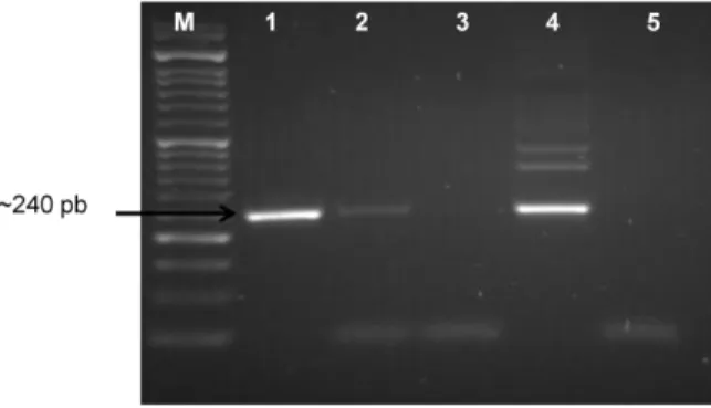
Figure 6 – Results of Trans-PCR in samples from a sheep fetus - analysed in agarose gel. ....23
Figure 7 – Image of an agarose gel (1.5%) electrophoresis corresponding to the amplification of
β-actin gene from DNA samples of small ruminants. ...24
Figure 8 – qRT-PCR amplification graphic and standard curve obtained with the C. burnetii
reference strain Nine Mile ATCC 616-VR. ...25
Figure 9 - Identification of the QpH1 (A) and QpRS (B) plasmids in the reference strain ATCC
616-VR...27
Figure 10 - Dendrogram obtained from this study. ...33 Figure 11 - Minimum spanning tree, showing a representation of the differences between the
MLVA genotypes found in nine animal clinical samples and the reference strain Nine Mile RSA 493. . ...34
iv
TABLES INDEX
Table 1 - Size of C. burnetii fragments amplified by nested PCR and sequences of the
oligonucleotides used in the 1st and 2nd reactions. . ...13
Table 2 - Oligonucleotides used for β-actin amplification reaction. . ...15
Table 3 - Oligonucleotides and probe sequences used in the Real-time PCR for quantifying C. burnetii DNA load. . ...16
Table 4 - Oligonucleotides used for plasmid-specific PCR. . ...17
Table 5 - PCR reaction conditions for plasmids QpH1 and QpRS ...17
Table 6 – Characteristics of each loci and set of primers used in MLVA typing ...19
Table 7 – Summary of all C. burnetii positive samples in this study, with reference to the isolation of other bacterial species, namely Brucella spp. ...30
Table 8 – Summary table. Clinical samples, geographical location, year of collection, Ct values and obtained MLVA genotypes. The discriminatory power index (D) values for the typing method and for each locus are presented. ...31
v
LIST OF ABBREVIATIONS
ACCM Acidified citrate cysteine medium
[CR] Cybergraphic reference
bp Base pair
CBA/FCUL Centro de Biologia Ambiental/Faculdade de Ciências de Lisboa CEVDI/INS
A
Centro de Estudos de Vectores e Doenças Infecciosas/Instituto Nacional de Saúde Dr. Ricardo Jorge
CFT Complemt fixation test
Ct Cycle threshold
DNA Deoxyribonucleic acid
dNTPs Deoxyribonucleotide triphosphates
EDTA Ethylenediaminetetraacetic acid
ELISA Enzyme-linked immunosorbent assay
EP Estradas de Portugal
FAM 6-carboxyfluorescein
HEX Hexachlorofluorescein
IFA Immunofluorescence Antibody Assay
IHC Immunohistochemistry
INIAV Instituto Nacional de Investigação Agrária e Veterinária
LCV Large-cell-variant
LNIV Laboratório Nacional de Investigação Veterinária
LPS Lipopolyssacharide
MLVA Multiple locus variable-number of tandem-repeats analysis
MST Minimum spanning tree
N/A Not available
NADP Nicotinamide adenine dinucleotide phosphate
OIE Office International des Epizooties
PBS Phosphate buffered saline
PCR Polymerase chain reaction
qRT-PCR Quantitaitve real-time PCR
RNA Ribonucleic acid
SCV Small-cell-variant
TBE Tris borate EDTA
TD-PCR Touchdown PCR
Tm Melting temperature
U Units
USA United States of America
UV Ultra violet
V Volts
VERO African green monkey epithelial
1
1. INTRODUCTION
1.1. Q fever
Q fever is an ubiquitous zoonotic bacterial disease, capable of transmission from animals to humans. Coxiella burnetii is the causative agent of Q fever in humans and in animals (also referred as coxiellosis in animals). The disease was first described in 1935 in Queensland, Australia, during an outbreak of a febrile illness of unknown origin, among abattoir workers (Porter et al., 2011). As the illness was not previously described, it was named “Q” fever (for query fever). Simultaneously, in Rocky Mountain Laboratory in Montana, USA, in an experiment concerning Dermacentor andersoni ticks collected at the Nine Mile Creek, it was isolated an infectious agent with properties not identified before. The agent was infectious for guinea pigs, could pass filters, Gram-negative, and had an extracellular and intracellular pleomorphic, rickettsia-like appearance (Davies & Cox, 1938).
Q fever is presently recognized as being worldwide endemic, with the notable exception of New Zealand (Cutler et al., 2007), where seroepidemiological studies were negative for the presence of anti-C. burnetii antibodies in sera collected from the major ruminant population (cattle and sheep). New Zealand was considered to be free from coxiellosis and thus from human Q fever (Maurin and Raoult, 1999).
In a study to estimate the effect of Q fever used as a bioweapon, the WHO estimated that if 50 kg of C. burnetii were aerosolized over an urban area with 500,000 inhabitants, there would be 125,000 cases of acute illness, 9,000 cases of chronic Q fever and 150 fatalities (WHO, 1970) (Oyston and Davies, 2011).
Q fever can be a severe public health problem, and awareness of the disease must be promoted worldwide (Porter et al., 2011).
2
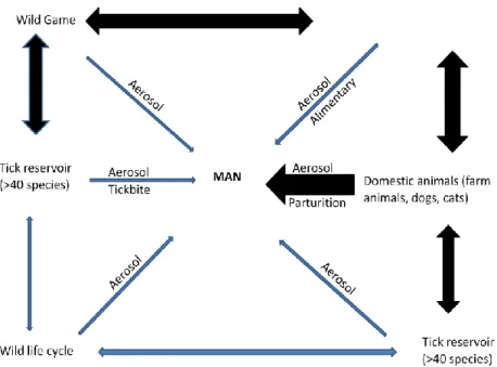
1.2. Sources of infectionFigure 1 - Transmission cycle of Coxiella burnetii. Potential mechanisms of human infection by C. Burnetii are shown. Heavy black arrows signify major infection pathways, and thin gray arrows represent minor infection pathways. Humans are considered dead-end hosts and human-to-human infections are very rare (Adapted from (Miller et al., 2006).
Ticks are considered to be the natural primary reservoir of Coxiella burnetii (Figure 1) and responsible for the spread of the infection in wild animals and for transmission to domestic animals (Kirkan et al., 2008). C. burnetii can be carried out by more than 40 naturally infected tick species, that transmit the agent both vertically (to their progeny) and horizontally (via bite or in faeces) to wild animals, especially birds and, very rarely, to humans (Miller et al., 2006).
Animals are assumed to become infected by inhalation or oral uptake of C. burnetii from the environment (Roest et al., 2012) and they contaminate the environment by shedding C. burnetii in milk, feces, urine, saliva, and very importantly, in vaginal secretions, placenta, amniotic fluids and other products of conception (Porter et al., 2011).
Domestic ruminants (cattle, sheep and goats) are considered to be the main reservoir for the pathogen which can infect a large variety of hosts: mammals (humans, ruminants, small rodents, dogs and cats) and also birds, fish, reptiles and arthropods.
3
1.3. Q fever in domestic and wild animalsC. burnetii infection in animals is generally asymptomatic (Roest et al., 2012). However, in cattle, sheep and goats, C. burnetii can cause abortion, premature birth, dead or delivery of weak offspring as it localizes in the female reproductive system (Panning et al., 2008; Agerholm, 2013).
In symptomatic individuals C. burnetii is excreted via faeces, vaginal mucus, milk and birth products.
Little is known on the distribution and incidence of C. burnetii in wild life, so the threat that infected animals pose to humans and domestic animals is uncertain (Astobiza et al., 2011). However, ticks may play an important role in transmission in the wild, for example between birds (Oyston and Davies, 2011).
1.4. Q fever in humans
Ruminants (sheep, goats and cattle) and pets, namely dogs and cats, are the main sources of human infection (Guatteo et al., 2006).
One of the largest outbreaks of Q fever in humans occurred in the Netherlands, between 2007 and 2010, with 4026 human cases reported (Roest et al., 2011). It happened during the event “lamb viewing days”, an event that occurs every year during lambing season and attracts thousands of people to visit farms. People could watch lambs being born and interact with them. At this time, the risk is greatest because it is when large numbers of highly infectious Coxiella strains are shedded. In the year 2007, the event “lamb open house” became “Q-fever open house”, due the high number of people that became infected with C. burnetii and, consequently, contracted Q fever [CR1].
In humans, Q fever infection usually occur via aerosol (Klee et al., 2006), contaminated dust (Panning et al., 2008) or ingestion of infected fresh milk and dairy products (Maurin and Raoult 1999).
Q fever is most often asymptomatic in humans (Kirkan et al., 2008). When existing, it can manifest as either: acute - a flu-like illness, self-limiting and easily treated with antibiotics; or chronic illness - more severe conditions such as endocarditis, pneumonia and hepatitis, that require prolonged antibiotic therapy (Maurin and Raoult, 1999).
Some studies have shown that men are more affected than women, which may be due to the different employment rates in certain risk professions. “At risk” occupations include, but are not limited to: veterinary personnel, stockyard workers, farmers, shearers, animal transporters and laboratory workers handling potentially infected veterinary samples or visiting abattoirs (Honarmand, 2012).
4
However, identifying individual farms as primary source for specific clusters of human cases remains a challenge, partly due to limited knowledge of the different C. burnetii strains circulating in livestock, the environment and humans (de Bruin et al., 2012).
1.5. Characteristics of Coxiella burnetii
C. burnetii is an obligate intracellular, filter-passing (0,3 μm), Gram-negative coccobacillus (Davies and Cox, 1938), belonging to the kingdom of Bacteria, phylum of Proteobacteria, class of the Gammaproteobacteria, order of the Legionellales, family of the Coxiellaceae, with the genus Coxiella and the only species, C. burnetii.
C. burnetii is highly infectious and can survive for long periods in the environment (Tilburg et al., 2010), due to extracellular spore-like forms (Klee et al., 2006), and even a single infective particle can initiate an infection in the animal model (Lorenz et al., 1998). Taking into account these facts, this microorganism was classified as a “Category B critical biological agent” by the Centre for Diseases Control and Prevention and is considered a potential weapon for bioterrorism (Porter et al., 2011).
The complete genome sequence of C. burnetii Nine Mile phase I RSA493 has been obtained and the circular chromosome was found to be 1,995,275 bp in length with a mol % G+C content of 42.6% (Seshadri et al., 2003). The Nine Mile strain also possesses a resident plasmid (QpH1) of 37,393 bp. The chromosome contains 29 insertion sequences, 21 being the unique transposon IS1111, all of them having more than 99% DNA identity, suggesting a recent introduction into the organism (Miller et al., 2006).
C. burnetii strains are known to vary in the type of plasmid carried: QpH1, QpRS, QpDG, QpDV or plasmidless.
The QpH1 plasmid was first obtained from a tick isolate and was also detected in most isolates originating from ticks, domestic animals (cattle, goats, sheep) and acute Q fever patients. The QpRS plasmid was detected in an isolate from an aborted goat and was then found in most isolates from patients with chronic Q fever. The QpDG plasmid was found in only a few isolates from wild rodents (Zhang et al. 1998) and the QpDV plasmid has been isolated in a strain from a human case of endocarditis (Porter et al., 2011). Plasmidless strains possess plasmid-homologous sequences integrated into the chromosome, implying a critical function for the core plasmid genes (Oyston and Davies, 2011).
The plasmids share significant regions of homology, but also have plasmid-specific sequences which can be used to differentiate them from each other. Generally, plasmids are of little interest for identification of microorganisms because they are not critical for survival and can infect a large variety of microorganisms (Porter et al., 2011). However, identification of Coxiella burnetii
5
plasmids may provide important information for the differential diagnosis of Q fever and for epidemiological investigations (Zhang et al., 1998).
1.6. Coxiella burnetii pathogenesis
The study of C. burnetii pathogenesis has benefited from two recent fundamental advances: improved genetic tools and the ability to grow the bacterium in extracellular media (Van Schaik et al., 2013), permitting a better understanding of the species invasion and host cell modulation, including the formation of replication-permissive Coxiella-containing vacuoles.
This microorganism has the unique ability to replicate within a large vacuolar compartment inside cells that resembles the acidic environment of a lysosome. Central to its pathogenesis is the delivery of bacterial effectors proteins into the host cell cytosol by a type IVB secretion system. These proteins can interact with and manipulate host factors, thereby leading to creation and maintenance of the vacuole where the bacteria grows (McDonough et al., 2013).
Once inhaled or ingested, the extracellular form of Coxiella burnetii (or SCV after small-cell-variant) attaches itself to a cell membrane and is internalized into the host cells. After phagolysosomal fusion, the acidity of the newly formed vacuole induces activation of SCV metabolism and its development into LCV, the metabolically active intracellular form of Coxiella burnetii (Porter et al., 2011).
Two different antigenic forms of Coxiella burnetii can be distinguished: phase I and phase II bacterial forms. The difference between these two phases resides in the variation of the surface lipopolysacharide (LPS) as classicaly described for enterobacteria.
Phase variants display different LPS lengths with phase I organisms producing a full-length LPS with O antigen sugars, and phase II organisms producing a truncated LPS without O antigen. This phase variation has been compared to the smooth and rough LPS variation found in Enterobacteriacae, with phase II equivalent to the rough LPS phase, and is often, but not always, associated with chromosomal deletion of genes involved in LPS biosynthesis. The phase I form is isolated from infected hosts, but not the phase II form (Oyston and Davies, 2011).
C. burnetii has adapted to the phagolysosomes of eukaryotic cells and is capable of multiplying in the acidic vacuoles, required to activate the metabolism of C. burnetii and initiate bacterial replication (Porter et al., 2011).
6
1.7. Isolation and culture of Coxiella burnetiiC. burnetii infection is usually diagnosed by serology and/or PCR detection; however, neither of these methods can determine the viability of the bacterium (Lockhart et al., 2012).
Cultivation of C. burnetii is very difficult and requires biosafety level 3 conditions, and only a small number of research and public health laboratories and limiting medical practitioners’ have access to C. burnetii culture for diagnostics.
In Portugal, one of authorised laboratories for isolation and culture of C. burnetii is Centro de Estudos de Vectores e Doenças Infecciosas, Instituto Nacional de Saúde Dr. Ricardo Jorge (CEVDI/INSA- Águas de Moura), a reference center for Q fever in human.
The microorganism has been first isolated in 1935, from infected ticks in Montana, USA (Davies and Cox 1938) and presently, continuous cell lines such as Vero (African green monkey epithelial cells) and DH82 (canine macrophages) are used for growing this microorganism. Embryonated chicken eggs and animal inoculation have been used for the isolation and growth of large number of cells of C. burnetii (Lockhart et al., 2012a; Lockhart et al., 2012b).
Recently, an axenic medium has been described for C. burnetii (Singh et al., 2013). It is an acidified citrate cysteine medium (ACCM), which supports host cell-free (axenic) growth of C. burnetii over 6 days in a microaerobic environment. A modified version of ACCM called ACCM-2, that supports improved growth of C. burnetii in liquid medium and as colonies in agarose plates, has been described (Omsland et al., 2011).
1.8. Diagnostic of Q fever: serological methods
At present, serological Enzyme-linked immunosorbent assay (ELISA), direct detection and quantification by PCR should be considered as methods of choice for laboratory diagnosis in animals (Office International des Epizooties - World Organization for Animal Health) [CR2]. The diagnosis should always include a differential investigation of other major abortive agents. Serological diagnosis of Q fever is usually performed by Immunofluorescent assay (IFA), Complement fixation test (CFT) or ELISA (Tilburg et al., 2010).
A skin test method was proposed to investigate the cellular response and to improve the detection of infected cows at herd level. The skin test consists in an intradermal injection of extremely diluted inactivated vaccine (Coxevac, CEVA-Santé Animale, Libourne, France). If the animal has previously been infected by Q fever, a nodule of variable size will appear at the site of injection. It is easily applied by rural practitioners (Porter et al., 2011).
At INIAV, the serological ELISA diagnostic is done using a “Q-fever Antibody Test Kit” (Idexx, Germany). This kit provides a rapid, simple, sensitive and specific method for detecting antibodies against Coxiella burnetii in serum, plasma and milk from ruminants.
7
In humans, indirect diagnostic methods identify specific humoral or cellular immunity in response to Coxiella burnetii infection. These tests are of limited use in the early phase of the disease, as it may take up to two weeks for a detectable immune response to develop (Tilburg et al., 2010). Antibodies cannot be detected during the early stage of the infection and it is difficult to discriminate between current and past infection by a test with a single serum sample. Also, serological tests cannot provide the ability to predict whether the patient has acute or chronic disease because they do not detect differences in C. burnetii isolates (Zhang et al., 1998). Indirect immunofluorescence assay is the diagnostic gold-standard method for serological detection of C. burnetii; however, a potential for serological cross-reactivity, with other pathogens, exists (Santos et al., 2008).
1.9. Diagnostic of Q fever: molecular methods based on PCR
Detection of shedders of Coxiella burnetii is one of the critical points for the control of its spread among animals and transmission from animals to humans. The characteristics of PCR, make it very useful for early diagnosis of infection during the period when antibodies are not yet present. Therefore, specific and sensitive diagnostic PCR systems have been used to detect even small numbers of coxiellae (Guatteo et al., 2006).
The prerequisite for a diagnostic PCR is a target sequence that is specific for C. burnetii, to exclude false positive results with other organisms, and that is conserved and present in all microorganisms, to prevent false negative results (Klee et al., 2006). Several PCR-based diagnostic methods, such as conventional PCR, nested PCR, or real-time PCR, have successfully been applied for the direct detection of C. burnetii DNA in clinical samples. The sequences targeted by these tests varied from plasmids (QpH1 or QpRS) to chromosomal genes, such as the isocitrate dehydrogenase gene (NADP) or the transposase gene of the C. burnetii IS1111 insertion element. The IS1111 insertion element is present in approximately 20 copies in the genome of the C. burnetii Nine Mile RSA 493 strain and, due to its multicopy nature, it provides a highly sensitive target for detection of C. burnetii DNA in clinical samples.
The PCR detects not only infectious agents but non-viable agents as well. It is more sensitive than capture ELISA and is much more rapid and convenient than cell culture, in which at least 6 days of examination is required for diagnostic results (Lorenz et al., 1998).
The usefulness of conventional PCR is limited by its inability to quantify the bacteria present. The development of quantitative real-time PCR (qRT-PCR) not only renders PCR a rapid diagnostic tool but also provides quantifiable information. qRT-PCR can be automated and thus can be used in large-scale studies.
In this work a nested PCR assay targeting the repetitive transposon-like element of C. burnetii insertion sequence (Trans-PCR), was performed with primers (Trans1/Trans2 and
8
Trans3/Trans4), based on the referred IS1111 region. Because it is so highly specific and sensitive it allows the detection of even very few copies of the specific target sequence in different clinical samples (Lorenz et al., 1998).
The specificity of PCR is determined by the specificity of the primers. To control for the possibilities of primers binding to more than one locus, nested primers are employed to ensure specificity: two pairs of PCR primers (Trans1/Trans2 and Trans3/Trans4) are used for a single locus. The first pair of primers amplify the locus, and the second pair of primers (nested primers) bind within the first PCR product, producing a second PCR product that will be shorter than the first one [CR3].
A Touchdown PCR (TD-PCR) increases specificity and sensitivity as it employs an initial annealing temperature above the projected melting temperature (Tm) of the primers being used,
then progressively transitions to a lower, more permissive annealing temperature over the course of successive cycles. The primer will anneal at the highest temperature, which is least permissive of nonspecific binding. Touchdown increases specificity of the reaction at higher temperatures and increases the efficiency towards the end by lowering the annealing temperature. Any difference in Tm between correct and incorrect annealing will produce an
exponential advantage of twofold per cycle (Korbie and Mattick, 2008).
1.10. Molecular typing
Molecular typing of pathogenic microorganisms is mainly used to study transmission routes and to assess sources of infection. Vaccination and use of antibiotics may also interfere on the structure of bacterial populations, and this can be evaluated by comparing molecular typing profiles of members of these populations. Genotypic characterization of Coxiella burnetii is a prerequisite for surveillance purposes and for epidemiological investigation of Q fever outbreaks. The information is necessary to evaluate the epidemiological link between the source of the outbreak and human cases, with the final objective of establishing control measures in potential animal hosts involved in the life cycle (Astobiza et al., 2012).
The epidemiology of Q fever is complex due to the worldwide distribution, reservoir and vector diversity, and lack of studies defining the dynamic interaction between these factors. In additionCoxiella is an agent that could be used as a bioterrorism weapon. Therefore, typing methods that can discriminate strains and be used to trace back infections to their source are of paramount importance. It is also particularly important to compare typing results of different C. burnetii strains but culture collections are rare, sparse and not easily transferred due to select agent regulations and biosecurity concerns (Hornstra et al., 2011).
According to Astobiza et al., (2012) several techniques have been used to genotype and characterize and differentiate C. burnetii isolates, based on the sequence analysis of certain
9
genes such as com1, icd or mucZ. Pulsed field electrophoresis was able to classify C. burnetii isolates in different groups; DNA restriction fingerprints and separation by SDS-PAGE differentiated genomic groups.
More recently, two DNA-based methods for typing C. burnetii have been described: multiple locus variable-number of tandem-repeats analysis (MLVA) and multispacer sequence typing. These techniques proved to be reliable, reproducible, and with a high discriminatory power. In addition, they do not require previous cultivation of the bacteria and can be implemented directly on clinical and/or environmental samples (Astobiza et al., 2012).
Multispacer sequence typing is based on DNA sequence variations in 10 short intergenic regions and can be performed on isolated C. burnetii strains or directly on extracted DNA from clinical samples.
Multiple locus variable-number of tandem-repeat analysis (MLVA) utilizes the naturally
occurring variation in the number of tandem repeat DNA sequences found in the microbial genome of most bacterial species. The first tandem repeats characterized were in eukaryote genomes. These tandem repeats cover megabases of DNA, and they represent a sufficiently large portion of the genome to be able to produce a “satellite” band on caesium chloride density gradients, so they were called satellite DNA. For this historical reason, the small tandem repeats (in the kilobase range), analyzed by Southern blotting were called minisatellites and later, even smaller structures were called microssatelites (Vergnaud and Pourcel, 2006).
Such tandem repeats may be perfect, but often imperfect repeats containing mutations are encountered (Figure 2).
Figure 2 -A tandem repeat locus with 8 base pair long tandem repeat units. In the second repeat unit a G has been substituted by a T. The blue boxes indicate the tandem repeats, flanked by DNA regions in gray [CR4].
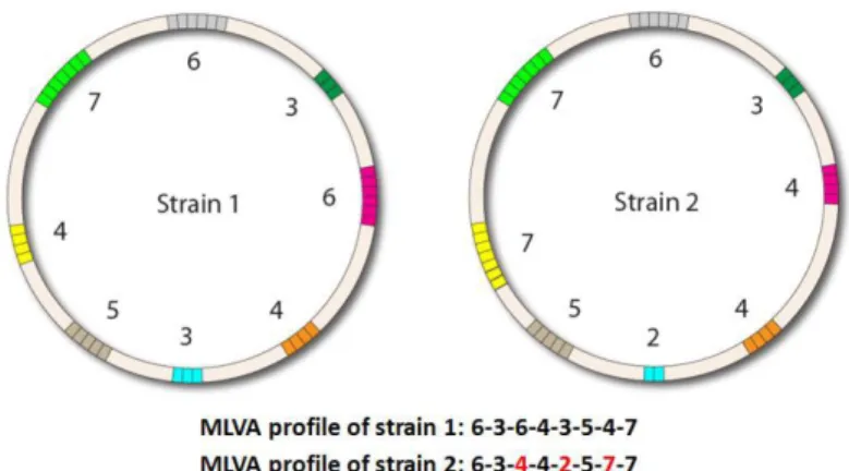
The number of tandem repeats in a particular locus may differ between different strains (Figure 3). Because of this variation, such loci are designated as variable number tandem repeat (VNTR) loci.
10
Figure 3 – Example of variation in the number of tandem repeats in multiple VNTR loci in two different strains, used to characterize them. The strains differ in three of the eight VNTR loci, marked in red [CR4].
Van Belkum et al., were the first in 1997 to report a study where they utilize VNTR loci in Haemophilus influenza to type this pathogen. In 2000 Keim et al., reported a similar approach to type Bacillus anthracis and introduced the terminology multiple locus variable-number of tandem-repeat analysis (MLVA). Since then many studies have been performed using MLVA. In 2006, for the first time, two publications (Svraka et al., and Arricau-bouvery et al.,) reported the development of MLVA for the characterization of C. burnetii. Nine unique MLVA types were detected in five laboratory variants and 16 C. burnetii isolates, from five different countries, using seven selected MLVA loci ( (Svraka et al., 2006). These MLVA types were grouped in five different clusters, proving the high discriminatory power of this method. In parallel, (Arricau-Bouvery et al., 2006) used 17 loci for MLVA and proposed two panels of MLVA markers: panel 1 composed of 10 minisatellite markers, that could be typed on agarose gel (repeat units longer than 9 bp) and panel 2, composed of 7 microsatellites markers (6 or 7 bp repeat units) with a higher discriminatory power. With these panels, 43 C. burnetii isolates could be differentiated in 36 different MLVA types. From there on, several studies were conducted applying this typing methods to both human and animal isolates, and also directly to several Coxiella DNA positive samples (Tilburg et al., 2010, 2011; Roest et al., 2012; Astobiza et al., 2012; Hilbert et al., 2012; Santos et al., 2012).
11
2. OBJECTIVES
Until two years ago the importance of Coxiella burnetii as a pathogenic and zoonotic agent was neglected in animals, mainly ruminants, and other abortive agents, like Brucella and Chlamydia, were considered more relevant. However, the human outbreaks of Q fever in 2007-2010 in The Netherlands brought a new focus in this disease and incremented studies on reservoirs, sources of infection and routes of transmission of C. burnetii.
Therefore, to have a first inside about the animal reservoirs and population structure of C. burnetii in Portugal, the main objectives of this study were:
3. To evaluate the presence of Coxiella burnetii DNA in a panel of clinical samples (tissues, faeces and swabs) from domestic and wild animals in Portugal;
4. To identify the MLVA genotypes of Coxiella burnetii that infect livestock in Portugal;
5. To compare them to other already identified genotypes, by assessing the genetic diversity of the agent;
6. To combine the results, in order to consider the relevance of the infection by Coxiella burnetii in animals, as a source and reservoir of Q fever for humans and as the causing agent of economic damages in animal production.
In order to achieve these goals, the following experimental plan was outlined:
DNA extraction from sample tissues of domestic ruminants and wild animals;
Screening of DNA tissue samples for the presence of Coxiella burnetii DNA by Trans-PCR targeting the transposase gene IS1111 insertion element;
Real-Time PCR of Coxiella burnetii positive samples in order to quantify the amount of Coxiella burnetii DNA present;
Molecular typing of the positive cases by plasmid characterization and MLVA (Multiple locus variable-number tandem-repeat analysis);
12
3. MATERIAL AND METHODS
3.1. Bacterial strains
DNA from Coxiella burnetii Nine Mile strain ATCC 616-VR, gently provided by CEVDI/INSA was used in this study. This strain is used for preparation of commercial antigen (Vircell, Spain). The complete genome sequence is available on NCBI/Genbank (accession nr. Y11502).
3.2. Clinical samples
This work was carried out using a total of 229 samples received at the National Institute of Agricultural and Veterinarian Research (INIAV).
One hundred and fifty five samples from production animals, considered to be relevant for detecting C. burnetii, were selected based on the animal species and the clinical history, mainly abortions. These samples were submitedto INIAV by veterinarians under their usual practice or by Animal Veterinary Authority (DGAV), under the eradication program for Brucellosis in ruminants. Authorization to use the samples for detection of Coxiella burnetii was previously granted.
To evaluate the existence of Coxiella reservoirs in the wild and possibility of expel in the environment, 55 DNA samples from wild carnivores tissuesand 19 vultures cloacal swabs, were also included. These samples were, respectively, available in the frame of two INIAV collaborations with CBA/FCUL Centro de Biologia Ambiental, Faculdade de Ciências de Lisboa (CBA/FCUL) and Quercos/Centros de Recuperação de Animais Selvagens (CERAS) of Castelo Branco.
Some of the 55 wild carnivores tissues were from road killed animals collected by the road maintenance technicians of ‘‘EP- Estradas de Portugal, S.A.’’ and donated for scientific purposes through a collaboration protocol, entitled “Monitoring of vertebrate mortality caused by road-kill in Portuguese roads" established between CBA/FCUL (Universidade de Lisboa, Centro de Biologia Ambiental, Faculdade de Ciêcias de Lisboa) and ‘‘EP- Estradas de Portugal, S.A.” Other samples were from animals killed in legal hunting sessions (following the Portuguese game legislation) by hunters with valid permits assigned by ‘‘Autoridade Florestal Nacional’’, and totally or partially donated for scientific purposes by the hunting associations/confederations responsible for managing the hunting journeys.
The samples were as follows:
155 tissue samples (liver, spleen, lymph node, mammary gland, uterus, testicles) from domestic animals (cattle, sheep, goat and pigs), with cases of miscarriage, stillbirth and/or suspected brucellosis;
55 DNA samples from wild carnivores (Egyptian mongoose, fox, weasel, genet and badger);
13
19 DNA samples obtained from cloacal swabs of vultures (Gyps fulvus).
3.3. DNA extraction
DNA extraction from organ samples of domestic animals was carried out using the High Pure PCR Template Preparation KitTM (Roche Diagnostics), according to the manufacturer’s instructions. Organ samples were cut into 50 mg sections and mechanically homogenized and disrupted in 200 μl of tissue lyses buffer with beads, using a FastPrep apparatus (Rebolyser), for 40 s at 6 V. Then, 200 μl of lyses buffer containing the disrupted cells were recovered and incubated overnight at 56ºC, with 75 μl of proteinase K (20 mg/ml) (Roche Diagnostics). During the final stage of preparation, the extracted DNA was eluted into 100 μl of elution buffer. Final DNA concentration was determined in a spectrophotometer (NanoDrop 2000 - Thermo Scientific) and DNA stored at -20ºC for further use in PCR assays and typing.
3.4. Trans-PCR assay
The PCR system used was both a nested and a touchdown PCR.
The sensitivity of the Trans-PCR assay for the detection of C. burnetii DNA was tested, using serial decimal dilutions (10-1 to 10-10), of a known DNA concentration (106.1 ng/μl) from C. burnetii Nine Mile strain ATCC 616-VR.
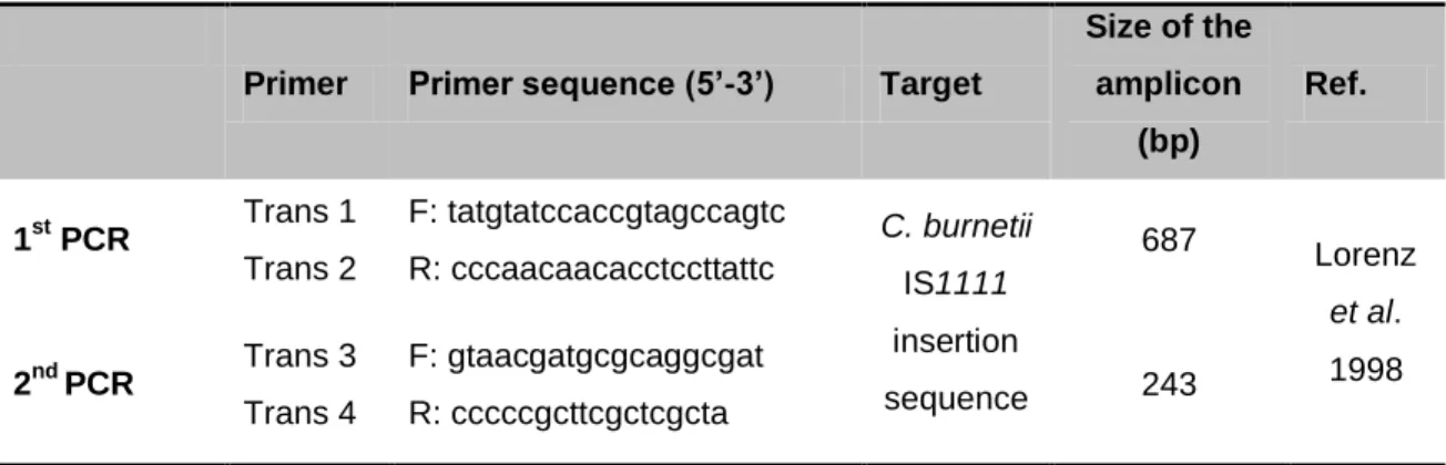
The Trans1/Trans2 and the Trans3/Trans4 primers pairs, were based on the published data sequence of the transposon-like repetitive region of the C. burnetii genome. This insertion element is a transposase gene, present in approximately 20 copies (Panning et al., 2008) in the genome of C. burnetii.
The sequences and direction of polymerization of each primer are detailed in table 1.
Table 1 - Size of C. burnetii fragments amplified by nested PCR and sequences of the
oligonucleotides used in the 1st and 2nd reactions. F and R indicate forward and reverse primers, respectively.
Primer Primer sequence (5’-3’) Target
Size of the amplicon (bp) Ref. 1st PCR Trans 1 Trans 2 F: tatgtatccaccgtagccagtc R: cccaacaacacctccttattc C. burnetii IS1111 insertion sequence 687 Lorenz et al. 1998 2nd PCR Trans 3 Trans 4 F: gtaacgatgcgcaggcgat R: cccccgcttcgctcgcta 243
14
Reactions were performed in a final volume of 25 μl, using 200 μM dNTPs (Promega), 3 mM MgCl2 (Promega), 0.08 μM of each primer (Invitrogen), 0.5 U of GoTaq® DNA polymerase, 1X
GoTaq® Flexi buffer (Promega) and 2.5 μl of each DNA dilution. For the second amplification reaction 2.5 μl of the product of the first reaction was used as template. Nine Mile reference strain was used as positive control and double distilled water as negative control.
Amplification was carried out in a Mastercycler gradient thermocycler® (Eppendorf, Germany), under the following conditions (adapted from Lorenz et al., 1998): five cycles consisting of denaturation at 94ºC for 30 s, annealing at 66 to 61ºC (the temperature was decreased by 1ºC between consecutive steps – touchdown PCR) for 60 s and extension at 72ºC for 60 s; followed by 40 cycles of denaturation at 94ºC for 30 s, annealing at 61ºC for 30 s and extension at 72ºC for 60 s. Both reactions (1st PCR and 2nd PCR) were performed under these same amplification conditions.
After standardization with the reference strain, field DNA samples were submitted to PCR, using the previously described primers sets (Trans1/Trans2 and Trans3/Trans4), targeting the transposon-like repetitive region of C. burnetii IS1111 insertion element, under the same reaction conditions. C. burnetii Nine Mile reference strain DNA was used as the positive control and sterile double distilled water as the negative control.
3.5. Agarose gel electrophoresis
Ten μl of the PCR reactions were analyzed in 1.5% agarose gel electrophoresis in TBE 1X (Tris Borate 40 mM, EDTA 1 mM, pH 8), stained with Ethidium bromide solution (2 mg/ml), and subjected to a constant current of 90 V. Electrophoresis was carried out in 1X TBE buffer, reconstituted from a 5X concentrate TBE solution. Molecular weight DNA markers III and IV from Bioline® were used.
The fragments obtained were analyzed and recorded using a UV transilumination system (BioDoc-ItTM Imaging System- UVP).
3.6. Amplification of the constitutive gene of β-actin
After testing all samples by Trans-PCR, all negative ones were tested for β-actin gene (a housekeeping gene, constitutively expressed at a constant level in all living beings), as a way to discard any inhibition of Taq DNA polymerase that may have occurred during both Trans-PCR reactions.
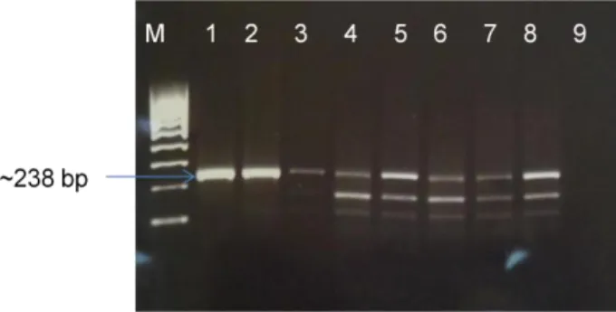
The primers used for this amplification were described by Robinson et al. 2007 (Table 2) and they flank a gene segment of about 238 bp.
15
Table 2 - Oligonucleotides used for β-actin amplification reaction. F and R indicate forward and
reerse sequences, respectively.
Primer Primer sequence (5’-3’) Target Reference
β-actinBov-F agcaagcaggagtacgatgagt
β-actin gene Robinson et al. 2007 β-actinBov-R atccaaccgactgctgtca
The amplification reaction was performed in a final volume of 25 μl, with 200 μM dNTPs, 3 mM MgCl2, 0.08 μM of each primer, 0.5 U of GoTaq Flexi DNA polymerase (Promega) and 1X of the
respective buffer, and 2.5 μl of DNA sample. As negative control, double distilled water was used and, as positive control, DNA extracted from bovine liver.
The reaction was carried out in a Mastercycler gradient thermocycler (Eppendorf, Germany), according to the following conditions: denaturation at 95ºC for 10 min, followed by 35 cycles of 95ºC for 60 s, 50ºC for 40 s and 72ºC for 30 s, with a final extension step of 72ºC for 10 min.
From each PCR reaction, 10 μl of DNA were analyzed by agarose gel electrophoresis 1.5% in TBE 1X (Tris-Borate 40 mM, EDTA 1 mM, pH 8), stained with Gel Red (Biotium) or ethidium bromide solution (1 mg/ml), and subjected to a constant current of 90 V.
The fragment sizes were visualized under UV transilumination (BioDoc-ItTM Imaging System) and identified using molecular DNA markers III and VI from Bioline®.
3.7. Purification and sequencing of the PCR amplicons
The resulting final products (amplicons) from Trans-PCR were purified using the QIAquick PCR Purification Kit (Qiagen), according to the manufacturer’s instructions. This purification procedure was carried out with the aim of further sequencing of the amplicons, to confirm that they correspond to C. burnetii DNA. The purification procedure is schematized in Figure 4.
16
Purified amplicons sequenced in the Instituto Nacional de Saúde Dr. Ricardo Jorge (INSA) in an automated sequencer 3330 XL - Genetic Analyser of 16 capillaries (Applied Biosystems®), using the two internal primers Trans3 and Trans4. The resulting sequences were analysed with “Chromas Lite” version 2.1 (2012) Technelysium Pty Ltd (South Brisbane, Queensland, Australia).
3.8. Quantitative Real-time PCR (qRT-PCR)
A qReal-Time PCR (qRT-PCR) assay was performed in all Trans-PCR positive samples, with the aim of quantifying the amount of C. burnetii DNA present in each sample and, thereby, determine whether it was possible to proceed to MLVA typing.
In fact, it has been shown (Santos et al. 2012) that MLVA typing of Coxiella strains can only be fully achieved on samples with a Ct values within the PCR detection limit. In general, Ct <29
values are strong positive reactions indicative of abundant target DNA in the sample; Ct
between 30-37 are positive reactions indicative of moderate amounts of target DNA; Ct of 38-40
are weak reactions indicative of minimal amounts of target DNA [CR6].
A standard calibration curve, with serial dilutions of the reference strain Nine Mile: 10-1 to 10-6 was designed. Reaction conditions were as follows: 20 μl of total volume, consisting of 10 μl of supermix 1X (Bio-Rad) containing dNTPs, MgCl2, reaction buffer and DNA polymerase, 0.4 μM
of each primer (forward and reverse), 0.2 μM of probe and 5 μl of DNA sample. Primers and probe sequences are listed in Table 3.
Table 3 - Oligonucleotides and probe sequences used in the Real-time PCR for quantifying C.
burnetii DNA load. F and R indicate forward and reverse sequences, respectively. Primer Primer sequence (5’-3’)
IS1111aF IS1111aR
catcacattgccgcgtttac ggttggtccctcgacaacat
Probe IS1111a aatccccaacaacacctccttattcccac
Amplification was carried out in aC1000 Thermal cycler (Bio-Rad®), conditions were as follows (adapted from Roest et al., 2011): one cycle consisting of denaturation/activation at 95ºC for 120 s, followed by 40 cycles of denaturation at 95ºC for 10 s, and annealing at 60ºC for 30 s. Results (on Ct values) were generated with CFX Manager Software (Bio-Rad, Germany).
17
3.9. Plasmid-specific PCR to identify plasmids QpH1 and QpRSDifferentiation of C. burnetii plasmid types offers an important tool for epidemiological investigations; therefore, a plasmid-specific PCR assay was performed with the aim of determining which plasmid exists on the reference strain (Nine Mile ATCC 616-VR) used in this study, as well as, in all samples that were positive for C. burnetii in the Trans-PCR assay.
Specific primers (CB5-CB6 and QpRS1-QpRS2) used for this reaction are listed on table 4.
CB5-CB6 primers pair was drawn from a particular QpH1 plasmid gene (cbhE’) and they amplify a region of about 977 bp. Primers QpRS1-QpRS2 were designed from a single QpRS plasmid gene (cbbE'), amplifying a region of about 693 bp (Zhang et al. 1998).
Table 4 - Oligonucleotides used for plasmid-specific PCR. F and R indicate forward and reverse
primers, respectively.
Target Primer Sequence (5’-3’)
Gene detecte d Amplicon size (bp) Ref. Plasmid QpH1 CB5 F: ataatgagattagaacaaccaaga cbhE’ 977 Zhang, et al. 1998 CB6 R: tctttcttgttcattttctgagtc Plasmid QpRS QpRS1 F: ctcgtacccaaagactatgaatatatcc cbbE’ 693 QpRS2 R: aacaccgatcaatgcgactagccc
Amplification was performed in a final volume of 25 μl, using 200 mM dNTPs (Promega), 3 mM MgCl2 (Promega), 62.5 μM of each primer (Eurofins MWG), 0.5 U of GoTaq ® DNA polymerase
(Promega), 1X GoTaq ® Flexi buffer (Promega) and 2.5 μl of DNA sample.
Reaction conditions for both plasmids (QpH1 and QpRS) are listed on the table below.
Table 5 - PCR reaction conditions for plasmids QpH1 and QpRS Stage Temp. (ºC) Time (s) Cycles
Initial denaturation 94 120 1 Denaturation 94 120 35 Annealing 56 60 Extension 72 120 Final extension 72 60 1
18
After the PCR reaction, an agarose gel (1.5%) electrophoresis was performed under the same conditions described previously. The fragment sizes were identified under UV transilumination as above.
3.10. Multiple locus variable-number tandem-repeat analysis (MLVA) typing
In MLVA, the analysis of variation in the number of repeats in a set of VNTR loci is achieved by performing PCR, using primers flanking that region, and subsequent sizing of the PCR products on agarose gels, capillary systems or automated DNA sequencers.
MLVA typing was performed using three hexanucleotide repeat markers (Ms27, Ms28 and Ms34) and 3 heptanucleotide repeat markers (Ms23, Ms24 and Ms33) as target loci, directly in 19 DNA clinical samples (14 DNA samples from domestic ruminants and five DNA samples from wild carnivores), that had been positive for C. burnetii by Trans-PCR analysis.
The PCR reaction was performed in a Mastercycler gradient thermocycler® (Eppendorf, Germany), in a total volume of 25 μl containing 1 U of GoTaq®
DNA polymerase (Promega), 1X GoTaq ® Flexi buffer, 4 mM MgCl2, 0.2 mM of dNTPs, 0.5 μM of amplification primers (table 6)
and 5 μl of DNA template. DNA from the Nine Mile strain (ATCC 616-VR) was used as a positive control and sterile bidistilled water as a negative control.
Amplification cycles were adapted from Roest et al. 2011 and consisted of an initial step of denaturation/activation for 2 min at 95ºC, followed by 40 cycles of denaturation for 30 s at 95ºC, annealing for 30 s at 58ºC, elongation for 30 s at 72ºC, followed by a final extension step for 5 min at 72ºC.
19
Table 6 – Characteristics of each loci and set of primers used in MLVA typing
*Obtained by in silico analysis of genomic sequence of Nine Mile strain RSA493 (NCBI/GenBank accession number NC_002971) and according to Tilburg et al. (2012) and Klaassen et al. (2009). In loci Ms23, Ms24 and Ms28 the sequence of the motive is not conserved in all repeats, differing in one (Ms28) or two nucleotides (Ms23 and Ms24). **A consensus repeat motive is proposed for these loci, based on the International Union of Pure and Applied Chemistry (IUPAC), where R can be any purine base (A or G); ***in reference strain Nine Mile RSA493 using the above primers.
Locus Alias
Size (bp) /sequence of repeat motive*
Consensus
motive** Forward primer Reverse primer
Amplicon size (bp)/Nr.of repeats*** Reference Ms23 Cox6; Cbu0197 7 / GAGGGCA 7 / GAAGACA 7 / GAGGACA GARGRCA
HEX-CGCMTAGCGACACAACCAC GACGGGCTAAATTACACCTGCT 133/9 Tilburg et al.
2012
Ms24 Cox4; CbuO259
7 / GACGGAA 7 / GACAGAA
7 /GACAGAG GACRGAR
FAM-TGGAGGGACTCCGATTAAAA GCCACACAACTCTGTTTTCAG 261/27 Tilburg et al.
2012
Ms33 Cbu1435 7 / CTGTCTT -- NED-TCGCGTAGCGACACAACC GTAGCCCGTATGACGCGAAC 104/4 Tilburg, et al.
2012
Ms27 Cox2; CbuO838 6 /TGAAGA -- HEX-TCTTTATTTCAGGCCGGAGT GAACGACTCATTGAACACACG 89/4 Klaassen et al.
2009 Ms28 Cox5; CbuO839 6 / TAAGAA 6 / TAAGGA TAAGRA NED-AGCAAAGAAATGTGAGGATCG GCCAAAGGGATATTTTTGTCCTTC 111/6 Klaassen et al. 2009
Ms34 Cox1; Cbu1471 6 / GAAAAG
-- FAM-TTCTTCGGTGAGTTGCTGTG GCAATGACTATCAGCGACTCGAA 101/5
Klaassen et al. 2009
20
3.10.1. Capillary analysis of MLVA PCR productsVNTR PCR products can be separated and analysed in a DNA sequencer, to evaluate its size. Fluorescently labeled primers are used and each VNTR locus has its own fluorescent label indicated by different colors. The high resolution separation of the sequencer enables accurate and high throughput analysis (≤ 2 base resolution).
Each 19 PCR products, with different fluorescent dyes were mixed with 12 μl of Hi-Di formamide (Applied Biosystems) and 0.2 μl of Rox 500 Size Standard (Applied Biosystems). After denaturation for 5 min at 90ºC, the PCR products were separated in an ABI PRISM 310 Genetic Analyser (Applied Biosystems, Germany) with a 36 cm array by using POP7 polymer.
Electropherograms were analysed using ABI PRISM GeneScan 3.7 (Applied Biosystems). DNA from the Nine Mile strain (ATCC 616-VR) was used as reference.
3.10.2. Agarose gel eletrophoresis analysis of MLVA PCR products
The MLVA results were also analyzed by agarose gel electrophoresis, as agarose gels can be easily read manually, revealing the size variation of the PCR products (Vergnaud and Pourcel, 2006).
The PCR amplication products (10 μl) were analyzed by agarose gel electrophoresis 3% in TBE 1X (Tris Borate 40 mM, EDTA 1 mM, pH 8), stained with Ethidium bromide solution (2 mg/ml) and subjected to a constant current of 160 V for 3 h.
The fragment sizes were visualized under UV transilumination (UV transiluminator Bio Doc-ItTM Imaging System) and identified using a 20 bp molecular DNA marker from Bio-Rad®.
3.10.3. Assessing the number of tandem repeats
The PCR product size was used to calculate the number of repeat units in each locus. The flanking regions, analyzed in silico including the hybridization sites of the primers used, were subtracted from the PCR product size, resulting in the net size of the repeat region. Small inaccuracies in sizing may occur but mostly do not prevent the assessment of the number of repeats.
The accuracy of the sizing was determined by comparing sequence data from the reference strain, with the obtained fragment size from the capillary electrophoresis and agarose gel electrophoresis and corrected, if necessary. The number of repeats for each locus was determined on the basis of the published and corrected annotation of the various loci (Table 8) [CR7].
21
3.10.4. Analysis and clustering of MLVA dataThe calculated number of repeats of the VNTR loci was combined into a string, referred to as the MLVA profile. Each unique MLVA profile was given a MLVA type designation (M1 to M7). The copy number for each locus was managed as a character dataset using BioNumerics version 6.6 (Applied Maths, Belgium). Cluster analysis was based on the categorical coefficient and unweighted pair group method using arithmetic averages (UPGMA).
MLVA typing tools and databases with MLVA profiles of several organisms, including links to tandem repeats which have already been investigated, were accessed over the internet [CR7; CR8; CR9; CR10; CR11; CR12 and CR13].
3.10.5. Determination of the discriminatory power (D) of the MLVA typing
The Discriminatory Power (D) or Hunter & Gaston Discriminatory Index (HGDI) is determined by the number of types defined by the test method and the relative frequencies of these types. It is based on the probability that two strains chosen at random from a population of unrelated strains will be distinguished by a chosen typing method (Hunter and Gaston, 1988):
Where xj(xj-1) is the number of strains in the population which are indistinguishable from the jth strain and N is the number of strains in the population. Each strain in turn is compared with all other strains in the population to determine how many other strains are indistinguishable from it to give xj(xj-1) (Hunter 1990).
The discriminating power of the typing method and the genetic diversity of each locus were determined using the Hunter & Gaston diversity index (D) via an online tool available at the website [CR13].
We applied this equation to determine the discriminating power of the typing method and for each individual locus. Results are presented on the final summary table 8.
22
4. RESULTS AND DISCUSSION
4.1 Determination of the detection limit of the Trans-PCR assay
Primer pairs Trans1/Trans2 and Trans3/Trans4 were used to detect C. burnetii DNA (106.1 ng/μl) from the Nine Mile reference strain ATCC 616-VR, in different dilutions.
Figure 5- Sensitivity of Trans-PCR for the detection of C. burnetii Nine Mile strain ATCC 616-VR DNA
(specific 240 bp amplicon). Lane 1: DNA molecular marker (100-bp); lanes 2-11: different dilutions (10-1-
10-10) of the template DNA; lane 12: negative control (bidistilled sterile water).
The analytical sensitivity of the Trans-PCR was found to be 1 fg/μl (Figure 5 – lane 5). This result is compatible to that obtained (later on) with qRT-PCR, confirming the suitability of the Trans-PCR assay for routine diagnostic of C. burnetii.
4.2. Detection of C. burnetii DNA in clinical samples using Trans-PCR assay
Initially, we used 239 macerated, already processed samples of different organs (liver, spleen, uterus, mammary gland, testicles) from cattle, sheep and goats, received in INIAV – Bacteriology, Lisboa, for bacteriological diagnosis of Brucella spp.
Since all of them tested negative for C. burnetii, we decided to change strategy and use other samples where the extraction of DNA was done directly from the tissues and not from the macerates. These samples were sent by Bacteriology, Vairão.
We believe that the negative results we had achieved in the first set of samples, were not due to the lack of DNA from C. burnetii, but due to the dilution of samples because of maceration, even though it was a highly sensitive and specific PCR.
In the image below (Figure 6) are presented two examples of C. burnetii Trans-PCR positive results, in tissue samples (liver and spleen) obtained from an ovine aborted fetus, with the specific 240 bp amplified fragment.