COMMON TO MAN AND ANIMALS
Third Edition
Volume II
Chlamydioses, Rickettsioses,
and Viroses
Scientific and Technical Publication No. 580 PAN AMERICAN HEALTH ORGANIZATION Pan American Sanitary Bureau, Regional Office of the
WORLD HEALTH ORGANIZATION 525 Twenty-third Street, N.W. Washington, D.C. 20037 U.S.A.
Also published in Spanish (2003) with the title:
Zoonosis y enfermedades transmisibles comunes al hombre y a los animales: clamidiosis, rickettsiosis y virosis
ISBN 92 75 31991 X (3 volume set) ISBN 92 75 31992 8 (Vol. 2)
PAHO HQ Library Cataloguing-in-Publication Pan American Health Organization
Zoonoses and communicable diseases common to man and animals: chlamydioses, rickettsioses, and viroses
3rd ed. Washington, D.C.: PAHO, © 2003.
3 vol.—(Scientific and Technical Publication No. 580) ISBN 92 75 31991 0—3 volume set
ISBN 92 75 31992 9—Vol. 2 I. Title II. Series
1. ZOONOSES 2. VIROSES
3. RICKETTSIA INFECTIONS
4. COMMUNICABLE DISEASE CONTROL 5. FOOD CONTAMINATION
6. PUBLIC HEALTH VETERINARY NLM WC950.P187 2003 V.2 En
The Pan American Health Organization welcomes requests for permission to reproduce or translate its publications, in part or in full. Applications and inquiries should be addressed to Publications, Pan American Health Organization, Washington, D.C., U.S.A., which will be glad to provide the latest information on any changes made to the text, plans for new editions, and reprints and translations already available.
© Pan American Health Organization, 2003
Publications of the Pan American Health Organization enjoy copyright protection in accordance with the provisions of Protocol 2 of the Universal Copyright Convention. All rights are reserved.
The designations employed and the presentation of the material in this publication do not imply the expression of any opinion whatsoever on the part of the Secretariat of the Pan American Health Organization concerning the status of any country, ter-ritory, city or area or of its authorities, or concerning the delimitation of its frontiers or boundaries.
Prologue . . . vii
Preface to the First Edition . . . ix
Preface to the Second Edition . . . xi
Introduction. . . xv
PART I: CHLAMYDIOSES AND RICKETTSIOSES
Rickettsiaceae . . . 3Asian-Ixodo Rickettsiosis . . . 4
Boutonneuse Fever . . . 5
Flea-borne Typhus . . . 9
Infections Caused by Bartonella henselae. . . 13
Q Fever . . . 16
Queensland Tick Typhus. . . 27
Rickettsialpox . . . 29
Rocky Mountain Spotted Fever . . . 32
Scrub Typhus . . . 38
Zoonotic Chlamydiosis. . . 42
Zoonotic Typhus Caused by Rickettsia prowazekii. . . 51
PART II: VIROSES
Argentine Hemorrhagic Fever . . . 57Bovine Papular Stomatitis . . . 64
Brazilian Hemorrhagic Fever . . . 66
California Encephalitis . . . 68
Chikungunya Virus Disease . . . 73
Colorado Tick Fever . . . 77
Contagious Ecthyma . . . 80
Cowpox . . . 84
Buffalopox. . . 88
Crimean-Congo Hemorrhagic Fever . . . 88
Dengue . . . 94
Diseases Caused by Hantaviruses. . . 98
Eastern Equine Encephalitis . . . 110
Ebola Disease . . . 117
Encephalomyocarditis. . . 122
Epidemic Polyarthritis . . . 126
Fever Caused by Group C Bunyaviruses . . . 131
Foot-and-Mouth Disease . . . 133
Herpes Simplex (Type 1) . . . 146
Herpesvirus simiae. . . 149
Ilheus Fever. . . 154
Influenza . . . 155
iv CONTENTS
Japanese Encephalitis . . . 172
Kyasanur Forest Disease. . . 179
Lassa Fever . . . 183
Louping Ill . . . 189
Lymphocytic Choriomeningitis . . . 193
Machupo Hemorrhagic Fever . . . 200
Marburg Virus Disease . . . 205
Mayaro Fever . . . 208
Measles . . . 211
Murray Valley Encephalitis . . . 215
Newcastle Conjunctivitis . . . 218
Omsk Hemorrhagic Fever. . . 225
Oropouche Virus Disease . . . 227
Orungo Fever . . . 231
Powassan Encephalitis . . . 232
Poxes of Monkeys. . . 235
Monkeypox . . . 236
Tanapox . . . 239
Poxes Caused by Other Yatapoxviruses . . . 240
Pseudocowpox . . . 242
Sealpox . . . 245
Rabies . . . 246
Rift Valley Fever . . . 276
Rocio Encephalitis . . . 284
Rotaviral Gastroenteritis . . . 286
Russian and Central European Spring-Summer Encephalitis . . . 297
Sindbis Fever . . . 302
Spongiform Encephalopathies of Animals and Man . . . 304
Diseases of Animals . . . 305
Scrapie . . . 305
Transmissible Mink Encephalopathy. . . 308
Chronic Wasting Disease of Deer . . . 308
Bovine Spongiform Encephalopathy . . . 309
Spongiform Encephalopathies in Other Animal Species in the UK . . . 311
Human Diseases . . . 312
Kuru . . . 312
Creutzfeldt-Jakob Disease . . . 313
Gerstmann-Sträussler-Scheinker Syndrome . . . 315
St. Louis Encephalitis . . . 318
Swine Vesicular Disease . . . 326
Vaccinia Virus Infection . . . 330
Venezuelan Equine Encephalitis. . . 333
Venezuelan Hemorrhagic Fever . . . 345
Vesicular Stomatitis . . . 347
Viral Hepatitis of Man and Nonhuman Primates . . . 356
Wesselsbron Disease. . . 362
West Nile Fever . . . 372
Yellow Fever . . . 377
LIST OF TABLES AND ILLUSTRATIONS
Chlamydioses and Rickettsioses Figures 1. Flea-borne typhus (Rickettsia typhi). Transmission cycle. . . 112. Q fever. Transmission—wildlife cycle.. . . 21
3. Q fever. Transmission—domestic cycle.. . . 22
4. Rickettsialpox (Rickettsia akari). Transmission cycle. . . 30
5. Rocky Mountain spotted fever. Transmission cycle in the US. . . 34
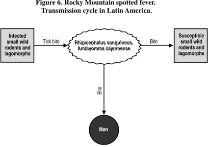
6. Rocky Mountain spotted fever. Transmission cycle in Latin America. . . 35
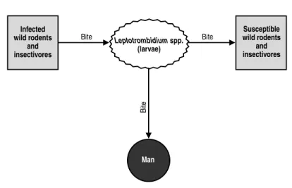
7. Scrub typhus (Rickettsia tsutsugamushi). Transmission cycle. . . 40
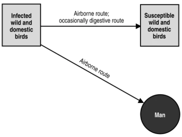
8. Zoonotic chlamydiosis (psittacosis, ornithosis). Transmission cycle. . . 47
Viroses Tables 1. Members of the genus Hantavirus, family Bunyaviridae, associated with human disease. . . 100
2. Reported cases of hantavirus pulmonary syndrome (HPS) in South America, 1993–2001.. . . 103
3. Distinguishing clinical characteristics for hemorrhagic fever with renal syndrome (HFRS) and hantavirus pulmonary syndrome (HPS). . . 104
4. Classification of the group C bunyaviruses. . . 131
5. Average annual number of reported cases of human rabies in the Americas, by subregion and by country, 1980–2000. . . 250
6. Average annual number of reported cases of canine rabies in Latin America and the Latin Caribbean, by subregion and by country, 1990–2000.. . . 251
7. Number of cases of bovine rabies in Central and South America, 1990, 1991, and 1999. . . 253
8. General and specific treatment of rabies. . . 268
Figures 9. Argentine hemorrhagic fever. Probable transmission cycle of the Junín virus. . . 59
10. California encephalitis (La Crosse virus). Transmission cycle. . . 69
11. Colorado tick fever. Transmission cycle. . . 78
12. Contagious ecthyma. Transmission cycle. . . 82
13. Eastern equine encephalitis. Transmission cycle in the US.. . . 113
vi
15. Herpesvirus simiae. Transmission cycle. . . 151
16. Japanese encephalitis. Transmission cycle. . . 175
17. Louping ill. Transmission cycle. . . 191
18. Lymphocytic choriomeningitis. Transmission cycle. . . 197
19. Machupo hemorrhagic fever. Transmission cycle. . . 202
20. Measles. Transmission cycle. . . 213
21. Newcastle disease virus. Transmission cycle.. . . 221
22. Oropouche fever. Possible circulation of the virus. . . 229
23. Powassan encephalitis. Transmission cycle. . . 234
24. Pseudocowpox. Mode of transmission.. . . 244
25. Urban rabies. Transmission cycle. . . 258
26. Russian and central European spring-summer encephalitis. Transmission cycle. . . 299
27. St. Louis encephalitis. Probable cycle of the virus. . . 321
28. Venezuelan equine encephalitis. Enzootic wildlife cycle (variants D, E, and F of virus subtype I and subtypes II, III, IV, V, and VI). . . 338
29. Venezuelan equine encephalitis. Epidemic/epizootic cycle (AB and C variants of virus subtype I).. . . 339
30. Hepatitis A transmitted by nonhuman primates. Probable transmission cycle. . . 359
31. Western equine encephalitis. Transmission cycle. . . 368
32. West Nile fever. Transmission cycle. . . 375
33. Jungle yellow fever in the Americas. Transmission cycle. . . 382
vii
In recent years, zoonoses and communicable diseases common to man and ani-mals have gained increasing attention worldwide. Human diseases that have their origins in infected animals, such as AIDS or Creutzfeldt-Jakob, have highlighted the need for a better understanding of animal diseases in terms of their epidemiology, mechanism of transmission to man, diagnosis, prevention, and control. Social and demographic changes have also contributed to the importance of gaining and dis-seminating knowledge about zoonoses. For instance, as people encroach further and further on ecological areas with which they had little contact and whose fauna may not be well known, their exposure to animals—and the infections they transmit— has increased. New knowledge is also being developed in the area of urban ecology. The ease and speed of modern travel also facilitates the spread of diseases once con-fined to specific geographic areas, as recently occurred with severe acute respiratory syndrome (SARS). Animal migration and trade pose a similar threat, as was shown by the outbreaks in the United States of West Nile fever, and most recently, mon-keypox—two diseases not previously known in the Western Hemisphere. Each of these examples highlights the need for improved knowledge and surveillance of and response to zoonoses.
The negative effects of zoonoses are far reaching. High incidence rates continue to cause significant morbidity and mortality in both humans and animals. Their eco-nomic impact is seen in lost labor productivity due to illness; reduced travel and tourism to affected areas; reduced livestock and food production; death and destruc-tion of affected animals; and restricdestruc-tions on and reducdestruc-tions in internadestruc-tional trade. Zoonoses can be a serious drain on a country’s economy, which in turn can have wide repercussions for a society’s health.
To help solve these problems, the Pan American Health Organization (PAHO)— an international public health organization that has devoted itself to improving the health and living conditions of the people of the Americas for over one hundred years—established the Veterinary Public Health Unit. The Unit’s overall objective is to collaborate with PAHO’s Member Governments in the development, implemen-tation, and evaluation of policies and programs that lead to food safety and protec-tion and to the prevenprotec-tion, control, or eradicaprotec-tion of zoonoses, among them foot-and-mouth disease.
To this end, PAHO’s Veterinary Public Health Unit has two specialized regional centers: the Pan American Foot-and-Mouth Disease Center (PANAFTOSA), created in 1951 in Rio de Janeiro, Brazil, and the Pan American Institute for Food Protection and Zoonoses (INPPAZ), established on November 15, 1991, in Buenos Aires, Argentina. INPPAZ’s precursor was the Pan American Zoonoses Center (CEPANZO), which was created through an agreement with the Government of Argentina to help the countries of the Americas combat zoonoses, and which oper-ated from 1956 until 1990.
viii PROLOGUE
animals, which cause high morbidity, disability, and mortality in vulnerable human populations. PAHO has also collaborated in the strengthening of preventive medi-cine and public health through the promotion of veterinary health education in learn-ing, research, and health care centers. An example of this work is the preparation of several publications, among which the two previous Spanish and English editions of Zoonoses and Communicable Diseases Common to Man and Animalsstand out.
Scientific knowledge has progressed since the last edition was published in 1986. Also, the countries of the Americas have modified their livestock production strate-gies in recent years, which has affected the transmission of zoonotic infections and their distribution. The publication of this third edition is an attempt to address these changes. The third edition is presented in three volumes: the first contains bacte-rioses and mycoses; the second, chlamydioses, rickettsioses, and viroses; and the third, parasitoses.
We believe that this new edition will continue to be useful for professors and stu-dents of public health, medicine, veterinary medicine, and rural development; work-ers in public health and animal health institutions; and veterinarians, researchwork-ers, and others interested in the subject. We also hope that this publication is a useful tool in the elaboration of national zoonosis control or eradication policies and programs, as well as in risk evaluation and in the design of epidemiological surveillance sys-tems for the prevention and timely control of emerging and reemerging zoonoses. In summary, we are confident that this book will contribute to the application of the knowledge and resources of the veterinary sciences for the protection and improve-ment of public health.
ix
This book considers two groups of communicable diseases: those transmitted from vertebrate animals to man, which are—strictly speaking—zoonoses; and those common to man and animals. In the first group, animals play an essential role in maintaining the infection in nature, and man is only an accidental host. In the sec-ond group, both animals and man generally contract the infection from the same sources, such as soil, water, invertebrate animals, and plants; as a rule, however, animals do not play an essential role in the life cycle of the etiologic agent, but may contribute in varying degrees to the distribution and actual transmission of infections.
No attempt has been made to include all infections and diseases comprised in these two groups. A selection has been made of some 150 that are of principal inter-est, for various reasons, in the field of public health. The number of listed zoonoses is increasing as new biomedical knowledge is acquired. Moreover, as human activ-ity extends into unexplored territories containing natural foci of infection, new zoonotic diseases are continually being recognized. In addition, improved health services and better differential diagnostic methods have distinguished zoonoses pre-viously confused with other, more common diseases. A number of diseases described in this book have only recently been recognized, examples of which include the Argentine and Bolivian hemorrhagic fevers, angiostrongyliasis, rotaviral enteritis, Lassa fever, Marburg disease, and babesiosis.
The principal objective in writing this book was to provide the medical profes-sions a source of information on the zoonoses and communicable diseases common to man and animals. Toward that end, both medical and veterinary aspects, which have traditionally been dealt with separately in different texts, have been combined in a single, comprehensive volume. As a result, physicians, veterinarians, epidemi-ologists, and biologists can all gain an overview of these diseases from one source. This book, like most scientific works, is the product of many books, texts, mono-graphs, and journal articles. Many sources of literature in medicine, veterinary med-icine, virology, bacteriology, mycology, and parasitology were consulted, as were a large number of reports from different biomedical disciplines, in order to provide up-to-date and concise information on each disease. It is expected that any errors or omissions that may have been committed can, with the collaboration of the readers, be corrected in a future edition.
Where possible, explanations were attempted with special emphasis on the Americas, particularly Latin America. An effort was made, one which was not always successful, to collect available information on diseases in this Region. Data on the incidence of many zoonoses are fragmentary and frequently not reliable. It is hoped that the establishment of control programs in various countries will lead to improved epidemiologic surveillance and disease reporting.
More space has been devoted to those zoonoses having greatest impact on public health and on the economy of the countries of the Americas, but information is also included on those regionally less important or exotic diseases.
x PREFACE TO THE FIRST EDITION
public health and animal health administrators, physicians, and veterinarians must be familiar with the geographic distribution and pathologic manifestations of the various infectious agents so that they can recognize and prevent the introduction of exotic diseases.
We, the authors, would like to give special recognition to Dr. Joe R. Held, Assistant Surgeon-General of the United States Public Health Service and Director of the Division of Research Services of the U.S. National Institutes of Health, who gave impetus to the English translation and reviewed the bacterioses sections.
We would also like to express our utmost appreciation to the experts who reviewed various portions of this book and offered their suggestions for improving the text. These include: Dr. Jeffrey F. Williams, Professor in the Department of Microbiology and Public Health, Michigan State University, who reviewed the chapters dealing with parasitic zoonoses; Dr. James Bond, PAHO/WHO Regional Adviser in Viral Diseases, who read the viroses; Dr. Antonio Pío, formerly PAHO/WHO Regional Adviser in Tuberculosis and presently with WHO in Geneva, and Dr. James H. Rust, PAHO/WHO Regional Adviser in Enteric Diseases, both of whom reviewed the bacterioses; and Dr. F. J. López Antuñano, PAHO/WHO Regional Adviser in Parasitic Diseases, who read the metazooses.
We would like to thank Dr. James Cocozza, PAHO/WHO Veterinary Adviser, for his review of the translation and Dr. Judith Navarro, Editor in the Office of Publications of PAHO, for her valuable collaboration in the editorial revision and composition of the book.
xi
The fine reception accorded the Spanish, English, and French versions of this book has motivated us to revise it in order that it still may serve the purpose for which it was written: to provide an up-to-date source of information to the medical profession and allied fields. This book has undoubtedly filled a void, judging by its wide use in schools of public health, medicine, and veterinary medicine, as well as by bureaus of public and animal health.
The present edition has been considerably enlarged. In the seven years since the first edition was published, our knowledge of zoonoses has increased broadly and rapidly, and new zoonotic diseases have emerged. Consequently, most of the dis-cussions have been largely rewritten, and 28 new diseases have been added to the original 148. Some of these new diseases are emerging zoonoses; others are patho-logic entities that have been known for a long time, but for which the epidemiopatho-logic connection between man and animal has been unclear until recently.
The use this book has had outside the Western Hemisphere has caused us to aban-don the previous emphasis on the Americas in favor of a wider scope and geomed-ical view. Moreover, wars and other conflicts have given rise to the migration of populations from one country or continent to another. A patient with a disease heretofore known only in Asia may now turn up in Amsterdam, London, or New York. The physician must be aware of these diseases in order to diagnose and treat them. “Exotic” animal diseases have been introduced from Africa to Europe, the Caribbean, and South America, causing great damage. The veterinary physician must learn to recognize them to be able to prevent and eradicate them before they become entrenched. It must be remembered that parasites, viruses, bacteria, and other agents of zoonotic infection can take up residence in any territory where they find suitable ecologic conditions. Ignorance, economic or personal interests, and human customs and needs also favor the spread of these diseases.
xii PREFACE TO THE SECOND EDITION
field of medicine to another. In view of worldwide concern over acquired immuno-deficiency syndrome (AIDS), a brief section on retroviruses has also been added, in which the relationship between the human disease and feline and simian AIDS is noted. Another topic deeply interesting to researchers is the mystery of the radical antigenic changes of type A influenza virus, a cause of explosive pandemics that affect millions of persons around the world. Evidence is mounting that these changes result from recombination with a virus of animal origin (see Influenza). That this should occur is not surprising, given the constant interaction between man and animals. As a rule, zoonoses are transmitted from animal to man, but the reverse may also occur, as is pointed out in the chapters on hepatitis, herpes simplex, and measles. The victims in these cases are nonhuman primates, which may in turn retransmit the infection to man under certain circumstances.
Among emerging zoonoses we cite Lyme disease, which was defined as a clinical entity in 1977; the etiologic agent was found to be a spirochete (isolated in 1982), for which the name Borrelia burgdorferi was recently proposed. Emerging viral zoonoses of note in Latin America are Rocio encephalitis and Oropouche fever; the latter has caused multiple epidemics with thousands of victims in northeast Brazil. Outstanding among new viral disease problems in Africa are the emergence of Ebola disease and the spread of Rift Valley fever virus, which has caused tens of thousands of human cases along with great havoc in the cattle industry of Egypt and has evoked alarm around the world. Similarly, the protozoan Cryptosporidiumis emerging as one of the numerous agents of diarrheal diseases among man and animals, and prob-ably has a worldwide distribution.
As the English edition was being prepared, reports came to light of two animal diseases not previously confirmed in humans. Three cases of human pseudorabies virus infection were recognized between 1983 and 1986 in two men and one woman who had all had close contact with cats and other domestic animals. In 1986, sero-logic testing confirmed infection by Ehrlichia canisin a 51-year-old man who had been suspected of having Rocky Mountain spotted fever. This is the first known occurrence of E. canisinfection in a human. These two diseases bear watching as possible emerging zoonoses.
The space given to each zoonosis is in proportion to its importance. Some diseases that deserve their own monographs were given more detailed treatment, but no attempt was made to cover the topic exhaustively.
We, the authors, would like to give special recognition to Dr. Donald C. Blenden, Professor in the Department of Medicine and Infectious Diseases, School of Medicine, and Head of the Department of Veterinary Microbiology, College of Veterinary Medicine, University of Missouri; and to Dr. Manuel J. Torres, Professor of Epidemiology and Public Health, Department of Veterinary Microbiology, College of Veterinary Medicine, University of Missouri, for their thorough review of and valuable contributions to the English translation of this book.
We are most grateful to Dr. F. L. Bryan for his generous permission to adapt his monograph “Diseases Transmitted by Foods” as an Appendix to this book.
Mr. Carlos Larranaga, Chief of the Audiovisual Unit at the Pan American Zoonosis Center, deserves our special thanks for the book’s artwork, as do Ms. Iris Elliot and Mr. William A. Stapp for providing the translation into English. We would like to express our most sincere gratitude and recognition to Ms. Donna J. Reynolds, editor in the PAHO Editorial Service, for her valuable collaboration in the scientific editorial revision of the book.
INTRODUCTION
This new edition of Zoonoses and Communicable Diseases Common to Man and Animals is published in three volumes: I. Bacterioses and mycoses; II. Chlamydioses and rickettsioses, and viroses; and III. Parasitoses. Each of the five parts corresponds to the location of the etiologic agents in the biological classifica-tion; for practical purposes, chlamydias and rickettsias are grouped together.
In each part, the diseases are listed in alphabetical order to facilitate reader searches. There is also an alphabetical index, which includes synonyms of the dis-eases and the etiologic agents’ names.
In this edition, the numbers and names of the diseases according to the International Statistical Classification of Diseases and Related Health Problems, Tenth Revision (ICD-10), are listed below the disease title. However, some zoonoses are not included in ICD-10 and are difficult to classify within the current scheme.
In addition, for each disease or infection, elements such as synonyms; etiology; geographical distribution; occurrence in man and animals; the disease in man and animals; source of infection and mode of transmission; role of animals in the epi-demiology; diagnosis; and control are addressed. Patient treatment (for man or other species) is beyond the scope of this work; however, recommended medicines are indicated for many diseases, especially where they are applicable to prophylaxis. Special attention is paid to the epidemiological and ecological aspects so that the reader can begin to understand the determining factors of the infection or disease. Some topics include simple illustrations of the etiologic agent’s mode of transmis-sion, showing the animals that maintain the cycle of infection in nature. Similarly, other graphics and tables are included to provide additional information on the geo-graphical distribution or prevalence of certain zoonoses.
The data on the occurrence of the infection in man and animals, along with data on the geographical distribution, may help the reader judge the relative impact that each disease has on public health and the livestock economy in the different regions of the world, given that the importance of different zoonoses varies greatly. For example, foot-and-mouth disease is extremely important from an economic stand-point, but of little importance in terms of public health, if animal protein losses are not considered. In contrast, Argentine and Machupo hemorrhagic fevers are impor-tant human diseases, but their economic impact is minimal, if treatment costs and loss of man-hours are not taken into account. Many other diseases, such as brucel-losis, leptospirosis, salmonelbrucel-losis, and equine encephalitis, are important from both a public health and an economic standpoint.
Finally, each disease entry includes an alphabetical bibliography, which includes both the works cited and other relevant works that the reader may consult for more information about the disease.
RICKETTSIACEAE
This family includes the tribes Rickettsieaeand Ehrlichieae. When human ehrli-chiosis was recognized in 1986, the disease was considered to be a zoonosis caused by Ehrlichia canis. However, findings in 1991 established that the human agent, although similar to E. canis, is actually a distinct species (Dawson et al., 1991). For this reason, ehrlichiosis falls outside the scope of the present volume.
Rickettsiae, like bacteria, are prokaryotic intracellular organisms. However, because they lack certain enzymes, they are dependent on a eukaryotic cell of the host. An exception within the tribe Rickettsieaeis the genus Rochalimaea, which can be cultured in an axenic environment. Rickettsiae reproduce by binary fission within the cells of an arthropod or a human or animal host; both their DNA and RNA can be synthesized, and they are sensitive to antibiotics. They measure approximately 0.5 by 0.3 microns and may be either rod-shaped or spherical. They show up well with Gimenez and Macchiavellos stains but not as well with Gram stain (Weiss and Moulder, 1984; Mettler, 1991).
In addition to the genus Rickettsia, within the tribe Rickettsieae the genera
Coxiellaand Rochalimaeaare also of interest.
Organisms of the genus Rickettsiamay be divided into the following three groups: spotted fevers, typhus, and scrub typhus.
The diseases in the spotted fever group are clinically similar and caused by related rickettsiae, and they are all transmitted by ticks.
Bibliography
Dawson, J.E., B.E. Anderson, D.B. Fishbein,et al. Isolation and characterization of an Ehrlichia sp. from a patient diagnosed with human ehrlichiosis. J Clin Microbiol 29:2741–2745, 1991.
Mettler, N.E. Rickettsiales. In: Carballal G., J.R. Oubiño, eds. Virología médica. Buenos Aires: El Ateneo; 1991.
Weiss, E., J.W. Moulder. Order 1. Rickettsiales Gieszczkiewicz 1939. In: Krieg, N.R., J.G. Holt, eds. Vol. 1: Bergey’s Manual of Systematic Bacteriology. Baltimore: Williams & Wilkins; 1984.
ASIAN IXODO-RICKETTSIOSIS
ICD-10 A77.2 Spotted fever due to Rickettsia sibirica
Synonyms:North Asian tick fever, Siberian tick typhus.
Etiology:Rickettsia sibirica (Dermacentroxenus sibiricus). This agent belongs to the spotted fever group of rickettsiae. Dermacentor marginatus, a variety of R. sibir-icathat was isolated from ticks in the former Czechoslovakia, is serologically dis-tinct from R. slovaca, although the differences may not be sufficient to warrant establishing a separate species (Weiss and Moulder, 1984).
Geographic Distribution: Armenia, Kazakhstan, Kyrgyzstan, northern China, Mongolia, Siberia, and various islands in the Sea of Japan. R. sibirica has also been isolated from ticks and mammals in the former Czechoslovakia and in Pakistan.
Occurrence in Man:Sporadic. The disease occurs mainly in farmers, hunters, forestry workers, and people who enter the disease’s natural foci in steppe and mon-tane regions. Ticks may be carried from natural foci to populated areas via the fur of domestic animals, firewood, or by other means, and thus increase the possibility of infection.
Occurrence in Animals:The etiologic agent has been isolated from at least 18 wild rodent species that live in the disease’s natural foci.
The Disease in Man:This is an acute, febrile, benign disease clinically similar to boutonneuse fever. It may also resemble the serious or moderate forms of Rocky Mountain spotted fever. It has an incubation period of two to seven days, and is treated with tetracycline.
The Disease in Animals:No information is available on the natural course of the disease in wild rodents or other species from which the rickettsia has been isolated; it is probably asymptomatic.
Source of Infection and Mode of Transmission:Man contracts the infection through tick bites. The principal vectors are ticks of the genera Dermacentor,
Haemaphysalis, andRhipicephalus. Nine species of naturally infected ticks have been found, and transovarial transmission has been confirmed in seven of them. The etiologic agent survives in the tick during hibernation. The continuous circulation of rickettsiae in natural foci is ensured by transovarial transmission from one arthropod generation to the next and by the presence of the infection in a wide variety of small mammal species.
At the end of hibernation and before egg laying, the ticks attach themselves to large domestic and wild mammals, and, accidentally, to humans who enter their habitat. Accordingly, the highest incidence of human disease occurs in spring, which is the period of greatest adult tick activity. It is usually the adult tick that attacks man, but larvae and nymphs of Dermacentor nuttalli and Haemaphysalis concinna
Role of Animals in the Epidemiology of the Disease:Man is an accidental host; the reservoir consists of wild rodents and ticks. The latter play a key role in main-taining and transmitting the infection. Transstadial and transovarial transmission of
R. sibirica has been confirmed in D. marginatus over a period of at least five years (Harwood and James, 1979). Domestic animals (cattle, horses, dogs) can serve as hosts for adult ticks.
Diagnosis: As with other spotted fevers, laboratory confirmation is obtained using such serological tests as complement fixation and microimmunofluorescence. The agent can be isolated in embryonated eggs or by inoculation in laboratory ani-mals (guinea pigs, rats, hamsters).
Control:Control measures are directed against the vectors. They include the use of tickicides on domestic animals and in their environment, as well as the reduction of rodent populations, since rodents are the principal hosts of larvae and nymphs. Individuals who enter natural foci should wear protective clothing and use tick repellents.
Bibliography
Bosler, E.M., J.L. Coleman, J.L. Benach, D.A. Massey, J.P. Hanrahan, W. Burgdorfer,et al. Natural distribution of the Ixodes dammini spirochete. Science220:321–322, 1983.
Burgdorfer, W. North Asian tick typhus. In: Hubbert, W.T., W.F. McCulloch, P.R. Schnurrenberger, eds. Diseases Transmitted from Animals to Man, 6th ed. Springfield: Thomas; 1975.
Harwood, R.F., M.T. James. Entomology in Human and Animal Health, 7th ed. New York: McMillan; 1979.
Weiss, E., J.W. Moulder. Rickettsiales Gieszczkiewicz, 1939. In: Krieg, N.R., J.G. Holt, eds. Vol. 1: Bergey’s Manual of Systematic Bacteriology. Baltimore: Williams & Wilkins; 1984.
Zdrodovskii, P.F., H.M. Golinevich. The Rickettsial Diseases. Oxford: Pergamon Press; 1960.
BOUTONNEUSE FEVER
ICD-10 A77.1 Spotted fever due to Rickettsia conorii
Synonyms: Marseilles fever, Mediterranean spotted fever, Mediterranean tick fever, African tick typhus, Kenya tick typhus, India tick typhus.
Etiology: Rickettsia conorii (Dermacentroxenus conorii). This microorganism belongs to the spotted fever group of rickettsiae. It can be differentiated from others in the group by serological and cross-immunity tests.
Geographic Distribution:The disease occurs in much of Africa, Southeast Asia, India, and areas of Europe and the Middle East adjacent to the Caspian, Mediterranean, and Black Seas.
Occurrence in Man:Sporadic. This is the most common rickettsial disease in South Africa. In Spain, the endemic area of Talavera de la Reina had 85 diagnosed cases in 1982 (España, Ministerio de Sanidad y Consumo, 1983). In Soria (Spain), 5% of 298 human sera samples were serologically positive for R. conorii. More than 90% of the cases were from the eastern part of the province, and 20% of the posi-tive cases were found in a small area (Saz et al., 1993). Similar results were obtained in Croatia on the Adriatic coast. The number of human cases in the Mediterranean basin has increased since the early 1980s, especially in Spain, France, Israel, and Italy. The number of cases in Italy rose from only 87 in 1974 to 1,128 in 1993 (per-sonal communication, G. Federico, cited in Mansueto et al., 1985). Most of the cases in the Mediterranean basin occur in summer, when ticks are most active.
Occurrence in Animals:In some areas, such as Kenya, serological studies have revealed a high proportion of reactors in several species of wild rodents (Heisch et al., 1962). R. conorii has been isolated from many rodent species in South Africa and Kenya. Antibodies for spotted fever group rickettsiae were detected in a small sampling of sheep and goat sera examined in Ethiopia (Philip et al., 1966) and also in nonhuman primates at the Kruger Reserve in South Africa (Kaschula et al., 1978). The dog, principal host of the ixodid tick Rhipicephalus sanguineus(brown dog tick), has been the subject of seroepidemiological studies because the tick is both reservoir and vector of R. conoriidisease in man. In western Sicily (Italy), where there are several endemic areas of boutonneuse fever, 81.5% of dogs examined were reactors in the indirect immunofluorescence test (Tringali et al., 1986). In the south of France, this same test was used to examine the sera of 481 dogs; 80% were pos-itive at a dilution of 1:32, and 45% at 1:128. The lower titers may indicate an old infection, and the higher titers, a recent infection. These data confirm that the dis-ease is endemic in the south of France (Raoult et al., 1985). In Israel, when sera from 92 dogs were examined using both the immunofluorescence and enzyme-linked immunosorbent assay tests on each sample, 30% were found to be positive. The prevalence of antibodies in dogs from two small communities where there had been cases of human disease caused by R. conoriiwas 2.8 times higher (82%–84%) (Keysary et al., 1988).
disease was characterized by irreversible shock, encephalopathy, renal failure, hemor-rhagic tendency, and death within 24 hours of hospital admission. None of the children were known to have been bitten by a tick, nor was the black scab (tâche noire) observed. One child had no cutaneous eruption, and two had no antibodies. The diag-nosis was based on isolation of R. conorii in the patients’ blood or tissues, either by cell culture or inoculation in laboratory animals. These cases show that there is a grave form of boutonneuse fever in Israel (Yagupsky and Wolach, 1993).
Some investigators have attributed the spotted fever in Israel to a different species,
Rickettsia sharonii, which would be antigenically different from the other rickettsiae in the spotted fever group and also from R. conorii (Goldwasser et al., 1974). A clin-ical difference has also been pointed out, namely, the absence of the black scab in the Israeli patients.
The recommended treatment is tetracycline.
The Disease in Animals:Dogs infested with R. sanguineus, the main vector in the Mediterranean region, may have rickettsemia but show no clinical infection. Elsewhere, in wild rodents from which the agent has been isolated, the natural course of infection is unknown, but it is probably asymptomatic.
Source of Infection and Mode of Transmission:The vector of the infection in the Caspian, Mediterranean, and Black Sea basins is R. sanguineus. This tick is responsible for the focal nature of boutonneuse fever. All the human cases in this region correspond to the distribution of R. sanguineus. The tick completes its entire life cycle near human dwellings. R. sanguineus always prefers a dog as its host and only occasionally bites man, which would explain the small number of human cases of the disease despite the abundance of infected ticks. The causal agent is transmit-ted trans-ovarially from one tick generation to the next, so that the arthropod serves as both vector and reservoir. Dogs and their ticks are the main source of infection in man; wild rodents and their ticks are the reservoir in natural foci. In South Africa, the dog ticks Haemaphysalis leachi and R. sanguineusare the principal vectors of human infection. The agent has been isolated from many other tick species in their natural habitat, and they are probably involved in its primary life cycle in the wild. Studies carried out in Kenya and Malaysia confirm that in natural foci the agent circulates in a basic cycle between small wild animals and ticks. When ticks are crushed with the hand, the agent can penetrate via the conjunctival mucosa or the skin.
Role of Animals in the Epidemiology of the Disease:Man is an accidental host. The infection is maintained in nature by wild rodents and their ticks. Dogs play a very important role by introducing infected ticks into the human environment.
Diagnosis: Serologic tests are used for laboratory confirmation; the test used most often is microimmunofluorescence. A technique that could be performed eas-ily is latex agglutination with R. conorii antigen, in much the same way as labora-tories in the US use R. rickettsiiantigen to diagnose Rocky Mountain spotted fever. Cell culture (chick embryo fibroblasts, mouse L-cells, BHK-21, etc.) can be used to isolate R. conoriias well as other rickettsiae in the group. Recent infections can be distinguished from past ones by using specific anti-IgM and anti-IgG sera in the immunofluorescence test (Edlinger, 1979). A nested polymerase chain reaction assay on serum and tissue samples is useful for diagnosis, particularly in fatal cases (Leitner et al., 2002).
Control: Control measures are directed against the vector and consist of using tickicides on dogs and their environment.
It is recommended that ticks not be crushed when they are detached.
Bibliography
Benenson, A.S., ed. Control of Communicable Diseases in Man, 15th ed. An official report of the American Public Health Association. Washington, D.C.: American Public Health Association; 1990.
Burgdorfer, W. Boutonneuse fever. In: Hubbert, W.T., W.F. McCulloch, P.R. Schnurrenberger, eds. Diseases Transmitted from Animals to Man, 6th ed. Springfield: Thomas; 1975.
Edlinger, E. Serological diagnosis of Mediterranean spotted fever. Ann Microbiol 130:203–211, 1979.
España, Ministerio de Sanidad y Consumo. Vigilancia de la fiebre exantemática mediter-ránea. Bol Epidemiol Sem588:137–138, 1983.
Goldwasser, R.A., Y. Steiman, W. Klingberg,et al.The isolation of strains of rickettsiae of the spotted fever group in Israel and their differentiation from other members of the group by immunofluorescence methods. Scand J Infect Dis6:53–62, 1974.
Heisch, R.B., W.E. Grainger, A.E.C. Harvey, G. Lister. Feral aspects of rickettsial infections in Kenya. Trans Roy Soc Trop Med Hyg56:272–282, 1962.
Kaschula, V.R., A.F. Van Dellen, V. de Vos. Some infectious diseases of wild vervet mon-keys (Cercopithecus aethiops pygerythrus) in South Africa. J S Afr Vet Assoc49:223–227, 1978.
Keysary, A., D.N. Torten, E.M. Gross, M. Torten. Prevalence of antibodies to Rickettsia conorii in dogs in Israel and its relation to outbreaks in man. Isr J Vet Med44:103–107, 1988. Leitner, M., S. Yitzhaki, S. Rzotkiewicz, A. Keysary. Polymerase chain reaction-based diag-nosis of Mediterranean spotted fever in serum and tissue samples. Am J Trop Med Hyg 67(2):166–169, 2002.
Mansueto, S., G. Vitale, M. Scalise,et al.Indegini siero-epidemiologiche sulla febre bot-tonosa in Sicilia Occidentale. III Ricerca di anticorpi anti R. conorii in sieri umani e canini dell’isola di Ustica. Clin Vet108:56–60, 1985.
Philip, C.B., H. Hoogstraal, R. Reiss-Gutfreund, C.M. Clifford. Evidence of rickettsial dis-ease agents in ticks from Ethiopian cattle. Bull World Health Organ35:127–131, 1966.
Raoult, D., B. Toga, S. Dunan,et al.Mediterranean spotted fever in the South of France; serosurvey of dogs. Trop Geogr Med37:258–260, 1985.
Raoult, D., P. Zuchelli, P.J. Weiller,et al.Incidence, clinical observations and risk factors in the severe form of Mediterranean spotted fever among patients admitted to hospital in Marseilles 1983–1984. J Infect12:111–116, 1986.
Saz, J.V., F. Bacellar, F.J. Merino, A. Filipe. Seroprevalencia de la infección por Coxiella burnetii y Rickettsia conorii en la provincia de Soria. Enferm Infecc Microbiol Clin 11:469–473, 1993.
Tringali, G., V. Intonazzo, A.M. Perna,et al.Epidemiology of boutonneuse fever in west-ern Sicily. Distribution and prevalence of spotted fever group rickettsial infection in dog ticks (Rhipicephalus sanguineus). Am J Epidemiol123:721–727, 1986.
Yagupsky, P., B. Wolach. Fatal Israeli spotted fever in children. Clin Infect Dis17:850–853, 1993.
FLEA-BORNE TYPHUS
ICD-10 A75.2 Typhus fever due toRickettsia typhi
Synonyms:Murine typhus, endemic typhus, urban typhus.
Etiology: Rickettsia typhi (R. mooseri), which belongs to the same group as R. prowazekii, the agent of endemic louse-borne typhus, and R. canada (not pathogenic to man), isolated from the tick Haemaphysalis leporispalustris. DNA:DNA hybridiza-tion between R. typhi and R. prowazekii is 70% to 79% (Myers and Wisseman, 1980).
R. typhiis more virulent than R. prowazekiiin guinea pigs. While some of the antigens are common to both species, others are species-specific. Immunologically, the two species can be differentiated by a cross-challenge of vaccinated guinea pigs or by the toxin neutralization test in mice (Weiss and Moulder, 1984).
Geographic Distribution:There are endemic areas throughout the world.
Occurrence in Man:Sporadic. Between 1963 and 1967, the average number of cases reported annually in the Americas was 241. Countries that reported cases during this period were Argentina, Brazil, Chile, Colombia (more than one-third of the total number of cases), Costa Rica, Ecuador, Mexico, Peru, USA, and Venezuela. In the US, there were some 42,000 cases between 1931 and 1946; after 1946, the incidence began to decline. There are fewer than 80 cases a year (Chin, 2000). Occurrence of the dis-ease is associated with rat infestation. Although incidence of the disdis-ease has fallen sharply, especially in the developed countries, enzootic areas continue to exist on all the continents. In Texas (USA), there were 200 human cases between 1980 and 1984: 74% of the patients lived in the southern part of the state, and 85% had to be hospitalized (Taylor et al., 1986). The island of Evia (Greece) is an endemic area; 49 cases were diagnosed at the general hospital in its capital city in 1985 (Tselentis et al., 1992). A case of murine typhus appeared in Australia, after 30 years with no diagnosis of the dis-ease (Graves et al., 1992). In Kuwait, there were 254 cases between April and August 1978, most of them among the poorest members of the population, 80% of whose homes were rat-infested (Al-Awadi et al., 1982). In southeast Asia, flea-borne typhus is an urban disease, since it is in the cities that man and rats, along with their fleas, share the same habitat. Scrub typhus, on the other hand, is endemic in rural areas. In Thailand, where murine typhus is endemic, a refugee camp was set up in 1985 to accommodate Khmers fleeing the civil war in neighboring Cambodia. Only eight months after the camp was constructed, 170 cases, including some of scrub typhus, were diagnosed at the camp hospital within a period of four months. At the same time, there was a sharp increase in the population of the rat Rattus exulans (Brown et al., 1988). In Africa, Ethiopia is an endemic country, as is Myanmar (Burma) in Asia.
The incidence is greatest in summer and fall, when rat fleas are most active.
Occurrence in Animals: The most important reservoirs of infection are the domestic rats Rattus norvegicus,R. rattus, and R. exulans. The principal vector is the eastern rat flea Xenopsylla cheopis. The basic transmission cycle of the infection is rat-flea-rat and, accidentally, rat-flea-man. Many other species of wild and domes-tic animals, as well as some of their ectoparasites, have been found to be naturally infected or experimentally susceptible, but their role in the epidemiology of endemic
typhus does not appear to be important. Nevertheless, there are indications that there may be an independent cycle of the agent in addition to the basic cycle. Such would be the case of infestation of the cat and opossum by the flea C. felis. This flea often parasitizes the opossum in suburban and rural areas of southern California (USA), where the classic vector X. cheopisis absent and rats are serologically negative.
The infection rate in rats varies greatly from one enzootic focus to another.
The Disease in Man:The incubation period is 6 to 14 days. The symptomatol-ogy of the disease is similar to that of epidemic louse-borne typhus, but its course is shorter and more benign. It begins with fever, severe cephalalgia, and generalized pains. Five or six days after the onset of fever a macular eruption appears, first on the trunk and then on the extremities, but it does not affect the palms of the hands, the soles of the feet, or the face. The symptomatology also includes coughing, nerv-ousness, nausea, and vomiting. In the refugee camp in Thailand, the main symptoms were persistent fever, retroorbital cephalalgia, and myalgia. In the 200 cases that occurred in southern Texas, only 58.1% of the patients manifested a cutaneous erup-tion, and only 44.9% experienced nausea. Complications are rare. When patients are not treated, convalescence can last several months. Case fatality increases with age; in the US, the rate is currently under 1% for all ages.
Treatment consists of administration of tetracycline or its long-acting analogs, such as doxycycline or minocycline. With this treatment, the fever subsides in a few days.
The Disease in Animals:Rickettsemia occurs in rats during the first week of infection. The agent remains viable in the brain and other organs for long periods. The infection is asymptomatic.
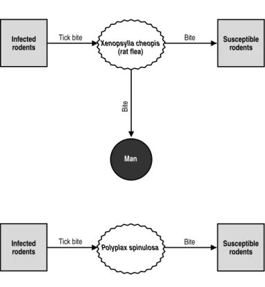
Source of Infection and Mode of Transmission (Figure 1):The most important reservoir of R. typhi is the rat, and the main vector is its flea, X. cheopis. Fleas become infected by feeding on the host when it has rickettsemia. The agent multi-plies in the flea’s intestine and Malpighian tubules without causing any apparent damage. The vector eliminates R. typhi in its feces throughout its lifetime, but not in its saliva. X. cheopis does not transmit the infection to its progeny, and the infection of new generations of fleas requires that they feed on a rickettsemic host. In other species of fleas, the infection follows the same pattern.
The infection is transmitted from rat to rat by means of the flea X. cheopis and the louse Polyplax spinulosa. The agent can survive for a long time in flea feces and in contaminated rodent burrows. The infection can be produced by contact with the mucous membranes of the conjunctiva and the mouth, or by inhalation.
Man becomes infected when the rat flea, or another flea, such as C. felis,bites him and defecates on his skin. When he scratches himself, he can introduce the contam-inated fecal matter through the bite or some other skin abrasion. He exposes himself to the same process if he swats a flea against his skin. Man can probably acquire the infection by other routes as well, such as the conjunctiva or by inhalation, though these modes of transmission are of little importance.
Role of Animals in the Epidemiology of the Disease:This is an infection in rats that is accidentally transmitted to man by fleas. Cats and opossums can also carry the infected flea C. felis into the human environment. The infection is not transmit-ted from one person to another.
Diagnosis:The agent can be isolated by inoculating the blood of a febrile patient into male guinea pigs and embryonated eggs. In guinea pigs, the infection produces the Neil-Mooser reaction (adhesion of the tunica vaginalis testis that prevents rein-troduction of the testicles into the abdomen). This reaction occurs both with the agent of murine typhus and also with those of the spotted fever group.
The complement fixation and indirect immunofluorescence tests are both very useful, though the latter is employed more often. The disadvantage of the comple-ment fixation test is the appearance of anticomplecomple-mentary sera. Also, the immuno-fluorescence test has the advantage that it can be adapted to distinguish IgM and IgG antibodies (Wisseman, 1982). The antibodies appear at the end of the second week of the disease, reach their peak two weeks later, and then gradually decline (Elisberg and Bozeman, 1979). Group specificity is good, although with human patients it is difficult to distinguish murine typhus from epidemic typhus, which is not the case in rodents. This distinction can be made with the complement fixation test if washed species-specific antigens are used.
FLEA-BORNE TYPHUS 11
Xenopsylla cheopis
(rat flea)
Tick bite Bite
Man
Bite
Infected rodents
Figure 1. Flea-borne typhus (Rickettsia typhi). Transmission cycle.
Susceptible rodents
Polyplax spinulosa Bite
Infected rodents
Susceptible rodents
Control:Control measures should be directed first against the vector and then against rodents. To decrease the number of fleas on rats, residual action insecticides are applied to rat runs, nests, and holes. Once the fleas have been dealt with, the next step is to control the rat population through the application of raticides. In addition, environmental sanitation measures can be taken, such as the elimination of rat holes and possible sources of food, as well as the construction of rat-proof buildings.
Bibliography
Adams, W.H., R.W. Emmons, J.E. Brooks. The changing ecology of murine (endemic) typhus in Southern California. Am J Trop Med Hyg19:311–318, 1970.
Al-Awadi, A.R., N. Al-Kazemi, G. Ezzat, A.J. Saah, C. Shepard, T. Zaghloul,et al.Murine typhus in Kuwait in 1978. Bull World Health Organ60:283–289, 1982.
Benenson, A.S., ed. Control of Communicable Diseases in Man, 15th ed. An official report of the American Public Health Association. Washington, D.C.: American Public Health Association; 1990.
Brown, A.E., S.R. Meek, N. Maneechai, G.E. Lewis. Murine typhus among Khmers living at an evacuation site on the Thai-Kampuchean border. Am J Trop Med Hyg38:168–171, 1988. Burgdorfer, W. Murine (flea-borne) typhus fever. In: Hubbert, W.T., W.F. McCulloch, P.R. Schnurrenberger, eds. Diseases Transmitted from Animals to Man, 6th ed. Springfield: Thomas; 1975.
Chin, J., ed. Control of Communicable Diseases Manual, 17th ed. An official report of the American Public Health Association. Washington, D.C.: American Public Health Association; 2000.
Elisberg, B.L., F.M. Bozeman. The rickettsiae. In: Lennette, E.H., N.J. Schmidt, eds. Diagnostic Procedures for Viral, Rickettsial and Chlamydial Infections. Washington, D.C.: American Public Health Association; 1979.
Graves, S.R., J. Banks, B. Dwyer, G.K. King. A case of murine typhus in Queensland. Med J Aust156:650–651, 1992.
Myers, W.F., C.L. Wisseman, Jr. Genetic relatedness among the typhus group of rickettsiae. Int J Syst Bacteriol30:143–150, 1980.
Pan American Health Organization (PAHO). Reported Cases of Notifiable Diseases in the Americas, 1967. Washington, D.C.: PAHO; 1970. (Scientific Publication 199).
Snyder, J.C. The typhus group. In: Beeson, P.B., W. McDermott, J.B. Wyngaarden, eds. Cecil Textbook of Medicine. 15th ed. Philadelphia: Saunders; 1979.
Taylor, J.P., T.G. Betz, J.A. Rawlings. Epidemiology of murine typhus in Texas. 1980 through 1984. JAMA255:2173–2176, 1986.
Tselentis, Y., T.L. Babalis, D. Chrysanths,et al.Clinicoepidemiological study of murine typhus on the Greek island of Evia. Eur J Epidemiol8:268–272, 1992.
Weiss, E., J.W. Moulder. Rickettsiales Gieszczkiewicz 1939. In: Krieg, N.R., J.G. Holt, eds. Vol. 1:Bergey’s Manual of Systematic Bacteriology. Baltimore: Williams & Wilkins; 1984.
Wisseman, Jr., C.L. Rickettsial disease. In: Wyngaarden, J.B., L.H. Smith, Jr., eds. Cecil Textbook of Medicine, 16th ed. Philadelphia: Saunders; 1982.
INFECTIONS CAUSED BY
BARTONELLA HENSELAE
Etiology:Bartonella henselae is a recently described species belonging to the family Rickettsiaceae. The genera Rickettsia and Bartonellaare genetically related, as demonstrated by the fact that there is 25% to 33% DNA hybridization between B. henselaeand Rickettsia prowazekii. The type species of the genus Bartonellais B. quintana, the agent of trench fever, which affected approximately 1 million soldiers in World War I and reemerged to a more limited extent in World War II. Some spo-radic cases still occur. It is believed that the primary reservoir of B. quintanawas a vole, probably Arvicola terrestris,which became independent of the zoonotic cycle and began to circulate between man and the louse Pediculus humanus(Weiss and Moulder, 1984).
The genus Bartonellacurrently includes four species:B. quintana,B. vinsonii,B.
elizabethae, and B. henselae(Groves and Harrington, 1994). The species of greatest interest as an emerging agent of new zoonotic diseases is B. henselae. This rickettsia is bacilliform, curved slightly inward, and measures 1 to 2 microns long by 0.5 to 0.6 microns in diameter. It is gram-negative and stains well with Gimenez stain. The genus Bartonella differs from the genus Rickettsia most notably by the fact that it does not need eukaryotic cells in order to develop. It can be grown in noncellular culture media such as tryptose soy agar or brain-heart infusion agar containing 5% sheep’s blood incubated at 35°C in a humidified stove in an atmosphere of 5% car-bon dioxide. The first culture develops slowly and may take as long as five weeks (Welch et al., 1992; Regnery et al., 1992a).
The reservoir of B. henselaeis the domestic cat, and the diseases that it causes are bacillary angiomatosis, bacillary parenchymatous peliosis, cat-scratch disease (CSD), and recurrent rickettsemia.
Geographic Distribution:Unknown, except for CSD, which appears to occur worldwide (Benenson, 1990).
Occurrence in Man and Cats:It is estimated that in the US, some 22,000 cases of CSD are diagnosed each year, and that more than 2,000 patients are hospitalized (Jackson et al., 1993). The etiologic agent of CSD is not yet known for certain, but there is definite evidence that B. henselaeplays an important role. It is still difficult to deter-mine the relative role played by B. henselaeand Afipia felisin the etiology of the dis-ease (see “Cat-scratch Disdis-ease” in Volume I: Bacterioses and Mycoses). However, research points to B. henselaeas the causative agent. In one serologic study, 88% of 41 patients were positive for B. henselaein the indirect immunofluorescence test, whereas only 25% of the same group reacted positively to Afipia felis(Regnery et al., 1992b).
The number of cases of bacillary angiomatosis is unknown. Bacillary peliosis has been observed in isolation and in association with angiomatosis. As of 1982, there were approximately 100 cases of this condition on record (García et al., 1982).
Researchers at the University of California at San Francisco (USA) conducted an epidemiologic study of four patients with bacillary angiomatosis in an effort to dis-cover the source of infection. The four patients had been in contact with seven cats, and B. henselaewas isolated from both the cats’ blood and their fleas. Blood samples were taken from 61 cats in the San Francisco metropolitan area, living both in homes and at an animal shelter, and B. henselaewas isolated from 41% of the samples.
The Disease in Man:B. henselaeinfection produces a broad range of clinical and pathological varieties: CSD (see “Cat-scratch Disease” in Volume I: Bacterioses and Mycoses), recurrent rickettsemia, bacillary angiomatosis, and bacillary peliosis.
Bacillary angiomatosis is a vasoproliferative reaction observed in histological sections taken from lesions of the skin, bones, lymph nodes, and brain. The presence of a large number of bacillary forms in the lesions can be detected with Warthin-Starry argentic stain or an electron microscope. Although the disease is seen most often in immunodeficient patients, especially those infected with the human immun-odeficiency virus (HIV), it also occurs in immunocompetent patients. The most common skin lesions are painful, angiomatous papules, which can be mistaken for Kaposi’s sarcoma, but which histologically resemble epitheloid hemangiomas. In the disseminated form of bacillary angiomatosis, patients experience fever, weight loss, discomfort, and increased volume of the affected organs (Koehler et al., 1992; Groves and Harrington, 1994). The etiology of bacillary angiomatosis is apparently shared between B. henselaeand B. quintana. Koehler et al.(1992) isolated B. quin-tanafrom three patients with cutaneous and osseous lesions of bacillary angiomato-sis. A DNA:DNA hybridization assay with the type species demonstrated 99% to 100% relatedness (strains with over 70% relatedness are considered to belong to the same species) (Koehler and Brenner, 1993).
Bacillary peliosis is a pathological entity specific to the solid internal organs (liver, spleen, abdominal lymph nodes, and bone marrow), which is expressed in the form of small blood-filled cysts. In some cases it can also affect the kidneys, pan-creas, and lungs. Most cases are seen in individuals who are weak and chronically ill, such as HIV-infected tuberculosis patients, those with cancer, and those on sys-temic anabolic steroids. The clinical symptoms are fever, weight loss, nausea, diar-rhea, abdominal pain, and lymphadenopathy.
In a group of 48 patients with bacillary angiomatosis or peliosis studied by Tappero et al.(1993), 42 were HIV-positive.
Another clinical form is recurrent rickettsemia, which is rare. In immunocompe-tent individuals, the rickettsemia is recognized clinically by its sudden onset, fever, muscle and joint pains, and sometimes, headache, meningism, and photophobia (Lucey et al., 1992). In immunodeficient patients, the disease develops slowly, with manifestations of fatigue, asthenia, discomfort, and weight loss. In AIDS patients,B. henselaecan cause inflammatory disease without angiomatosis or peliosis, which can be demonstrated using immunocytochemical techniques on autopsy specimens of infected tissue (Slater et al., 1994). The authors describe three cases of AIDS patients without neoangiogenic lesions on their organs but whose pathological changes were caused by B. henselae, as was demonstrated by immunocytochemistry.
The recommended treatment for bacillary angiomatosis, bacillary peliosis, and recurrent rickettsemia is the administration of erythromycin, rifampicin, or doxycy-cline for six weeks. In bacillary angiomatosis, if the lesions are limited to the skin, surgical excision alone is sufficient. With recurrent rickettsemia, the recommended treatment is intravenous gentamicin and ceftriaxone, followed by oral ciprofloxacin (Groves and Harrington, 1994). For the treatment of CSD, see “Cat-scratch Disease” in Volume I: Bacterioses and Mycoses.
Source of Infection and Mode of Transmission:The reservoir is the domestic cat. In a study carried out in California (USA),B. henselaewas isolated from blood in 25 (41%) of 61 cats from family homes and animal shelters (Koehler et al., 1994). It was also demonstrated that the rickettsemia is prolonged: the agent was isolated from a naturally infected cat for 18 weeks after the infection was first detected sero-logically (Regnery et al., 1992b). These data indicate that immunocompetent indi-viduals are not very susceptible to B. henselaeinfection and that other factors lower their resistance and contribute to the development of bacillary angiomatosis or peliosis. On the other hand, the agent was not observed to be opportunistic in CSD. In CSD, the causal link to the scratch or bite of a cat, especially one under 12 months old, is a salient fact in the epidemiology of this disease. With other human diseases caused by B. henselae, except for recurrent rickettsemia, it is clear that they are contracted directly from the scratch or bite of a young cat, or via their fleas (Groves and Harrington, 1994). Little is known about cat-to-cat transmission, but it is assumed to be through fleas, bites and scratches during play among young cats, or fights between tomcats.
Diagnosis:The most certain method of diagnosis is isolation of the agent in cul-ture media (see the section on etiology), but this technique takes too long; serolog-ical methods are more practserolog-ical. An indirect immunofluorescence test (Regnery et al., 1992b) has been developed for diagnosis of CSD. The test showed that CSD patients had high titers to B. henselaeantigens. Of 41 CSD patients, 88% tested pos-itive, whereas in a group of 107 controls only 3% were positive. A diagnosis can also be obtained using immunocytochemistry on pathological specimens (Slater et al., 1994).
Control:The epidemiology of diseases caused by B. henselaeis just beginning to be understood and there are still many areas to explore before any rational founda-tion can be established for their prevenfounda-tion and control. Transmission to man could be reduced by controlling cat fleas and perhaps by treating infected cats with antibi-otics. Any wound inflicted by a cat should be promptly washed with soap and water and disinfected.
Bibliography
Benenson, A.S., ed. Control of Communicable Diseases in Man, 15th ed. An official report of the American Public Health Association. Washington, D.C.: American Public Health Association; 1990.
García, R.L., M.K. Khan, R.B. Berlin. Peliosis of the spleen with rupture. Hum Pathol 13:177–179, 1982.
Groves, M.G., K.S. Harrington. Rochalimaea henselae infections: Newly recognized zoonoses transmitted by domestic cats. J Am Vet Med Assoc204:267–271, 1994.
Jackson, L.A., B.A. Perkins, J.D. Wenger. Cat scratch disease in the United States: An analysis of three national databases. Am J Public Health83:1707–1711, 1993.
Koehler, J.E., D.J. Brenner. Isolation of Rochalimaeaspecies. [Author reply]. N Engl J Med 328:1422–1423, 1993.
Koehler, J.E., C.A. Glaser, J.W. Tappero. Rochalimaea henselaeinfection. A new zoonosis with the domestic cat as reservoir. JAMA271:531–535, 1994.
Koehler, J.E., F.D. Quinn, T.G. Berger,et al.Isolation of Rochalimaeaspecies from cuta-neous and osseous lesions of bacillary angiomatosis. N Engl J Med327:1625–1631, 1992.
Leong, S.S., R.A. Cazen, G.S. Yu,et al.Abdominal visceral peliosis associated with bacil-lary angiomatosis. Ultrastructural evidence of endothelial destruction by bacilli. Arch Pathol Lab Med116:866–871, 1992.
Lucey, D., M.J. Dolan, C.W. Moss,et al.Relapsing illness due to Rochalimaea henselaein immunocompetent hosts: Implication for therapy and new epidemiological associations. Clin Infect Dis14:683–688, 1992.
Regnery, R.L., B.E. Anderson, J.E. Clarridge III, et al. Characterization of a novel Rochalimaeaspecies,R. henselaesp. nov. isolated from blood of a febrile, human immuno-deficiency virus-positive patient. J Clin Microbiol30:265–274, 1992a.
Regnery, R.L., J.G. Olson, B.A. Perkins, W. Bibb. Serological response to Rochalimaea henselaeantigen in suspected cat-scratch disease. Lancet339:1443–1445, 1992b.
Slater, L.N., J.V. Pitha, L. Herrera, et al. Rochalimaea henselaeinfection in acquired immunodeficiency syndrome causing inflammatory disease without angiomatosis or peliosis. Demonstration by immunocytochemistry and corroboration by DNA amplification. Arch Pathol Lab Med118:33–38, 1994.
Slater, L.N., D.F. Welch, D. Hensel, D.W. Coody. A newly recognized fastidious gram-neg-ative pathogen as a cause of fever and bacteremia. N Engl J Med323:1587–1593, 1990.
Tappero, J.W., J. Mohle-Boetani, J.E. Koehler, et al. The epidemiology of bacillary angiomatosis and bacillary peliosis. JAMA269:770–775, 1993.
Weiss, E., J.W. Moulder. The Rickettsias and Chlamydias. In: Krieg, N.R., J.G. Holt, eds. Vol. 1:Bergey’s Manual of Systematic Bacteriology. Baltimore: Williams & Wilkins; 1984.
Welch, D.F., D.A. Pickett, L.N. Slater,et al. Rochalimaea henselaesp. nov., a cause of sep-ticemia, bacillary angiomatosis, and parenchymal bacillary peliosis. J Clin Microbiol 30:275–280, 1992.
Zangwill, K.M., D.H. Hamilton, B.A. Perkins,et al.Cat scratch disease in Connecticut. Epidemiology, risk factors, and evaluation of a new diagnostic test. N Engl J Med329:8–13, 1993.
Q FEVER
ICD-10 A78
Synonyms: Pneumorickettsiosis, Balkan influenza, coxiellosis, abattoir fever, Australian Q fever, hiberno-vernal bronchopneumonia, nine-mile fever, quadrilat-eral fever, infection due to Coxiella burnetii.
C. burnetii is bacilliform and measures 0.4–1 by 0.2–0.4 microns. In order to develop, it requires the presence of eukaryotic cells, where it tends to take up resi-dence in the phagolysosomes, rather than the cytoplasm or the nucleus as the
Rickettsia species do. It shows up well with Gimenez stain (Weiss and Moulder, 1984). It has been found to have several different plasmids, the functions of which are not yet understood.
C. burnetiican be highly pleomorphic when it reproduces inside the phagolyso-somes of an invaded host cell. Two different forms can be distinguished under an electron microscope: one, large and bacilliform, and the other, coccoid, which devel-ops from the former and has greater electronic density (McCaul and Williams, 1981). A third form appears in the large cells after passage through embryonated eggs or BGM cell cultures when they have been kept in suboptimal temperature con-ditions or fresh medium has not been added. These small, high-density forms are similar to spores (Aitken et al., 1987). The morphogenesis is comparable, but not identical, to cell differentiation in the formation of endospores. These small forms are responsible for the high resistance of the Q fever agent to environmental factors and many disinfectants.
C. burnetiihas two antigenic phases (I and II), much like the S-to-R variation in salmonellae or brucellae. When harbored in the animal or tick organism, it is in phase I. After several passages through the yolk sac of embryonated eggs, it converts to phase II, which is avirulent. This antigenic variation is important for diagnosis and prophylaxis.
Geographic Distribution: Worldwide. The infection is endemic in many areas and its presence has been confirmed in at least 51 countries. Although the Nordic countries were previously believed to be free of Q fever and that the few cases seen there were imported, in the 1990s, the disease was recognized as endemic in Sweden.
Occurrence in Man:Q fever appears in the form of sporadic cases or outbreaks. The human infection is often asymptomatic, and its mild form can be mistaken for other febrile diseases. For this reason, sporadic cases often go undiagnosed and the true incidence of the disease is unknown. Moreover, the indiscriminate use of antibi-otics in febrile patients hampers the clinical identification of Q fever as well as other rickettsioses and bacterioses. In Australia, which is considered an endemic area, there were some 2,000 cases in 1979–1980 (Hunt et al., 1983), and the United Kingdom has at least 100 laboratory-confirmed cases each year (Heard et al., 1985). Several epidemic outbreaks have occurred in abattoirs and wool-processing plants. In Uruguay, 310 of 630 workers and veterinary inspection personnel in a meat-packing plant fell ill in a single month in a 1976 epidemic. Cases were most concentrated among workers involved in bone-milling and the collection of animal wastes, such as placentas, fetuses, and viscera. The outbreak was attributed to aerosols, probably generated by the handling of placentas and amniotic fluid. Three more outbreaks occurred, apparently in that same meat-packing plant, in August and October 1981 and in 1984, with 25, 17, and 46 cases, respectively. Most of the affected personnel worked in slaughtering and deboning (Ortiz Molina et al., 1987). According to the same authors, there have been 15 more outbreaks in abattoirs since 1976, mostly involving cattle. Epidemics have occurred among slaughterhouse workers in other parts of the world as well. In Quebec (Canada), an outbreak in the 1950s affected 62 employees (36.5% of the company’s total workforce) within a