DEPARTAMENTO DE BIOLOGIA VEGETAL
Regulation of alternative splicing
of the small GTPase Rac1
Vânia Marina Cristóvão Gonçalves
Doutoramento em Biologia
(Biologia Molecular)
2009
Tese orientada por:
Doutor Peter Jordan (Instituto Nacional de Saúde Dr. Ricardo Jorge)
Professor Doutor Rui Artur Paiva Loureiro Gomes (Faculdade de Ciências da Universidade de Lisboa)
As opiniões expressas nesta publicação são da exclusiva responsabilidade do seu autor
Every experiment proves something. If it doesn't prove what you wanted it to prove, it proves something else.
Prefácio
Esta tese de Doutoramento é o resultado do trabalho de investigação realizado no Laboratório de Oncobiologia do Departamento de Genética do Instituto Nacional de Saúde Dr. Ricardo Jorge, em Lisboa, entre Janeiro de 2005 e Janeiro de 2009, sob a orientação do Doutor Peter Jordan. Este trabalho foi ainda co-orientado pelo Professor Doutor Rui Artur Paiva Loureiro Gomes, membro da Faculdade de Ciências da Universidade de Lisboa onde esta tese será submetida.
O trabalho aqui apresentado teve como objectivo fundamental a análise molecular da regulação do splicing alternativo da pequena GTPase Rac1, cuja variante de splicing designada Rac1b se encontra sobre-expressa em certos tumores colo-rectais podendo por isso ser um bom alvo terapêutico e de diagnóstico.
De acordo com o disposto no Artigo 41º do Regulamento dos Estudos Pós-Graduados da Universidade de Lisboa, Deliberação nº 1506/2006, publicada no Diário da República, 2ª série — Nº 209 — 30 de Outubro de 2006, a presente dissertação encontra-se redigida em língua inglesa, contendo um resumo alargado (mais de 1200 palavras) em língua portuguesa (Resumo).
Ainda de acordo com o disposto no mesmo artigo, foram utilizados nesta dissertação resultados de trabalhos de colaboração, estando a minha contribuição pessoal devidamente indicada, tendo estes permitido a elaboração de dois artigos, um dos quais já publicado e o outro já submetido para publicação:
Gonçalves V, Matos P and Jordan P. 2008. The beta-catenin/TCF4 pathway modifies alternative splicing through modulation of SRp20 expression. RNA. 14:
Gonçalves V, Matos P and Jordan P. 2009. Antagonistic SR proteins regulate alternative splicing of tumour-related Rac1b downstream of the PI3-kinase and Wnt pathways. (submetido).
Ainda de referir que no âmbito do trabalho presente nesta dissertação foi ainda publicado um capítulo de um livro e um artigo:
Jordan P, Gonçalves V and Matos P. 2006. Alternative splicing changes regulation
and signalling properties of the small GTPase Rac1. In: Alternative Splicing in
Cancer, Editor: Julian P. Venables. Chapter 3. Transworld Research Network, India
Gonçalves V, Theisen P, Antunes O, Medeira A, Ramos JS, Jordan P, Isidro G.
2009. A missense mutation in the APC tumor suppressor gene disrupts an ASF/SF2
splicing enhancer motif and causes pathogenic skipping of exon 14. Mutat Res. 662:
33-36
Gostaria ainda de salientar que a realização deste trabalho só foi possível graças ao apoio financeiro da Fundação para a Ciência e a Tecnologia (FCT) e do
Fundo Social Europeu (FSE), através do projecto POCTI/47546/02, do Programa
de Financiamento Plurianual do CIGMH, e sob a forma de uma Bolsa de Doutoramento com a referência BD/18262/2004.
Vânia Marina Cristóvão Gonçalves Lisboa, 19 de Junho de 2009
Resumo
A expressão de genes eucarióticos é um processo com várias etapas que incluem a transcrição do gene, o splicing e a poliadenilação do transcrito, e ainda o transporte do RNA mensageiro para o citoplasma (Singer e Green 1997).
Na etapa inicial da formação do spliceossoma são reconhecidas sequências consenso nos locais de splicing 5! e 3!, por componentes da maquinaria de splicing. Foram identificadas também sequências adicionais que promovem (splice enhancer) ou reprimem (splice silencer) o reconhecimento e inclusão de determinado exão, podendo estas localizar-se nesse mesmo exão ou nos intrões adjacentes (Cartegni et al. 2002). Estes elementos são específicos de cada gene e reconhecidos por proteínas adicionais de ligação ao pré-RNA mensageiro, como por exemplo membros da família de proteínas SR ou hnRNP, que promovem ou impedem as interacções proteína-proteína necessárias à reacção de splicing. A associação destas interacções numa dada célula define se, ou em que extensão, um exão é reconhecido e processado pela maquinaria de splicing (Black 2003). Neste sentido,
splicing alternativo significa regulação da escolha dos locais de splicing.
Um gene humano alvo do splicing alternativo é o RAC1. A proteína Rac1 é um membro da família das pequenas GTPases Rho que estimula as vias de sinalização envolvidas no controlo da dinâmica dos filamentos de actina e da activação transcricional. Rac1 alterna entre um estado inactivo ligado a GDP e um activo ligado a GTP sendo esta transição controlada na célula por três classes de proteínas: GEFs, GAPs e GDIs. Uma variante de splicing alternativo, designada Rac1b foi identificada em tumores colo-rectais e da mama e contém 19 aminoácidos adicionais devido à inclusão do exão 3b, o qual é geralmente excluído. A presença destes aminoácidos adicionais altera profundamente a regulação e as propriedades de sinalização da GTPase. Apesar da proteína Rac1b ser expressa nas células em níveis reduzidos, está maioritariamente no seu estado activo e portanto ligado ao GTP. De facto, Rac1b é incapaz de interagir com Rho-GDIs, ficando assim comprometido um mecanismo importante de regulação negativa. Adicionalmente, diversas vias de sinalização clássicas de Rac1, tais como a formação de
lamellipodia ou a activação das cinases PAK ou JNK, não são estimuladas pelo
Rac1b activo. No entanto, Rac1b estimula a activação do NF!B mediada por espécies reactivas de oxigénio e promove mecanismos de transformação celular tais como a progressão do ciclo celular, a sobrevivência celular e a transição epitelial-mesênquimal. Estes dados demonstram como o splicing alternativo pode afectar profundamente a função da proteína. Estudos recentes sugerem que as alterações no nível de expressão de Rac1b contribuem para a tumorigénese (Singh et al. 2004, Radisky et al. 2005, Matos e Jordan 2006, Esufali et al. 2007, Matos e Jordan 2008). Por exemplo, Rac1b e B-RafV600E (uma mutação activadora do B-Raf presente em alguns tumores do cólon) cooperam funcionalmente para manter a sobrevivência de células de tumores colo-rectais (Matos et al. 2008). Assim, a expressão de Rac1b promove a sobrevivência de células tumorais do cólon e por isso, compreender o mecanismo molecular subjacente a este evento de splicing alternativo é de interesse terapêutico. O trabalho apresentado nesta tese descreve estudos com vista a elucidar os mecanismos que regulam o splicing alternativo de Rac1.
Dados experimentais de linhas celulares e de tecidos normais revelaram que o exão 3b geralmente se encontra excluído, sendo apenas incluído numa pequena fracção do transcrito total (Jordan et al. 1999, Radisky et al. 2005). Este exão tem características típicas de exões alternativos tais como o seu pequeno tamanho, de 57 nucleótidos, e um polypyrimidine tract pouco conservado. Com estas limitações impostas pela sequência genómica quanto à inclusão do exão 3b de RAC1, as modificações no splicing alternativo observadas em tumores colo-rectais podem ser o resultado de mutações pontuais na sequência consenso dos elementos conservados de splicing. Muitas mutações patogénicas que causam doenças hereditárias interferem com o splicing normal do pré-RNA mensageiro (Faustino e Cooper 2003). As mutações genómicas subjacentes podem afectar os motivos conservados de sequências consenso necessárias à formação do complexo spliceossomal funcional, ou criar/destruir elementos enhancer ou silencer (Cartegni et al. 2002, Pagani et al. 2004). Assim, amplificámos a sequência genómica de RAC1 de três linhas celulares colo-rectais com diferentes níveis de expressão de Rac1b e
a montante e 224 nucleótidos a jusante do mesmo. Nenhuma das três linhas celulares apresentava alguma mutação neste fragmento do gene que pudesse explicar as diferenças observadas no splicing.
A ausência de mutações genómicas nas linhas celulares analisadas sugere que as diferenças na expressão de Rac1b são baseadas num evento de splicing alternativo regulado, como já descrito em numerosos exemplos de alterações no
splicing alternativo durante o desenvolvimento embrionário, entre diferentes tecidos
e também em determinados tipos de tumores.
Neste trabalho demonstramos que o splicing alternativo de Rac1 é regulado pelas proteínas SR ASF/SF2 e SRp20 em células colo-rectais. Para analisar os mecanismos que regulam o splicing alternativo de Rac1b, construímos um minigene RAC1 que reproduz as decisões de splicing endógenas em linhas celulares colo-rectais. Assim, testámos vários factores de splicing candidatos quanto ao seu efeito no minigene e identificámos um factor que promove a expressão de Rac1b, o ASF/SF2, e outro que a suprime, o SRp20. Os seus papéis fisiológicos no splicing alternativo de Rac1 em células de tumores colo-rectais foram confirmados ao nível endógeno por experiências de interferência de RNA. Adicionalmente, estes factores ligam-se a sondas transcritas in vitro a partir do exão 3b, em experiências de alterações na mobilidade electroforética (EMSA). Em conjunto, os nossos dados levam-nos a propor que o exão 3b contém um enhancer exónico reconhecido pelo ASF/SF2 junto a um silencer exónico reconhecido pelo SRp20. Alguns programas bioinformáticos prevêem a existência de tais elementos reguladores que servem como locais de ligação para aqueles factores de splicing, corroborando os resultados obtidos.
No nosso sistema experimental de linhas celulares colo-rectais, observámos que as concentrações relativas da proteína ASF/SF2 versus SRp20 determinavam a proporção de splicing alternativo. Não só os níveis endógenos da proteína de ambos os factores se correlacionavam com a quantidade de Rac1b expresso em diferentes linhas celulares, como também a manipulação experimental dos níveis de expressão de ASF/SF2 e SRp20 através de sobre-expressão e/ou RNAi, modificou o
splicing alternativo do exão 3b. Vários estudos documentam que a abundância
relativa de factores de splicing antagonistas, incluindo o ASF/SF2 e o SRp20, pode afectar decisões de splicing (por exemplo, Jumaa e Nielsen 1997, Mayeda et al. 1993, Galiana-Arnoux et al. 2003). É portanto reconhecido que os níveis de expressão dos factores de splicing são um mecanismo central para despoletar diferenças nos padrões de splicing alternativo.
Relativamente aos eventos a montante que regulem as concentrações relativas das proteínas ASF/SF2 versus SRp20, identificámos duas vias de sinalização que afectam a sua expressão promovendo um aumento ou uma diminuição correspondente em Rac1b. A via da Wnt activa o complexo transcricional "-catenina/TCF4, sendo o gene SFSR3, que codifica o SRp20, um alvo directo
(Gonçalves et al. 2008). A estimulação da via aumentou os níveis de SRp20 que descobrimos actuar como um silencer no exão 3b, conduzindo à expressão reduzida de Rac1b. Por outro lado, encontrámos a via da PI3-cinase envolvida na regulação do factor antagonístico ASF/SF2. A inibição desta via aumentou a expressão dos níveis do transcrito e da proteína ASF/SF2, que age como um enhancer da inclusão do exão 3b, levando a uma expressão aumentada de Rac1b. Em conjunto, estes dados mostram que diferentes vias de sinalização influenciam simultaneamente factores de splicing independentes a fim de controlar o splicing alternativo de Rac1b. É possível que a fosforilação das proteínas ASF/SF2 ou SRp20 esteja também envolvida na regulação do splicing alternativo de Rac1b. Sabe-se que as funções nucleares e citoplasmáticas do factor ASF/SF2 são moduladas por fosforilação (Xiao e Manley 1997, Sanford et al. 2005) e recentemente, a proteína AKT foi descrita como capaz de fosforilar in vitro o ASF/SF2 (Blaustein et al. 2005). Neste sentido, observámos que a sobre-expressão de PTEN promove um aumento na expressão de ASF/SF2 enquanto a de um mutante cinase-inactivo de AKT não, apesar de ambas as circunstâncias induzirem um aumento em Rac1b, o que sugere a existência de mecanismos de regulação por fosforilação. Adicionalmente, a inibição da PI3-cinase foi já implicada na regulação de outros eventos de splicing alternativo, como nos genes da fibronectina e do PKC "II (Blaustein et al. 2004,
Pelisch et al. 2005, Patel et al. 2001), embora os detalhes mecanísticos estejam ainda por caracterizar. Serão necessárias experiências adicionais para esclarecer exactamente de que modo a via da PI3-cinase e AKT afectam os níveis e/ou a fosforilação do ASF/SF2. É possível que a via controle outros reguladores de proteínas SR, como a família de proteínas cinase SRPK e Clk/Sty que funcionam no citosol (Gui et al. 1994) podendo contrariar o efeito da AKT nas proteínas SR
(Blaustein et al. 2005).
A nossa observação que a sinalização da PI3-cinase e da "-catenina inibem a expressão de Rac1b ajuda esclarecer as vias genéticas que levam à tumorigénese colo-rectal. Um grupo de tumores que apresenta mutações oncogénicas no gene KRAS aumenta a proliferação celular através da sinalização de ERK e fornece um estímulo de sobrevivência através da via PI3-cinase/Rac1 (Qiu et al. 1995). Este grupo é representado pela linha celular SW480, que não expressa Rac1b endógeno, sendo isto compatível com a presença de K-Ras mutante que activa fortemente a PI3-cinase, conduzindo assim à diminuição da expressão de ASF/SF2. Por outro lado, acumulam-se quantidades consideráveis de "-catenina no núcleo das células SW480, causando o aumento da expressão de SRp20 que contribui também para a inexistência de Rac1b nesta linha celular.
Em contraste, as células HT29 acumulam pequenas quantidades de "-catenina nuclear, conduzindo assim à diminuição da expressão de SRp20 e a um aumento de Rac1b. Além disso, nesta linha celular o gene KRAS é selvagem mas existe, em alternativa, uma mutação oncogénica do gene BRAF (Matos et al. 2008). A proteína B-Raf funciona a jusante de K-Ras na via da ERK e assim não pode estimular a PI3-cinase directamente, o que está de acordo com o facto das células HT29 expressarem mais ASF/SF2 e mais Rac1b. Estas células representam um outro grupo de tumores colo-rectais em que a mutação oncogénica B-RafV600E ocorre associada à sobre-expressão de Rac1b, e onde B-RafV600E estimula a proliferação celular, enquanto a sinalização de Rac1b mantém a sobrevivência celular (Matos et al. 2008). No entanto, os detalhes moleculares que conduzem à sobre-expressão de Rac1b permaneceram imprecisos. Os dados aqui apresentados
permitem agora propor um mecanismo molecular para a sobre-expressão de Rac1b: lesões genéticas que iniciam a tumorigénese colo-rectal sem activar a sinalização da PI3-cinase e da "-catenina, promovem a selecção de contextos celulares com níveis mais elevados de ASF/SF2 relativamente a SRp20. Este cenário celular favorecerá a sobre-expressão de Rac1b que conduz ao aumento da sobrevivência celular e à subsequente progressão tumoral.
Outra grande contribuição deste trabalho é a demonstração que diferentes vias de sinalização celular actuam em conjunto para regular um evento de splicing alternativo específico. No caso estudado, sincronizando a expressão de duas proteínas SR com papéis antagónicos, ASF/SF2 e SRp20, as vias da PI3-cinase e da Wnt regulam a inclusão ou exclusão do exão alternativo 3b do pré-RNA mensageiro de Rac1.
Diversas evidências suportam a nossa conclusão que a via de transdução de sinal da "-catenina/TCF4 estimula directamente a transcrição génica do factor de
splicing SRp20. Demonstrámos que os níveis endógenos do transcrito e da proteína
SRp20 se correlacionam com a actividade transcricional da "-catenina em diferentes linhas celulares colo-rectais. Além disso, a estimulação ou inibição experimental da sinalização da "-catenina/TCF4 afectam os níveis de SRp20, e o promotor do gene
SFSR3 (que codifica para o factor SRp20) responde a essa mesma estimulação e
inibição. Mostrámos ainda que o aumento dos níveis da proteína SRp20, estimulado pela "-catenina, é suficiente para promover decisões de splicing alternativo em células colo-rectais, quer num minigene repórter quer no RAC1 e ainda num outro gene endógeno, o CD44.
Em geral, estes resultados demonstram que a via da "-catenina/TCF estimula não só a transcrição génica, mas também a formação de um subconjunto de transcritos variantes por splicing alternativo. Assim, são afectados dois níveis do programa de expressão génica celular pela sinalização da "-catenina/TCF: a activação transcricional do factor de splicing SRp20 codificado pelo gene SFSR3, e como consequência do aumento dos níveis desta proteína, a produção de variantes de splicing alternativas. Estes dados suportam a noção recente de que a transcrição
e o splicing alternativo representam dois níveis diferentes de regulação da expressão génica e que diversas vias de sinalização agem coordenadamente sobre o conjunto de transcritos resultantes.
Em conclusão, este trabalho contribui para a compreensão dos mecanismos moleculares responsáveis pela expressão alterada de alguns factores de splicing observada em vários tipos de tumores (Stickeler et al. 1999, Venables 2004) e apresenta uma explicação mecanística de como alguns sinais celulares podem regular o splicing alternativo afectando a actividade de um factor de transcrição, o qual controla directamente o nível da transcrição de factores de splicing em resposta a mudanças nas vias de sinalização respectivas. Elucidámos também parte do mecanismo de tumorigénese que ocorre num subtipo de tumores do cólon caracterizando a sinalização envolvida na regulação do evento de splicing alternativo que origina a variante Rac1b. Isto permitiu-nos propor um mecanismo para a sobre-expressão de Rac1b, o qual faz parte de uma via alternativa de transformação maligna, para além das mutações oncogénicas clássicas já descritas em tumores colo-rectais. Adicionalmente, os nossos dados abrem a possibilidade do desenvolvimento de metodologias terapêuticas específicas devido à identificação dos factores de splicing envolvidos e das sequências alvo no exão 3b.
Summary
Tumours develop through the stepwise acquisition of genetic changes including those affecting signalling pathways that control cell proliferation and survival. The small GTPase Rac1 regulates signalling pathways controlling actin filament dynamics and transcriptional activation. An alternative splicing variant Rac1b contains 19 additional amino acids due to inclusion of a usually skipped exon 3b and is overexpressed in a subset of colorectal tumours. Rac1b is required to sustain colorectal tumour cell survival and understanding the molecular mechanism behind this alternative splicing event is of therapeutic interest. Here we describe that antagonistic SR proteins ASF/SF2 and SRp20 regulate Rac1 alternative splicing in colorectal cells. Using a Rac1 minigene we identified that SRp20 increased skipping of alternative exon 3b in HT29 cells, while ASF/SF2 increased its inclusion. Depletion of endogenous expression of these splicing factors by specific siRNAs confirmed that ASF/SF2 enhances, whereas SRp20 silences endogenous Rac1b splicing. Moreover, we found that both splicing factors bound to Rac1 exon 3b sequences and were regulated by upstream signalling pathways: inhibition of PI3-kinase pathway increased ASF/SF2 expression and promoted Rac1b, whereas activation of "-catenin/TCF4 increased SRp20 expression and inhibited Rac1b generation. We further found that "-catenin/TCF4 directly stimulates gene transcription of SRp20 and generates a subset of transcript variants through alternative splicing. This supports the recent notion that transcription and alternative splicing represent two different layers of gene expression and that signalling pathways act upon a coordinated network of transcripts in each layer. A major contribution of this work is the demonstration that different cellular signalling pathways act in concert to regulate a specific alternative splicing event. The results predict that overexpression of Rac1b can occur in tumours without enhanced stimulation of PI3-kinase and Wnt pathways, which synchronize the expression of two antagonistic SR proteins, ASF/SF2 and SRp20, regulating alternative splicing of the Rac1 pre-mRNA.
Palavras-chave
Splicing alternativo Rac1b ASF/SF2 SRp20 "-catenina PI3-cinase Tumorigénese colo-rectalKeywords
Alternative splicing Rac1b ASF/SF2 SRp20 "-catenin PI3-kinase Colorectal tumorigenesisAbbreviations
A adenosine
aa amino acid
AKT / PKB protein kinase B
APC adenomatous polyposis coli AS alternative splicing
ATP adenosine triphosphate
BAC bacterial artificial chromosome
BGHpa bovine growth hormone polyadenylation signal BK calcium-activated potassium channels
bp base pairs
BPS branch point sequence BSA bovine serum albumin
C cytosine
CaMK calcium/calmodulin-dependent kinases cAMP cyclic adenosine monophosphate cDNA mRNA-complementary DNA Clk/Sty Cdc2-like kinase
CMV cytomegalovirus
CRIB Cdc42/Rac interactive binding CTD C-terminal domain
C-terminal carboxyl-terminal CTP cytosine triphosphate
CV consensus value
DAPI 4,6-diamidino-2-phenylindole DMD Duchenne!s muscular dystrophy DMEM Dulbecco's Modified Eagle's Medium DMSO dimethyl sulfoxide
DNA deoxyribonucleic acid DTT dithiothreitol
ECM extracellular matrix
EDTA Ethylenediamine tetraacetic acid EMSA electrophoretic mobility shit assay EMT epithelial to mesenchymal transition ERK extracellular signal-regulated kinase ESE exonic splicing enhancer
ESR exonic splicing regulatory element ESS exonic splicing silencer
EST expressed sequence tag
Flag epitope tag from the gene-10 product of bacteriophage T7
G guanosine
GDI guanine nucleotide dissociation inhibitor GDP guanosine diphosphate
GEF guanine nucleotide exchange factor GFP green fluorescent protein
GSK3 glycogen synthase kinase 3" GTP guanosine triphosphate
HA epitope tag from Hemagglutinin of the influenza virus HIV human immunodeficiency virus
hnRNP heterogeneous nuclear RNP I#B$ inhibitor of #B$
IKK$ I#B$ kinase complex IP immunoprecipitation ISE intronic splicing enhancer ISS intronic splicing silencer JNK c-Jun N-terminal kinase kb kilo base pairs
kDa kilodalton
MAPK mitogen-activated protein kinase MEK MAPK kinase
MMP matrix metalloprotease mRNA messenger RNA MW molecular weight
Myc epitope tag derived from the c-myc gene product NE nuclear extracts
NF#B nuclear factor kappa-light-chain-enhancer of activated B cells
nt nucleotide
ORF open reading frame N-terminal amino-terminal
PAGE polyacrylamide gel electrophoresis PAK p21 activating kinase
PBS phosphate buffered saline PCR polymerase chain reaction PI3K phosphatidylinositol 3 kinase PKC protein kinase C
Pol II RNA Polymerase II PolyA poly-adenilate
PTB polypyrimidine tract binding protein PTEN phosphatase and tensin homolog RNA ribonucleic acid
RNAi ribonucleic acid interference RNAsin ribonuclease inhibitor RNP ribonucleoprotein ROS reactive oxygen species RT-PCR reverse transcription PCR
SCF Skp, Cullin, F-box containing SDS sodium dodecyl sulphate siRNA small interfering RNA SMA spinal muscular atrophy snRNA small nuclear RNA snRNP small nuclear RNP SRPK SR protein kinase
SR protein serine/arginine-rich protein
ss splice site
STREX stress axis-regulated exon
T thimidine
T7 epitope tag from T7 bacteriophage gene10 TBE Tris/Borate/EDTA
TBP TATA binding protein TCF4 transcription factor 4
TCF/LEF T-cell factor/lymphoid enhancer factor TIMP tissue-specific inhibitor of MMP Tris tris(hydroxymethyl)aminomethane TRITC Tetramethyl Rhodamine Iso-Thiocyanate tRNA transfer RNA
U uridine
UTR untranslated region
WB western blot
Wnt wingless
Table of contents
Prefácio v Resumo vii Summary xv Palavras-chave/Keywords xvii Abbreviations xixTable of contents xxiii
Chapter I – General Introduction 27 Pre-mRNA splicing, constitutive and alternative splicing 29
Dynamic assembly of the spliceosome 30
Regulation of splice site recognition 32
Splice site strength 32
Splicing enhancers and silencers 33
Exon/Intron architecture 35
RNA secondary structure 37
Pre-mRNA synthesis by RNA polymerase II 38
Combinatorial control of exon recognition and alternative splicing 39
Types of alternative splicing events 40
Effects of alternative splicing 42
Signal-mediated alternative splicing control 43
Global analysis of alternative splicing 45
Splicing regulatory network as a subnetwork of gene regulation 47
Alternative splicing and cancer 48
Small GTPases (Guanosine TriPhosphatases) and cancer 49
The Rho family of small GTPases 50
Regulators, effectors and functions 51
Association with cancer 53
The small GTPase Rac1 55
Rac1b, the alternative splice variant of Rac1 58 Altered regulation and downstream signalling 59
Role of Rac1b in tumorigenesis 62
Objectives 65
Chapter II – Antagonistic SR proteins regulate alternative splicing of tumour-related Rac1b downstream of the PI3-kinase and Wnt pathways 67
Summary 69
Introduction 69
Experimental procedures 72
Cell culture and transfection 72
DNA constructs 73
Nucleic acid amplifications, semi-quantitative and Real-Time
RT-PCR 74
SDS-PAGE, Western blotting and PAK-CRIB domain pull down
assay 75
Electrophoretic mobility shit assays (EMSA 76
Results 77
A Rac1 minigene approach identifies candidate regulatory splicing
factors 77
Depletion of ASF/SF2 or SRp20 affects endogenous Rac1/Rac1b
splicing ratios 81
The Rac1 exon 3b sequence contains binding sites for ASF/SF2 and
SRp20 83
PI3-kinase and "-catenin signalling pathways affect Rac1b
expression 87
Discussion 90
Acknowledgements 93
Chapter III – The "-catenin/TCF4 pathway modifies alternative splicing through
modulation of SRp20 expression 99
Summary 101
Introduction 101
Experimental procedures 104
Cell culture and transfection 104
DNA plasmids and constructs 105
Analysis of transcript expression and semi-quantitative RT-PCR 105
DNA/protein co-immunoprecipitation 106
SDS-PAGE and Western blotting 108
Cell fractionation 108
Confocal immunofluorescence microscopy 109
Luciferase Reporter Assay 109
Results 110
SRp20 protein levels correlate in colorectal cell lines with the extent of endogenous "-catenin/TCF4 signalling 110 "-catenin/TCF4 signalling modulates SRp20 expression 111 The SFSR3 promoter responds to "-catenin/TCF4 signalling 113 "-catenin signalling modifies alternative splicing decisions 118 "-catenin signalling induces the tumour-associated splice variant
CD44E 121
Discussion 123
Acknowledgements 126
Supplemental data 126
Chapter IV – General Discussion 129 Alternative splicing of Rac1 is a regulated event 131 Molecular and bioinformatic approaches to elucidate alternative
Alternative splicing regulation through changes in splicing factor
expression 135
Implications for colorectal tumorigenesis 139
References 145
Agradecimentos 163
Cells of a multicellular organism are genetically homogeneous but can become structurally and functionally heterogeneous owing to the differential expression of genes (Jaenisch and Bird 2003), allowing them to have a selective expression in different tissues and at different times according to their needs.
Expression of eukaryotic genes is a multistep process that includes transcription of the gene, splicing and polyadenylation of the primary transcript, and transport of the fully processed mRNA to the cytoplasm (Singer and Green 1997), where it can be translated into protein. How the controlled expression of the tens of thousands of genes in a genome is orchestrated is a difficult question to answer (van Driel et al. 2003). To date, it is known that regulation can occur at any point in this pathway; specifically, at the levels of chromatin domains, transcription, transcriptional modification, RNA transport, translation, mRNA degradation and post-translational modifications. Pre-mRNA splicing is a part of the post-transcriptional regulation of gene expression and will be considered in more detail in the following.
Pre-mRNA splicing, constitutive and alternative splicing
One critical step in the expression of nearly all eukaryotic genes is pre-mRNA splicing, in which intron sequences are removed and exons are joined together to generate a mature protein-coding mRNA transcript. The chemistry of the splicing reaction is mediated by the “spliceosome”, an RNA-based machine containing five snRNAs and numerous associated proteins (Jurica and Moore 2003). Both the snRNA and protein components of the spliceosome interact with defined sequences at the exon/intron boundaries to direct RNA excision and ligation at these “splice sites” (Fig. 1.1). In addition, several of the snRNAs interact with one another to ensure the correct juxtaposition of distant regions of the substrate required for splicing catalysis
(House and Lynch 2008).
Although the spliceosome catalyzes RNA cleavage and ligation with high fidelity, the inherent flexibility of this enzymatic complex allows it to be highly
sensitive to regulation. A frequent consequence of spliceosome regulation is the differential inclusion or exclusion of exons in the final mRNA product in a process known as alternative splicing. Alternative splicing is predicted to occur in the vast majority of mammalian genes and is a primary mechanism by which complex organisms can regulate protein expression and generate a diverse proteome from a relatively limited genome (Black 2003, Ben-Dov et al. 2008). Although initial studies of alternative splicing suggested that regulation occurred predominantly at the earliest steps of spliceosome assembly, more recent studies have demonstrated regulation of splicing patterns at many points throughout the assembly pathway (House and Lynch 2008).
Figure 1.1: Consensus sequences that define exon/intron boundaries. Y=U or C; R=G or A. The
term for the sequences is shown below; ss: splice site, BPS: branch point sequence, PPT: polypyrimidine tract. (adapted from House and Lynch 2008)
Dynamic assembly of the spliceosome
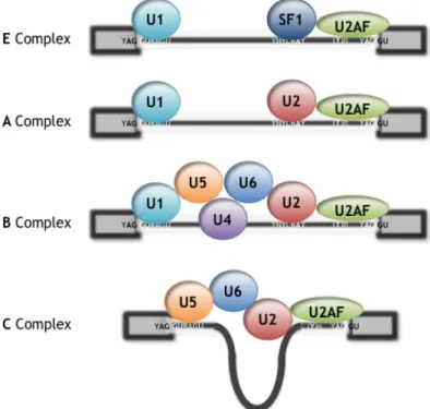
Three sites, the 5! splice site (5!ss), the 3! splice site (3!ss), and the branch point sequence (BPS), are present in every intron and are known as the core splicing signals (Fig. 1.1). These signals are recognized multiple times during spliceosome assembly (Wang and Burge 2008). Each of the snRNAs that compose the spliceosome (U1, U2, U4, U5, and U6 snRNAs) associates with a large number of proteins to form a ribonucleoparticle called an “snRNP.” The catalytic conformation of the spliceosome (so-called “C” complex) forms in a highly dynamic process best described by a stepwise pathway involving several intermediate complexes (E-A-B) that have been identified and characterized in vitro and in vivo (Fig. 1.2) (Black 2003, Tardiff and Rosbash 2006). The earliest known complex committed to the splicing pathway (E) is defined by U1 snRNP base-paired to a 5!ss, with the 3!ss recognized by binding of the U2AF heterodimer (U2AF65/35) to the polypyrimidine tract and
3!-terminal AG, respectively, and association of the protein SF1 with the BPS. The E complex is chased into the pre-spliceosome A complex by the ATP-dependent addition of U2 snRNP at the 3!ss facilitated by base pairing between the U2 snRNA and BPS. Recruitment and addition of the U4#U6/U5 tri-snRNP results in formation of the B complex. Finally, the C complex forms by extensive remodelling of both the snRNA and the protein components that are present in the B complex, including loss of both the U4 and U1 snRNPs, to produce an active site that is capable of catalyzing the transesterification chemistry required for exon ligation and lariat release (Bessonov et al. 2008). The release of U1 and U4 snRNPs, as well as many other molecular rearrangements required for assembly, is promoted by the action of a series of DEX(D/H) box ATPase proteins (House and Lynch 2008).
Figure 1.2: Schematic representation of the spliceosomal complexes. See text for details.
Since each molecular rearrangement and transition during spliceosome assembly represents a potential point of regulation, a more detailed characterization of spliceosome assembly will ultimately lead to a deeper understanding of the mechanisms of alternative splicing (House and Lynch 2008).
Regulation of splice site recognition
Although the splice sites within the pre-mRNA function to direct the splicing machinery, these sequence elements in higher eukaryotes are highly degenerate and often imbedded within introns that are significantly longer than exons (Black 2003). Therefore, it is not surprising that sequence elements outside of the splice sites can strongly affect metazoan pre-mRNA splicing. Use of most exons is now believed to be under the combinatorial control of multiple regulatory RNA elements as well as the inherent strength or weakness of the flanking splice sites (Smith and Valcarcel 2000, Singh and Valcarcel 2005, Matlin et al. 2005, Hertel 2008, House and Lynch 2008).
Splice site strength
A critical step in pre-mRNA splicing is the recognition and pairing of 5!ss and 3!ss (Fig. 1.1). Whereas the 5!ss junction is defined by a single element of 9 nt, the 3!ss is defined by three sequence elements usually contained within 40 nt upstream of the exon/3!-intron junction (Reed 1996). These elements are the branch point sequence, the polypyrimidine tract, and the exon/3!-intron junction. Initial recognition of exon/intron junctions is based on direct interactions between U1 snRNP with the 5!ss, the U2 auxiliary factor with the polypyrimidine tract, and U2 snRNP with the branch point sequence. Because the sequence specificity of these interactions is driven by pre-mRNA/snRNA interactions and the U2 auxiliary factor binding preference for polypyrimidines, splice sites are classified by their complementarity to U1 snRNA (5!ss) and the extent of the polypyrimidine tract (3!ss). Greater complementarity with U1 snRNA and longer polypyrimidine tracts translate into higher affinity binding sites for these spliceosomal components and thus more efficient exon recognition (Hertel 2008). Splice site consensus values (CVs) reflects the strength of the splice site and are usually calculated using previously described matrices (Shapiro and Senapathy 1987), obtained through the analysis of splice sites from different organisms. There are several on-line tools for calculating splice sites
CVs that have been improving with the increasing knowledge in this specific area and the development of bioinformatics. Some examples of these tools are: Splice Site Prediction by Neural Network, http://www.fruitfly.org/seq_tools/splice.html (Reese et al. 1997); Analyzer Splice Tool, http://ast.bioinfo.tau.ac.il/SpliceSiteFrame.htm
(Carmel et al. 2004); and Human Splicing Finder, http://www.umd.be/HSF/HSF.html
(Hubbard et al. 2006). These tools are especially useful when the effect of mutations in genetic disorders is analyzed.
Splicing enhancers and silencers
Although the splice sites within the pre-mRNA function to direct the splicing machinery, these sequence elements in higher eukaryotes are highly degenerate and often imbedded within introns that are significantly longer than exons (Black 2003). Therefore, it is not surprising that sequence elements outside of the splice sites can strongly affect metazoan pre-mRNA splicing. Cis-acting auxiliary sequences occur within both exonic and intronic regions and can either promote recruitment of the spliceosome and exon inclusion (splicing enhancers) or disrupt assembly of the splicing machinery and cause exon skipping (splicing silencers). In general, these splicing regulatory elements function by recruiting trans-acting splicing factors that activate or suppress splice site recognition or spliceosome assembly by various mechanisms (Matlin et al. 2005).
The best characterized of the regulatory elements, exonic splicing enhancers (ESEs), are usually recognized by a family of proteins known as SR proteins, which contain an RNA-binding domain and a region rich in arginine-serine dipeptides (RS domain). Interestingly, SR protein-binding sites are present not only within alternatively spliced exons, but also within the exons of constitutively spliced pre-mRNAs (Schaal and Maniatis 1999). It is therefore expected that SR proteins bind to sequences found in most, if not all, exons.
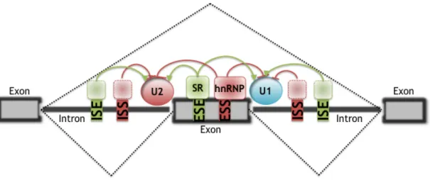
However, regulation of pre-mRNA splicing is much more complex than the simple ESE recruitment model. Intronic splicing enhancers (ISEs) and splicing
silencers, either exonic (ESS) or intronic (ISS), occur frequently and influence splice site selection (Fig. 1.3) (Black 2003, Pagani and Baralle 2004). Several mechanisms have been proposed for ESS- or ISS-mediated splicing repression. Heterogeneous nuclear RNP-bound splicing silencers have been shown to repress spliceosomal assembly through multimerization along exons (Zhu et al. 2001), through blocking the recruitment of snRNPs (Tange et al. 2001), or by looping out exons (Martinez-Contreras et al. 2006).
Figure 1.3: A schematic of regulated splicing. Dashed lines: Two alternative splicing pathways,
with the middle exon either included or excluded. Splicing is regulated by cis-elements (ESE, ESS, ISS, and ISE) and trans-acting splicing factors (SR proteins, hnRNP, and unknown factors). (adapted from Wang and Burge 2008)
Typically, silencers and enhancers are present within the vicinity of potential exon/intron junctions, suggesting that the interplay between activating and repressing cis-acting elements modulates the probability of exon inclusion (Hertel 2008). These observations suggest that the recognition of every exon is influenced by multiple distinct cis-acting elements, a notion strongly supported by computational analyses (Zhang and Chasin 2004, Wang et al. 2004). According to this concept, a large number of diseases are now known to result from intronic or exonic mutations that disrupt normal splicing patterns. This adds to diseases caused by missense, nonsense, and frame-shifts mutations in the open reading frame, which give rise to defective proteins (Cooper et al. 2009). Up to 25% of synonymous (in terms of amino acid coding) substitutions can disrupt normal splicing, as can nonsynonymous and
termination codons (Pagani et al. 2005), emphasizing the importance of considering silent mutations as mediators of pathogenic effects. For the studied genes, up to 50% of point mutations within exons affect splicing, and it has been hypothesized that more than half of known disease-causing mutations disrupt splicing (Lopez-Bigas et al. 2005).
Generally, the splicing regulatory elements are relatively short (4-18 nucleotides) (Cartegni et al. 2002, Fairbrother et al. 2002) and degenerate, so that an accurate understanding of their sequence context is still lacking. Some recent studies employed computational methods to predict sequence motifs for this elements, for example, ESEfinder program (Cartegni et al. 2003, available at http://rulai.cshl.edu/cgi-bin/tools/ESE3/esefinder.cgi?process=home), PESX: Putative Exonic Splicing Enhancers/Silencers (Zhang and Chasin 2004, Zhang et al. 2005, available at http://cubweb.biology.columbia.edu/pesx/), ESR search (Goren et al. 2006; available at http://ast.bioinfo.tau.ac.il/ESR.htm) and Splicing Rainbow (Stamm et al. 2006, available at http://www.ebi.ac.uk/asd-srv/wb.cgi?method=8). Several groups of ESEs are known, including purine-rich and AC-rich elements, as well as some with more complex composition (Graveley 2000, Zheng 2004). ESS sequences have higher content of T (38%) and G (36%) and reduced levels of A (17%) and C (9%) (Wang et al. 2004). However, up to now it is not possible to reliably identify these elements and predict the effect of a genomic mutation on the splicing process.
Exon/Intron architecture
The exon/intron architecture has been shown to have an influence on splice site recognition (Berget 1995). For example, increasing the size of mammalian exons results in exon skipping. However, the same enlarged exons were included when the flanking introns were small (Sterner et al. 1996). Thus, splice site recognition is more efficient when introns or exons are small. These early observations suggested that splice sites are recognized across an optimal nucleotide length and predicted that intron length significantly influences the efficiency of pre-mRNA splicing and
alternative splice site choice. This is an important hypothesis because of the divergent distribution of intron length in the human genome and because it had been proposed that, the spliceosome uses two modes of recognition to define splice sites: intron and exon definition (Berget 1995). These two models are still unproven, and all the indications for their existence are circumstantial. However, intron definition is presumably the ancient one, in which the splicing machinery recognizes an intronic unit and places the basal machinery across introns. Therefore, the size of the intron is under selection. Indeed in S. cerevisiae and S. pombe, almost all introns are less than 350 nt long, and all the information for accurate splicing is within the intron sequences (Guthrie 1991). This suggests that intron definition is the only system that directs the splicing machinery in these organisms (Berget 1995). In the second mechanism, exon definition, the basal splicing machinery is placed across exons. The length of exons must not exceed 300 nt. It was postulated that during evolution the enlargement of intronic sequences forced the splicing machinery to shift from the recognition of short intronic sequences to the selection of short exonic sequences – from intron to exon definition. This could explain the selective pressure to maintain short intronic sequences in yeast genes and short internal exons in the human genome (and other higher metazoans) (Robberson et al. 1990). Because the transesterification step of splicing occurs across the intron, a switch between exon and intron definition must exist to assemble the mature spliceosome. Recently, it was also demonstrated that disruption of the transition from exon-to-intron definition is a mechanism for alternative splicing regulation (Sharma et al. 2008).
Exon skipping is more likely to occur when exons are flanked by long introns in the human genome. Interestingly, experimental and computational analyses showed that the length of the upstream intron is more important in inducing alternative splicing than the length of the downstream intron, most likely reflecting the influence of RNA transcription on pre-mRNA splicing. These results showed that the exon/intron architecture defines baseline mechanisms of splice site recognition and influences the frequency of alternative pre-mRNA splicing (Fox-Walsh et al. 2005).
RNA secondary structure
Single-stranded RNA is known to adopt local secondary folds and tertiary interactions that may involve up to hundreds of nucleotides. Although pre-mRNAs are typically depicted in a linear fashion, we have to assume that higher order structures exist that maintains a good portion of the RNA double-stranded. Depending on the thermodynamic stability, these structures may persist long enough to interfere or modulate splice site recognition. In principle, these local structures can be inhibiting or activating spliceosomal assembly. This is because the recognition of splice sites, enhancers, and silencers usually depends on interactions between protein factors and a single-stranded portion of the pre-mRNA. Local RNA structures can interfere with spliceosomal assembly if they conceal splice sites or enhancer-binding sites within stable helices. On the other hand, local RNA structures can also promote spliceosomal assembly by masking splicing repressor-binding sites (Hertel 2008).
The importance of RNA secondary structure in modulating splice site selection has been documented frequently. For example, two classes of conserved RNA elements have been identified in the Dscam (Down syndrome cell adhesion molecule) exon 6 cluster, which contains 48 alternative exons 6. Each exon 6 variant contains a unique selector sequence that can base pair with a common upstream docking site (located in the intron downstream of constitutive exon 5) to form a secondary structure, thereby activating anddirecting mutually exclusive exon pairing
(Graveley 2005). An inhibitory role of RNA secondary structure was demonstrated for splice site recognition of SMN2 exon 7. The formation of an RNA hairpin close to the 5! splice site of SMN2 exon 7 interfered with its interaction with U1 snRNP, resulting in reduced exon inclusion levels (Singh et al. 2007).
Pre-mRNA synthesis by RNA polymerase II
It is widely accepted that the recognition and differential selection of splice sites occurs during transcription (Goldstrohm et al. 2001, Listerman et al. 2006). Furthermore, various studies have shown that transcription activators affect alternative splicing decisions. Such effects of transcription on splicing can be explained by two, non-mutually exclusive models: one model relies on the recruitment of splicing factors to the Pol II C-terminal domain (CTD); the second model is based on the kinetics of transcriptional elongation (Auboeuf et al. 2007). In mammals, the CTD consists of 52 repeats of the motif YS2(P)TS5(P)S, in which S2
and S5 are phosphorylated dynamically. During initiation, Pol II is phosphorylated first on S5 and then on S2, at which point Pol II becomes processive and interacts with various splicing factors (Goldstrohm et al. 2001, Howe 2002). The interactions with the CTD might enable an increased concentration of specific splicing factors in the proximity of the nascent transcript, and thereby influence splicing decisions. Thus, any physiological post-translational modification on the CTD that affects its binding properties could contribute to modulate the combinatorial regulation of splice-site selection (de la Mata and Kornblihtt 2006).
The kinetic model is based on the notion that, because of their relative weakness, splice sites in the alternative exons might need longer to interact with the spliceosome, and their use might be favoured by a low elongation rate of Pol II (de la Mata et al. 2003). The recruitment and elongation rate of Pol II are coordinated, probably because the recruitment of splicing factors and the elongation rate of Pol II also depends on phosphorylation of the CTD (Listerman et al. 2006, de la Mata and Kornblihtt 2006).
Combinatorial control of exon recognition and alternative splicing
Over the last few years, it has become increasingly clear that exon selection is influenced by a number of activating and inhibitory elements. Given the divergent sequence and architecture of genes, every exon has its specific set of identity elements that permit its recognition by the spliceosome. Each exon is flanked by a unique pair of splice site signals and contains a unique group of splicing enhancers, silencers and secondary structures. The sum of contributions from each of these identity elements then defines the overall recognition potential of an exon or the overall binding affinity for the spliceosome. Considering the variation in splice sites, exon/intron architecture, number of enhancers and silencers, and secondary structures, the potential for a given exon to become recognized is expected to span a wide range. Because the spliceosome assembles around splice sites, the binding potential of splice sites is crucial for efficient exon definition. The contributions of the other parameters will vary significantly from exon to exon or cell to cell, augmenting or reducing the overall affinity of the splicing machinery. As a consequence of the observed differences in the concentrations of spliceosomal components and splicing activator/repressors between different cell types or between distinct biological processes such as the cell cycle and development, it is anticipated that the same exon may display variable exon recognition potentials in these scenarios. As a result, exons that are alternatively included in one cell type can be alternatively excluded in another (Hertel 2008).
An implication of this combinatorial interpretation of the splicing code is that the precise expression level, activity or subcellular localization of regulatory proteins in any given cell can have a profound influence on the ultimate splicing pattern of a gene (House and Lynch 2008).
In the literature, alternative splicing is attributed mainly to the activities of splicing enhancers and repressors that allow transient interactions with splicing regulators (Black 2003). In most cases, the presence or absence of splicing regulatory proteins modulates the overall exon recognition to significantly tilt the
balance between exon inclusion and exclusion. Similarly, protein interactions within the pre-mRNA may induce or interfere with the formation of RNA secondary structures that modulate efficient spliceosomal assembly (Hertel 2008). Regulation of alternative splicing can be achieved through modulating any one of the exon recognition components (House and Lynch 2008). However, specific regulation requires the selected targeting of splicing activator/repressor combinations unique to particular exons. This is often mediated through changes in post-translational modifications that are essential for optimal activity of many splicing regulatory factors, such as alterations in the phosphorylation state of specific SR proteins
(Stamm 2008).
Invariable elements such as splice site sequences and exon/intron architecture have also the potential to mediate differential splicing. Based on the principle of mass action, fluctuations in snRNP levels can induce changes in the efficiency of splice site recognition, thus altering exon inclusion ratios. Such changes in the concentration of the general splicing factors could account for many of the alternative splicing events observed between different cell types (Hertel 2008).
In the cell, alternative splicing has also been attributed to promoter-dependent recruitment of specific splicing regulators or to changes in the kinetics of pre-mRNA synthesis (Kornblihtt 2005). Thus, modulating the recruitment of specific splicing factors or modulating the relative synthesis of competing splice sites can influence the selection of alternative splicing patterns. Alternatively, changes in the kinetics of RNA synthesis are able to influence the likelihood that local RNA secondary structures form that induces alternative splice site selection (Hertel 2008).
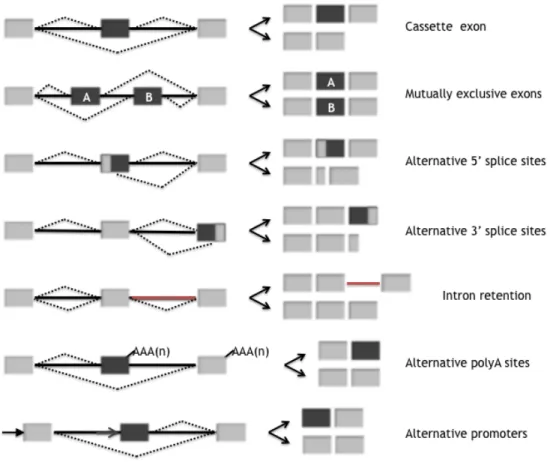
Types of alternative splicing events
Based on systematic analyses, especially from mammalian species, it is apparent that the most common type of alternative splicing, accounting for at least one-third of known alternative splicing events, involves cassette-type alternative exons. These exons, which are either skipped or included in the final message, are
flanked by intron sequences (Figure 1.4). Alternative selection of 5!ss or 3!ss within exon sequences are also frequent, together accounting for at least one-quarter of the known alternative splicing events (Figure 1.4). This type of alternative splicing is capable of introducing subtle changes into coding sequences, differing by as little as a single codon (Blencowe 2006). Other types of alternative splicing events include retained introns (Ohler et al. 2005) and exons that are spliced in a mutually exclusive fashion (Figure 1.4).
In addition to the alternative splicing mechanisms mentioned above, the exon composition of transcripts is often altered by differential selection of transcription initiation and 3! end processing/termination sites (Figure 1.4), and these events can impact on adjacent or distal alternative splicing events in the same transcript (Zavolan et al. 2003, Kornblihtt 2005).
Figure 1.4: Alternative Splicing Events in Metazoan Transcripts.Types of alternative splicing that are responsible for the generation of functionally distinct transcripts. Dashed lines represent different possibilities for splice site joining. Constitutive regions are shown in light gray while alternative ones in dark grey. (adapted from Blencowe 2006)
Finally, it should also be kept in mind that each of the types of alternative splicing summarized above and shown in Figure 1.4 can occur within both translated and untranslated regions (UTRs) of transcripts (Blencowe 2006). Alternative exons within 5!-untranslated and 3!-untranslated regions can either add or remove RNA regulatory motifs and, thereby, modulate the stability and translation of transcripts. Several examples of alternative UTRs have been characterized, including those found in AXIN2, FGF1 and BRCA1 (Hughes 2006).
Effects of alternative splicing
Alternative splicing is considered to be one of the main mechanisms by which proteome diversity is encoded by a limited number of genes. As most alternative splicing events occur in translated regions of mRNAs, they can affect the sequence of the encoded proteins. Changes in the primary structure might influence all aspects of protein function and properties, such as stability, intracellular localization, binding properties, enzymatic activity and post-translational modification. This yields a wide range of effects, from complete loss to subtle modulations of protein function. In some cases, protein isoforms that are generated by the same gene might have different functions (Kriventseva et al. 2003, Stamm et al. 2005, Blencowe 2006, Yura et al. 2006).
Exon selection also affects the levels of gene expression. For example, alternative exons within UTRs can modulate the stability and translation of transcripts (Hughes 2006). Furthermore, it has been estimated that about one-third of alternatively spliced exons introduce premature translation-termination codons (PTCs). The splicing variants that contain such PTCs are degraded through the nonsense-mediated mRNA-decay (NMD) pathway and do not produce proteins. It has been speculated that alternative splicing-coupled NMD could provide a mechanism for the regulation of gene expression (Lewis et al. 2003, Blencowe 2006). However, some studies have shown that most PTC-containing splice variants are not conserved between human and mouse (Baek and Green 2005, Pan et al 2006).
Moreover, a recent microarray analysis of alternative splicing events in mammalian cells and tissues suggested that most PTC-containing splice variants are produced at low levels independent of the action of NMD and are rarely subject to tissue-specific regulation (Pan et al. 2006). These results support the view that alternative splicing-coupled NMD may not play a widespread role in gene regulation. However, this process may serve to fine tune the levels of specific classes of genes, including subsets of splicing factors and other RNA binding proteins (Blencowe 2006). For example, it was recently found that all human SR protein genes have alternative splice forms that are degraded by NMD. Thus, this family of splicing factors might couple alternative splicing and NMD to regulate their own protein production, possibly by a mechanism of feedback regulation (Lareau et al. 2007).
Signal-mediated alternative splicing control
Extracellular signals impact eukaryotic gene expression at different levels, including pre-mRNA splicing (Wilson and Cerione 2000). A large body of evidence has been accumulated indicating that splicing regulation can be induced by various
extracellular signals such as cell growth/death factors,
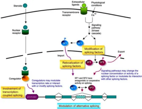
neurotransmitters/electrophysiological conditions, and environmental nutrients/stresses. Signal-mediated splicing regulation is primarily operated through activation of intricate networks of signal transduction pathway (Figure 1.5; Tarn 2007). Several signalling pathways and numerous splicing regulatory factors have now been implicated in the modulation of pre-mRNA splicing (Stamm 2002, Shin and Manley 2004).
Signalling pathways may converge on RNA-binding proteins that function as
trans-acting splicing regulatory factors (Wilson and Cerione 2000, Stamm 2002, Shin and Manley 2004). Signalling, largely through kinase cascades, leads to phosphorylation of target splicing factors and subsequently alters their activity or cellular localization
(Stamm 2002, Shin and Manley 2004). Such alterations may influence splicing efficiency or induce alternative splicing. Besides phosphorylation, the possibility that
other types of post-translational modifications also affect splicing activity still remains
(Tarn 2007). For example, dephosphorylation (Chalfant et al. 2001), methylation
(Bedford and Richard 2005), ubiquitination (Bellare et al. 2006) and sumoylation (Li et al. 2004) have already been reported to play a role in signal-mediated splicing control. However, the mechanistic details of the pathways involved, from cellular signals to alternative splicing events, lack comprehensive description and thus need further investigation. In addition, evidence begins to emerge recently, indicating that steroid hormone receptors or their cofactors may modulate the rate of transcription or directly interact with splicing factors and thereby affect splicing through a transcription-coupled splicing regulation (Auboeuf et al. 2002, Dowhan et al. 2005).
Several cellular signalling pathways have been implicated in splicing regulation, including Ras/MEK/ERK, Rac/JNK/p38-MAPK, Ras/PI3-kinase/AKT and Ca2+/calmodulin/CaMK IV (Stamm 2002, Shin and Manley 2004). For example, activation of the p38-MAP kinase pathway led to phosphorylation of hnRNP A1, resulting in its cytoplasmic sequestration and altered splicing of an adenovirus reporter gene (van der Houven van Oordt et al., 2000). Depolarisation of secretory cells repressed inclusion of the STREX exon in BK potassium channel transcripts through the action of Ca2+/calmodulin-dependent protein kinases (CaMKs), thereby changing the excitability of the channel (Xie and Black, 2001). Also, inclusion of exon v5 into the cell surface tumour marker CD44 was dependent on activation of the Ras-ERK pathway (Weg-Remers et al., 2001; Matter et al., 2002) and nucleo-cytoplasmic transport of the splicing regulator polypyrimidine tract-binding protein (PTB) is modulated by the cAMP dependent protein kinase (Xie et al., 2003). Finally, phosphorylation of SRp40 by AKT2 has been shown to regulate alternative splicing of protein kinase C "II (Patel et al., 2005; Jiang et al., 2008). Although many examples of signalling-induced splicing control have been described, their mechanistic details still remain largely unclear (Tarn 2007).
Figure 1.5: Alternative splicing regulation by cellular signals. Simplified models of
signalling-controlled alternative splicing are depicted. Extracellular signals may activate kinase cascades to phosphorylate splicing factors, and whereby change their subcellular localization or activity or alter their interaction with other cellular factors. Steroid hormones may modulate splicing through a cotranscriptional mechanism. Nuclear receptor coregulators may modulate the transcription rate, or directly recruit or post-translationally modify splicing factors, and whereby influence pre-mRNA splicing. Signalling pathways may concurrently target several splicing factors (SFs) to achieve an optimal control of alternative splicing. (from Tarn 2007)
Global analysis of alternative splicing
The availability of sequenced genomes and large databases of sequenced transcripts, primarily comprising expressed sequence tags (ESTs) and smaller numbers of cDNA sequences, has provided a rich source of information for the identification and analysis of alternative splicing events (Blencowe 2006).However, a major limitation of alternative splicing analyses employing transcript sequence data is that EST coverage is typically biased toward the 3! and 5! ends of transcripts, and in general there are insufficient numbers of sequenced transcripts to infer the frequency with which specific alternative exons are included or skipped in a given cell or tissue source or under particular experimental conditions (Johnson et al. 2003, Pan et al. 2004).
Some of the limitations inherent in the analysis of EST/cDNA have been overcome by the development of custom microarrays and computational tools, as well as differential hybridization techniques, which permit the large-scale profiling of alternative splicing. %%Exon junction arrays!! represent one early design, with high-density oligonucleotide probes targeted to the junctions between consecutive exons
(Johnson et al. 2003). Other designs include the use of probe sets to target bodies and junctions of constitutive and alternative exons (Pan et al. 2004, Sugnet et al. 2006), as well as the use of bead-based fiberoptic microarray platforms with high detection sensitivity (Yeakley et al. 2002). These designs have facilitated analyses of genome-wide alternative splicing in human, mouse, and chimp (Srinivasan et al. 2005, Calarco et al. 2007), as well as detection of the global impact of specific splicing factors or environmental stimuli on splicing regulation (Hung et al. 2007, Makeyev et al. 2007, Pleiss et al. 2007a).
The systematic identification of RNA targets for different trans-factors can also be achieved by some new approaches such as a cross-linking/immunoprecipitation (CLIP) (Ule et al. 2003), RNP immunoprecipitation (RIP) (Keene et al. 2006), and genomic SELEX (Lorenz et al. 2006). These analyses have the potential to identify regulatory targets of a factor and can be applied genome-wide when coupled with microarray or high-throughput sequencing technologies. Analysis of the target sequences will help to define the sequence determinants of binding, and may also help to identify cooperative or antagonistic relationships between different factors. Since only a subset of binding events confers regulatory activity, it is important to also have evidence of regulation. Such evidence can be obtained from knockout/knockdown or overexpression of the factor, which can be also applied on a genome-wide scale when coupled with high-throughput sequencing (Wang and Burge 2008).
In the post-genomic world, research into the mechanisms of splice site selection is leading toward the establishment of rules that will allow splice patterns to be predicted based on sequence information. Computational methods combined with laboratory experiments have already generated algorithms that predict splicing
regulatory sequences (Fairbrother et al. 2002, Wang et al. 2004, Zhang and Chasin 2004). This significant progress suggests the exciting possibility of crafting a “splicing code” that permits the prediction of exons and the probability of their inclusion in the most abundant mRNA isoform. Ideally, a splicing code should be able to differentiate between alternative splicing events in different tissues and different biological processes (Hertel 2008, Wang and Burge 2008).
Splicing regulatory network as a subnetwork of gene regulation
Recent analyses of sequence and microarray data on alternative splicing revealed that the majority of genes regulated in a tissue-specific manner by alternative splicing are different from those regulated in a tissue-specific manner at the transcriptional level. Moreover, it is emerging that groups of tissue-specific AS events may function in a coordinated manner in specific pathways or interaction networks, in much the same way as has been observed for groups of genes coregulated at the transcriptional level. This promotes the concept of “layers” of gene regulation and raises the interesting question as to the extent and nature of exon networks that may serve in parallel with other layers to coordinate gene activities and interactions so as to refine and/or expand cell- and tissue-type-specific functions
(Blencowe 2006).
The splicing regulatory network is part of a larger network of gene regulation with which it is linked both physically and functionally (Wang and Burge 2008). In fact, the abundance of protein isoforms results from regulatory events at various stages of gene expression, including chromatin remodelling, transcription initiation and elongation, pre-mRNA splicing and polyadenylation, mRNA export, and translation (Auboeuf 2007).
Although bioinformatics predictions on alternative splicing are evolving, it is important to understand they are incomplete. Currently, the determination of the splicing regulation of a particular splice form or whether it is actually carrying out an