Cytoplasmic Viral RNA-Dependent RNA Polymerase
Disrupts the Intracellular Splicing Machinery by Entering
the Nucleus and Interfering with Prp8
Yen-Chin Liu1,2, Rei-Lin Kuo1,2,3, Jing-Yi Lin4, Peng-Nien Huang1, Yi Huang5, Hsuan Liu5,
Jamine J. Arnold6, Shu-Jen Chen5, Robert Yung-Liang Wang1,7, Craig E. Cameron6, Shin-Ru Shih1,2,3,8*
1Research Center for Emerging Viral Infections, College of Medicine, Chang Gung University, Tao-Yuan, Taiwan,2Graduate Institute of Biomedical Sciences, College of Medicine, Chang Gung University, Tao-Yuan, Taiwan,3Department of Medical Biotechnology and Laboratory Science, College of Medicine, Chang Gung University, Tao-Yuan, Taiwan,4School of Medical Laboratory Science and Biotechnology, Taipei Medical University, Taipei, Taiwan,5Molecular Medicine Research Center, Chang Gung University, Tao-Yuan, Taiwan,6Department of Biochemistry and Molecular Biology, Pennsylvania State University, University Park, Pennsylvania, United States of America, 7Department of Biomedical Sciences and Graduate Institutes of Biomedical Sciences, College of Medicine, Chang Gung University, Tao-Yuan, Taiwan,8Clinical Virology Laboratory, Chang Gung Memorial Hospital, Tao-Yuan, Taiwan
Abstract
The primary role of cytoplasmic viral RNA-dependent RNA polymerase (RdRp) is viral genome replication in the cellular cytoplasm. However, picornaviral RdRp denoted 3D polymerase (3Dpol) also enters the host nucleus, where its function remains unclear. In this study, we describe a novel mechanism of viral attack in which 3Dpolenters the nucleus through the nuclear localization signal (NLS) and targets the pre-mRNA processing factor 8 (Prp8) to block pre-mRNA splicing and mRNA synthesis. The fingers domain of 3Dpolassociates with the C-terminal region of Prp8, which contains the Jab1/MPN domain, and interferes in the second catalytic step, resulting in the accumulation of the lariat form of the splicing intermediate. Endogenous pre-mRNAs trapped by the Prp8-3Dpolcomplex in enterovirus-infected cells were identified and classed into groups associated with cell growth, proliferation, and differentiation. Our results suggest that picornaviral RdRp disrupts pre-mRNA splicing processes, that differs from viral protease shutting off cellular transcription and translation which contributes to the pathogenesis of viral infection.
Citation:Liu Y-C, Kuo R-L, Lin J-Y, Huang P-N, Huang Y, et al. (2014) Cytoplasmic Viral RNA-Dependent RNA Polymerase Disrupts the Intracellular Splicing Machinery by Entering the Nucleus and Interfering with Prp8. PLOS Pathog 10(6): e1004199. doi:10.1371/journal.ppat.1004199
Editor:Mark T. Heise, University of North Carolina at Chapel Hill, United States of America ReceivedDecember 10, 2013;AcceptedMay 5, 2014;PublishedJune 26, 2014
Copyright:ß2014 Liu et al. This is an open-access article distributed under the terms of the Creative Commons Attribution License, which permits unrestricted use, distribution, and reproduction in any medium, provided the original author and source are credited.
Funding:This work was supported by NRPGM (NSC 102-2325-B-182-015). The funders had no role in study design, data collection and analysis, decision to publish, or preparation of the manuscript.
Competing Interests:The authors have declared that no competing interests exist.
* Email: [email protected]
Introduction
RNA viruses in general replicate in the cytoplasm and interfere host cellular gene expression by utilizing proteolytic destruction of cellular targets as the primary mechanism [1]. However, several viral proteins have been found in the nucleus and altered host gene expression [2]. For example, our previous finding shows that picornaviral 3C protease cleaves CstF-64 and inhibits cellular polyadenylation in the nucleus [3]. Besides the protease, the RNA-dependent RNA polymerase (RdRp) also appears in the nucleus, but the role of viral RNA polymerase in the nucleus remains unclear. This study utilized picornaviral polymerase to probe the function of RdRp in the nucleus.
Picornaviruses cause numerous diseases in humans and various animal species. The enteroviruses in the Picornaviridae family are critical human pathogens that typically cause hand, foot, and mouth disease (HFMD) and contribute to severe neurological complications, including aseptic meningitis, brainstem encephali-tis, poliomyeliencephali-tis, and even death [4,5]. Enterovirus 71 (EV71) has played an increasingly substantial role in emerging epidemics around the Asia Pacific region, and these infections are particularly life-threatening in young children [6–8].
Although picornaviral 3Dpolprimarily performs viral replication in the host cytoplasm, 3CD and 3Dpolare capable of entering the nucleus in virus-infected cells [2,19–22]. 3CD and 3Dpol of poliovirus (PV) enter the nucleus through a nuclear localization signal (NLS), KKKRD, which spans 125–129 amino acids (aa) within 3Dpol. The putative NLS in the 3Dpol coding region is partially contained within the sequence KKRD (126–129 aa), which is typical among all known picornaviral 3Dpol [2,22]. Picornaviral 3C is delivered into the nucleus through its precursor 3CD, which results in the cleavage of cellular transcriptional factors or regulators, such as CstF-64, polymerase I factor SL-1, TATA-box binding protein (TBP), cyclic AMP-responsive element binding protein (CREB), Octamer binding protein-1 (Oct-1), p53, histone H3, and RNA polymerase III transcription factor IIIC [2,3,23–28]. However, previous studies have only demonstrated that 3Dpolcontaining a NLS can transport 3CD to the nucleus, and the precise role of 3Dpol in the host cell nucleus remains unclear. In this study, we identified several nuclear target proteins in U5 small nuclear ribonucleoprotein particles (U5 snRNPs) that interact with 3Dpol.
U1, U2, U4, U5, and U6 snRNPs are essential components of the spliceosome in the pre-mRNA splicing process, which involves introns excision and exon ligation to form a mature mRNA. U5 snRNPs contain several functionalities crucial to pre-mRNA splicing factors, such as Prp8/220K, Brr2/200K, Snu114/116K, Prp6/102K, Prp28/100K, Lin1/52K, SNRNP40/40K, and Dib1/15K [29–31]. The human pre-mRNA processing factor 8 (Prp8), one of the largest and highly-conserved nuclear proteins, provides a large scaffold and occupies a central position in the catalytic core of a spliceosome. Prp8 contains 5 functional domains: NLS, RNA recognition motif (RRM), 39 splice site (39SS) fidelity region, 59 splice site (59SS) holding domain, and Jab1/MPN. The putative bipartite NLS of Prp8 enables Prp8 to enter the nucleus, assisted by the import-afamily, and the RRM domain provides an RNA binding center for pre-mRNA. The 59SS holding domain of Prp8 is believed to lock the 59SS-OH (exon) and load it onto the 39SS-associated region of Prp8. The Jab1/MPN domain in Prp8 represents a pseudo-enzyme that is
converted into a protein-protein interaction platform. Prp8 is the only spliceosomal protein that directly cross-links to the 59SS, 39SS, and branch point (BP) of the pre-mRNA substrate, as well as to U2, U5 and U6 snRNAs. Prp8 also interacts with several spliceosomal proteins, including the RNA helicase Brr2, the GTPase Snu114, and Prp6 of the U5 snRNP. The human Prp8 terminal fragment, the N-terminal region containing the NLS domain and the C-terminal region containing the Jab1/MPN domain also interact with each other, suggesting that these regions form an intermolecular bridge [32–38].
In pre-mRNA processing, the U1 snRNP initially binds to the 59SS, and the U2 snRNP associates with the BP of the pre-mRNA. The U5NU4/U6 tri-snRNPs assembly then forms the pre-spliceosome B-complex. Then, a major structural change occurs upon the release of the U1 and U4 snRNPs, transforming the B-complex into the catalytically active component (Bact-complex) of the spliceosome. The first catalytic step of the splicing process involves the Bact-complex catalyzing the first trans-esterification reaction, which is the attack of the branch site 29-OH on the 59SS. This produces exon1 and the lariat form (intron-exon2), which forms the C1-complex. The second catalytic step is to transition from the C1 to C2 complex for the secondtrans-esterification. The attack of the 39-OH of the 59exon on the 39SS results in 39SS cleavage and exon ligation to produce the mRNA and release the excised intron [39–41]. Prp8, U5 snRNA, and the nonspliceoso-mal proteins Prp16, Slu7, Prp18, and Prp22 are required for the second catalytic step. In addition, Arg1753 of Prp8 cooperates with Prp18 to stabilize the U5/exon contacts that are crucial for the second catalytic step and ensure that the Prp22 helicase disrupts the interactions between the U5 snRNP and mRNA to release the mRNA [36,42].
In this study, we describe a novel mechanism for picornavirus invasion of host cells that involves a previously unidentified function of 3Dpol that differs from its classic role in viral replication. Our results suggest that the 3Dpolenters the cellular
nucleus to associate with the core splicing factor Prp8. Further-more, 3Dpolaffects the normal function of Prp8 during the second catalytic splicing step, leading to the inhibition of pre-mRNA splicing, the accumulation of the lariat form, and a decrease in the resulting mRNA. This is the first study demonstrating that a cytoplasmic RNA virus can use its polymerase to alter cellular gene expression by hijacking the splicing machinery.
Results
3Dpolspecifically interacts with the nuclear protein Prp8
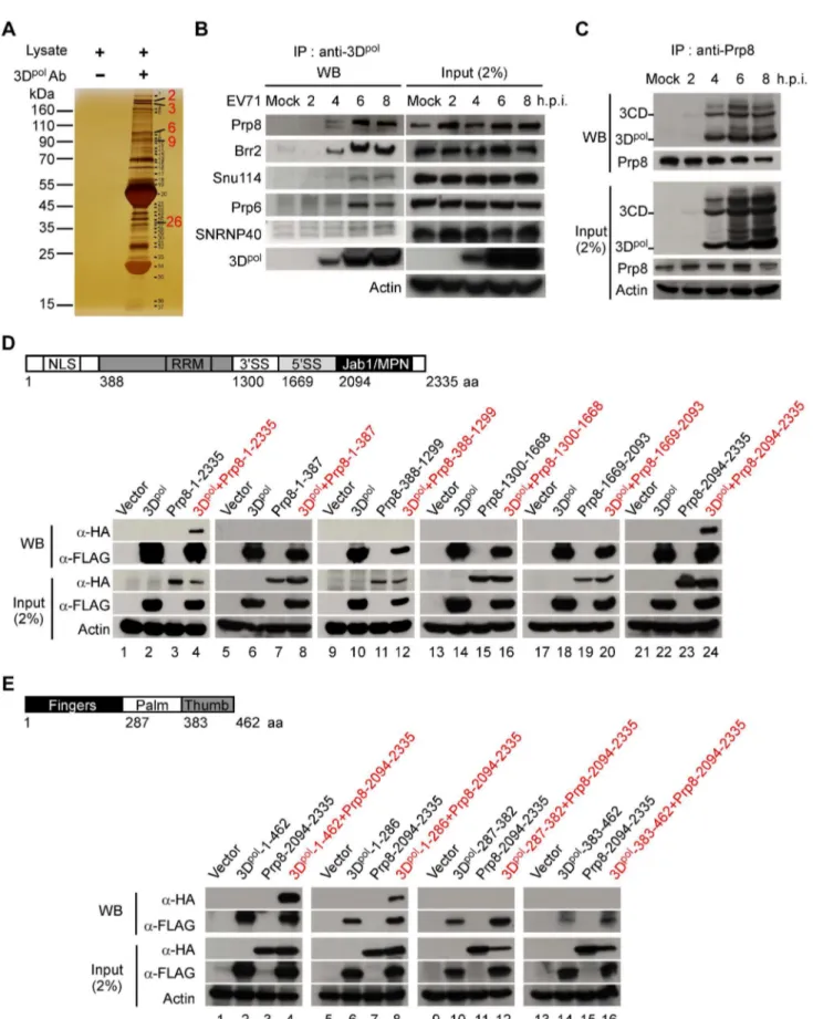
Picornaviral 3Dpol has been shown to enter the nucleus of infected cells [2,20,22]; however, the role of 3Dpolin the nucleus has not been explored. We generated an EV71 3Dpolmonoclonal antibody that could recognize 3Dpol in the lysates of RD cells infected with EV71 at a multiplicity of infection (MOI) of 40. To determine the role of 3Dpol in the host nucleus, the 3Dpol -interacting proteins were pulled down and detected using a one-dimensional sodium dodecylsulfate-polyacrylamide gel electropho-resis (1D SDS-PAGE) assay (Figure 1A). These potential target proteins were identified using matrix-assisted laser desorption ionization-time of flight mass spectrometry (MALDI-TOF MS) analysis, and the results are summarized in Table 1. These data indicated that 3Dpol may interact with numerous U5 snRNP nuclear proteins, including Prp8, Brr2, Snu114, Prp6, and SNRNP40. This interaction was further confirmed in EV71-infected RD cell lysates following RNase A treatment using co-immunoprecipitation (Co-IP) and Western blotting (WB) assays (Figure 1B). Prp8, which is the nuclear protein at the central
Author Summary
position in the catalytic core of the spliceosome, was selected for further study. The interaction of Prp8 and 3Dpol was further verified in EV71-infected RD cell lysates following RNase A treatment by Co-IP and WB assays with antibodies against endogenous Prp8 and viral 3Dpol, respectively. The result of these assays suggested that endogenous Prp8 interacts with EV71 3Dpol and 3CD between 4 to 8 h post-infection (h.p.i.), without intermediation of the RNA (Figure 1C). To discover the interacting domains of Prp8 and 3Dpol, we constructed tags that were fused with hemagglutinin (HA) or FLAG epitopes for the various truncated forms of Prp8 and 3Dpol, respectively, and the various fragments of Prp8 and 3Dpolwere cloned separately from each functional domain. To identify which Prp8 domain is responsible for the 3Dpolinteraction, plasmids containing various truncated forms of Prp8 fused with HA and the FLAG-tagged
full-length 3Dpolwere transfected into HEK293T cells, followed by anti-FLAG IP and WB assays. The results of these assays revealed that the full-length Prp8 (lane 4) and the C-terminal region (2094– 2335 aa) containing the Jab1/MPN domain of Prp8 (lane 24) interacted with full-length 3Dpol (Figure 1D). Furthermore, to determine which domain of 3Dpolinteracts with the C-terminal region of Prp8, we used different FLAG-tagged truncated forms containing the fingers, palm, and thumb domains, as well the HA-tagged C-terminal region, in anti-FLAG IP and WB assays. This mapping study revealed that the fingers domain in the N-terminal region (1–286 aa) of 3Dpolinteracts with the C-terminal region (2094–2335 aa) of Prp8 (lane 8) (Figure 1E). Here, we demonstrate that 3Dpolassociates with the nuclear protein Prp8 via a protein-protein interaction that involves at least one region between the finger domain of 3Dpoland the C-terminal domain of Prp8. RD cells at 2 to 8 h.p.i. were treated with RNase A (10mg/ml) and immunoprecipitated using an anti-3Dpolantibody. The 5 components of the U5 snRNPs that interacted with 3Dpolwere detected using a WB assay. The input samples were verified in the presence of 3Dpoland the five components of the U5 snRNPs in the lysates. Actin served as an internal control. (C) The core spliceosome splicing factor Prp8 can also pull down 3Dpoland 3CD. EV71-infected RD cell lysates from 2 to 8 h.p.i. were treated with RNase A (10mg/ml) and incubated with antibodies against the Prp8 probe. After the IP assay, 3Dpoland 3CD were analyzed using WB with an anti-3Dpolantibody. (D) 3Dpolassociates with the C-terminal domain of Prp8 containing the Jab1/MPN region. The functional domain architecture of human Prp8 is shown (upper panel). HEK293T cells were transfected with plasmids encoding full-length FLAG-3Dpol(lanes 2, 4, 6, 8, 10, 12, 14, 16, 18, 20, 22, and 24), various truncated forms of HA-Prp8 (lanes 3, 4, 7, 8, 11, 12, 15, 16, 19, 20, 23, and 24), and empty vectors (lanes 1, 5, 9, 13, 17, and 21). At 48 h after transfection, the lysates were treated with RNase A (10mg/ml) and immunoprecipitated with antibodies against FLAG. The truncated form of Prp8 that interacted with 3Dpolwas detected by WB using an antibody against HA. (E) The C-terminal domain of Prp8 interacts with the fingers domain of 3Dpol. The functional domain architecture of EV71 3Dpolis shown (upper panel). HEK293T cells were transfected with plasmids encoding HA-Prp8-2094-2335 (lanes 3, 4, 7, 8, 11, 12, 15, and 16), various truncated forms of FLAG-3Dpol(lanes 2, 4, 6, 8, 10, 12, 14, and 16), and empty vectors (lanes 1, 5, 9, and 13). The various truncated forms of 3Dpolwere pulled down by IP with an anti-FLAG antibody. The C-terminal domain containing the Jab1/MPN region of Prp8, which interacts with the truncated form of FLAG-3Dpol, was detected with an anti-HA antibody in a WB assay.
doi:10.1371/journal.ppat.1004199.g001
Table 1.Potential protein targets of EV71 3Dpolwere identified by MALDI-TOF MS analysis.
Band
Number Protein Name NCBI GI No. Score
Sequence
Coverage (%) Mass (Da)
2 Pre-mRNA processing factor 8 homolog (Prp8) (220 kDa U5 snRNP-specific protein)
gi|39963074 339 38 274719
3 200 kDa U5 snRNP-specific spliceosomal protein (Brr2)
gi|45861372 391 44 246032
6 U5-116KD (Snu114) gi|48145665 223 37 110360
9 U5 snRNP associated 102 kDa protein (Prp6) gi|119595584 146 39 101447
10 Aminopeptidase puromycin sensitive gi|119615217 72 27 93428
14 Sec23a24A HETERODIMER, Complexed With The Snare Protein Sec22b
gi|149242495 75 30 87320
15 78 kDa glucose-regulated protein gi|16507237 60 35 72402
16 Heat shock 70 kDa protein 8 isoform 1 gi|5729877 136 52 71082
17 Ras-GTPase-activating protein SH3-domain-binding protein
gi|119582066 91 48 56658
23 Selenium donor protein gi|1000284 75 26 42754
24 Eukaryotic translation initiation factor 3, subunit 3 gamma
gi|4503515 88 53 40076
26 U5 snRNP-specific 40 kDa protein (SNRNP40) gi|3820594 115 54 39730
27 Eukaryotic translation initiation factor 3, subunit 2 beta
gi|4503513 86 55 36878
32 40S ribosomal protein S3 gi|15718687 59 48 26842
33 Heat shock protein beta-1 gi|4504517 60 37 22826
Footnotes:
The score of the above-mentioned proteins was greater than 50. The band numbered 1, 4–5, 7–8, 11–13, 18–22, 25, 28–31, 34–37 did not achieve a significant score for protein identification and were not listed in the Table.
3Dpolenters the cellular nucleus and colocalizes with Prp8
The localization of 3Dpoland Prp8 in RD cells following a time-course of EV71 infection was studied using anti-3Dpol(green color) and anti-Prp8 (red color) antibodies in an immunofluorescence
assay (IFA) by confocal microscopy. The images revealed that EV71 3Dpolwas localized primarily in the cytoplasm; however, this polymerase partially entered the nucleus and was colocalized with Prp8 at 4 h.p.i. During the late stage, from 6 to 8 h.p.i., both 3Dpoland Prp8 were mainly present in the cytoplasm (Figure 2A).
Figure 2. 3Dpoland Prp8 are colocalized in the nucleus at 4 h.p.i.(A) The 3Dpol-Prp8 association is localized in the nucleus at 4 h.p.i. Mock- or EV71 40 MOI-infected RD cells were fixed and stained using antibodies against EV71 3Dpol(green color) and Prp8 (red color) at 2, 4, 6, and 8 h.p.i. The nuclei of RD cells were stained with Hoechst 33258 dye (blue color), and the merged images show the 3Dpoland Prp8 immunofluorescence signals. All immunofluorescence images were detected by confocal microscopy. Scale bar, 10 and 20mm. (B) 3CD, 3Dpol, and Prp8 appear in the nuclei of infected cells at 4 h.p.i. The cytoplasmic (C) and nuclear (N) fractions of EV71-infected RD cells at 2 to 4 h.p.i were extracted and loaded with the same percent-volume for SDS-PAGE. EV71 3CD, 3Dpol, and Prp8 were detected using anti-3Dpoland Prp8 antibodies in a WB assay. GAPDH and Lamin A/C were detected as cytoplasmic and nuclear protein controls, respectively. (C) EV71 3Dpolenters the nucleus through the KKKD amino acids of the NLS. FLAG-tagged constructs of 3Dpolcontaining the FLAG-tagged wt 126–129 aa NLS (KKKD) and mutant NLS (AAAA) were used to map the NLS on EV71 3Dpol. RD cells were transfected with these plasmids expressing FLAG-3Dpol-wt or FLAG-3Dpol-mut for 48 h and then stained using antibodies against FLAG (green color). The nuclei were stained with Hoechst 33258 dye (blue color). The immunofluorescence was visualized by confocal microscopy. Scale bar, 10 and 20mm.
doi:10.1371/journal.ppat.1004199.g002
We also examined the distribution of 3Dpol and Prp8 in the cytoplasmic (C) and nuclear (N) fractions of EV71-infected RD cells at 2, 3, and 4 h.p.i. using the same volume percent of the cytoplasmic and nuclear extracts for WB analysis. Prp8, 3CD, and 3Dpol were present in the nucleus of the RD cells at 4 h post-EV71-infection (Figure 2B, lane 12). To determine whether the NLS present within the 3Dpolsequence impacts the nuclear entry of EV71 3Dpol and 3CD, the wild-type (wt) NLS sequence containing aa 126–129 (KKKD) was mutated to AAAA. Then, RD cells were transfected with FLAG-tagged 3Dpolwith a wt or mutated NLS, and the resulting fluorescence was detected with an anti-FLAG antibody (green color) by IFA and confocal microsco-py. As shown in Figure 2C, the overexpressed FLAG-3Dpolwith the wt NLS was partially expressed in the nucleus, whereas the FLAG-3Dpol with the mutant NLS was only expressed in the cytoplasm of RD cells. The result indicated NLS mutation interferes with nuclear entry of FLAG-3Dpol.
3Dpolinterferes with the splicing process and promotes accumulation of the lariat form and inhibition of mRNA synthesis
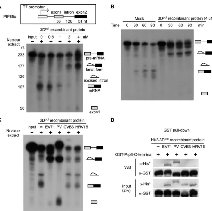
Whether picornaviral polymerase play a role in host pre-mRNA splicing remains unknown. Therefore, we first investigated whether 3Dpol affects the splicing process using in vitro splicing assays. PIP85a pre-mRNA was synthesized and labeled with32P as the substrate, and then mixed with nuclear extracts of HeLa cells and 0.5, 1, 2, or 4mM of EV71 3Dpol recombinant protein for
90 min. The splicing intermediates and products were analyzed by electrophoresis on urea-PAGE gels. The 3Dpolinhibited mRNA production and induced the accumulation of the lariat form in the nuclear extract, depending on the amount of EV71 3Dpol recombinant protein (Figure 3A). Moreover, in vitro splicing was evaluated using a mock treatment or treatment with 4mM of
EV71 3Dpol recombinant protein in a time-course study. The result showed that EV71 3Dpolsuppressed the splicing process and produced the lariat form as early as 30 min after the start of the reaction (Figure 3B). We also determined the inhibitory splicing effect of recombinant 3Dpol proteins from other picornaviruses, including poliovirus (PV), coxsackievirus B3 (CVB3), and human rhinovirus type 16 (HRV16), in which a NLS was identified. Following the same conditions used in thein vitrosplicing analysis, we discovered that PV 3Dpolinhibited the splicing process and led to a decrease in mRNA production and accumulation of the lariat form, similar to the results for EV71; however, neither the CVB3 nor the HRV16 3Dpolinhibited the pre-mRNA splicing process (Figure 3C). These data demonstrate that the 3Dpolof EV71 and PV blocked the second catalytic splicing step involving 39SS cleavage and exon ligation, leading to the accumulation of the lariat form and a decrease on mRNA levels. Because Prp8 is involved in the second catalytic step of pre-mRNA splicing, we next assessed whether the inhibitory effects of these picornaviruses on the splicing process were related to Prp8. Purified His+
-3Dpol from various picornaviruses and GST-Prp8-C-terminal region fusion proteins were mixed and subjected to GST pull-down and WB assays. The results of these assays revealed that the 3Dpol proteins of EV71 and PV directly associate with the C-terminal region of Prp8, whereas the CVB3 and HRV16 3Dpol do not (Figure 3D). Therefore, the 3Dpol-Prp8 interaction is required for inhibition of the second catalytic step. These results suggest that the 3Dpolof EV71 and PV are associated with the splicing factor Prp8 and affect the normal function of Prp8 during the second catalytic splicing step, leading to inhibition of pre-mRNA splicing, accumulation of the lariat form, and a decrease in the resulting mRNA.
3Dpolinhibits intracellular pre-mRNA splicing by interacting with Prp8
We next sought to assess the effects of pre-mRNA splicing upon picornaviral infection. First, we investigated whether EV71 affects the splicing process by interfering the core splicing factor Prp8. The splicing reporter pSV40-CAT(In1) containing human b-globin intron 1 [43,44], which encodes chloramphenicol acetyl transferase, was transfected to RD cells. After viral infection for 2 and 4 h, the RNA expression of the reporter plasmid was measured by reverse transcription quantitative PCR (RT-qPCR). EV71 infection signif-icantly inhibited the splicing of the reporter transcription at 4 h.p.i., resulting an accumulation of the pre-mRNA and a reduction of the mRNA (Figure 4A, lane 5 vs. 6). Consistently, the splicing activity of EV71-infected cells could be restored by the overexpression of HA-tagged Prp8 (Figure 4A, lane 6 vs. 8). To confirm whether EV71 interferes with the endogenous gene splicing process, we designed specific primers for the precursor and mature RNA of the endogenousNCLgene, which encodes the protein nucleolin. The RT-qPCR data revealed that EV71 could suppress endogenous nucleolin pre-mRNA splicing by reacting with Prp8 (Figure 4B), similar to the results obtained by using the exogenous reporter described above. To further confirm that EV71 inhibits the pre-mRNA splicing process through the interaction of viral 3Dpoland host protein Prp8, the pSV40-CAT(In1) reporter was co-transfected with FLAG-tagged 3Dpoland HA-tagged Prp8 to RD cells. 3Dpol alone inhibited the splicing process, and led to increased levels of pre-mRNA and decreased levels of mRNA (Figure 4C, lane 1 vs. 3), whereas overexpression of both 3Dpoland Prp8 restored the pre-mRNA splicing activity (Figure 4C, lane 3 vs. 4). These results demonstrate that EV71 affects the cellular splicing process through the interaction between 3Dpoland Prp8 at 4 h.p.i.
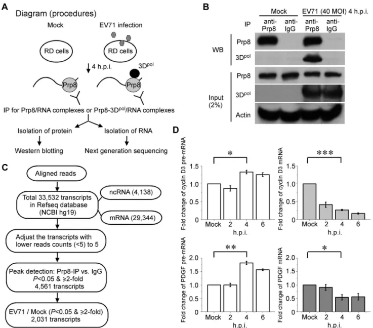
High-throughput sequence screening of the target pre-mRNA captured by Prp8 associated with 3Dpol
After demonstrating that 3Dpolassociates with Prp8 and affects its function during the second step of the splicing process, leading to the accumulation of pre-mRNA intermediates (Figure 3A and 3B), we further analyzed whether endogenous pre-mRNA substrates of Prp8 associate with 3Dpolin EV71-infected RD cells. A diagram for the experimental procedure is provided in Figure 5A. We isolated RD cell lysates that were either infected with EV71 at a MOI of 40 for 4 h or without infection, and then a Prp8 antibody was applied to pull-down the Prp8/RNA or Prp8-3Dpol/RNA complexes in the lysates, named RNA-binding protein IP (RIP) assay. After the Co-IP of protein-RNA complex, the proteins in the precipitates were analyzed by WB analysis. The RNAs in the precipitates were also isolated and subjected to next generation sequencing (NGS) analysis (Figure 5A). Firstly, we verified the binding of Prp8-3Dpol in EV71-infected cells by IP assay with the anti-Prp8 antibody. The Prp8 antibody specifically bound to Prp8, and the Prp8-3Dpolinteraction was detected in the Prp8-3Dpol/RNA complexes from EV71-infected cells. An anti-body against IgG was used as a negative control (Figure 5B). The selection of target RNA for the NGS analysis is illustrated in the flow chart. The sequences were aligned to the human reference genome (hg19, GRCh37) and the aligned regions were annotated as known transcripts based on the Reference Sequence Database (Refseq) of the National Center of Biotechnology Information (NCBI). There are total 33,532 transcripts in Refseq database, including 29,344 mRNA and 4,138 non-coding RNA sequences. We then adjusted the transcripts with read counts lower than 5 (,
forP,0.05 and$2-fold enrichment. Among the 4,561 transcripts, after filtering for P,0.05 and $2-fold, 2,031 transcripts were more highly expressed following EV71 infection than mock infection (Figure 5C). The differentially expressed transcripts were uploaded to DAVID Tools [45], and functional annotations were assigned to the KEGG pathways (Table 2). The comparison of the targeted RNA in EV71 infection and mock infection functionally associated with cell growth, proliferation, and differentiation, and
these associations were related to focal adhesion and the mitogen-activated protein kinase (MAPK) pathway (Figure S1A and S1B; red aster). To validate our RIP-Seq analysis, the intracellular targeted RNAs, such as cyclin D3 of focal adhesion pathway and platelet-derived growth factor (PDGF) of MAPK pathway, were further investigated to confirm the increased levels of pre-mRNA and decreased mRNA levels at 4 to 6 h.p.i. in EV71-infected RD cells by RT-qPCR (Figure 5D).
Figure 3. The EV71 and PV 3Dpolinterfere with the splicing process and inhibit mRNA synthesis.(A) Recombinant EV71 3Dpol inhibits mature mRNA production. Anin vitrosplicing assay was performed for 90 min using32P-labeled PIP85a pre-mRNA as the substrate, nuclear extracts of HeLa cells, and varying amounts of purified recombinant 3Dpol. The autoradiogram revealed the presence of different radioactive RNA forms, including pre-mRNA, the lariat form, excised intron, mature mRNA, and exon1. (B) Recombinant EV71 3Dpolstops the splicing process in the lariat form. Thein vitro splicing substrate,32P-labeled PIP85a pre-mRNA, was incubated with mock- or EV71 3Dpolrecombinant protein-containing nuclear extracts for varying time periods. The autoradiogram shows the different forms of RNAs in the splicing reaction. (C) Recombinant EV71 and PV 3Dpolinhibit the synthesis of mature mRNA. Thein vitrosplicing assay was performed using the same conditions described above, including a protein concentration of 4mM and a reaction time of 90 min, with recombinant 3Dpolproteins from EV71, PV, CVB3, and HRV16. (D) The EV71 and PV 3Dpolproteins directly associate with the C-terminal domain of Prp8.In vitropull-down assay, a total of 5mg of bacterially purified His+-3Dpolfrom EV71, PV, CVB3, or HRV16 was mixed with 5mg of the GST-Prp8-C-terminal domain fusion protein for 90-min reaction time, followed by GST pull-down and WB assays.
doi:10.1371/journal.ppat.1004199.g003
Discussion
Picornaviral 3Dpolplays a key role in viral genome replication in the cytoplasm. Both picornaviral 3CD and 3Dpolhave also been observed in the nucleus as a result of infection, and this localization is mediated through the NLS of 3Dpol. In the nucleus, mature 3C from the precursor 3CD shuts off host cell transcription
[2,19,22]. Although evidence for the entry of picornaviral 3Dpol into the nucleus was first reported approximately a decade ago, the precise role of 3Dpolin the host nucleus has remained unknown. A schematic model is provided in Figure 6. Our study uncovered a novel mechanism for picornaviral 3Dpolinvasion of host cells by its localization to the nucleus and association with the Prp8 protein, which is located at the center of the spliceosome. The viral 3Dpol
Figure 4. 3Dpolaffects cellular pre-mRNA splicing by interacting with Prp8.(A) EV71 inhibits cellular pre-mRNA splicing by interacting with Prp8. RD cells were transfected with pCMV-HA (lanes 1, 2, 5, and 6) or the vector encoding HA-tagged Prp8 (lanes 3, 4, 7, and 8). After 24 h, the exogenous reporter pSV40-CAT(In1), which encodes chloramphenicol acetyl transferase inserted by humanb-globin intron 1, was transfected into all of the samples for 24 h. The total RNA obtained from RD cells was subsequently harvested after EV71 40 MOI infection at 2 h.p.i. (lanes 2 and 4) and 4 h.p.i. (lanes 6 and 8) for RT-qPCR. The fold changes in the amount of pre-mRNA and mRNA were calculated. The overexpression of HA-tagged Prp8 and the level of viral 3Dpolin infected cells were detected using anti-HA and anti-EV71 3Dpolantibodies, respectively, in a WB assay. (B) To confirm the effects of EV71 on endogenous splicing, RNAs were isolated from EV71-infected cells at 2 to 4 h.p.i. and evaluated with a specific primer for nucleolin by RT-qPCR. (C) 3Dpolinhibits cellular pre-mRNA splicing by interacting with Prp8. RD cells were transfected with constructs encoding FLAG-tagged 3Dpol(lanes 3 and 4) or HA-tagged Prp8 (lanes 2 and 4). The vectors pFLAG-CMV2 and pCMV-HA were used as negative controls (lane 1). The exogenous reporter pSV40-CAT(In1) was transfected into all of the samples for 24 h, and the total RNA obtained was subsequently harvested from RD cells for RT-qPCR. The fold changes in the amount of pre-mRNA and mRNA were calculated. In a WB assay, the overexpression of HA-tagged Prp8 and the level of FLAG-tagged 3Dpolwere detected using anti-HA and anti-FLAG antibodies, respectively. Error bars, mean
6SD (n = 3). The statistical significance was analyzed using attest.***p,0.001;**p,0.01.
affects the splicing function of Prp8 in the C1-complex and inhibits the second step of the splicing process, resulting in accumulation of the lariat form and a reduction on mRNA levels. The intracellular targeted RNAs that are trapped by the Prp8-3Dpolcomplexes are primarily responsible for cell growth, proliferation, and differen-tiation.
The host nuclear protein Sam68 has been shown to interact with PV 3Dpolusing a yeast two-hybrid system, but the function of this Sam68-3Dpolinteraction remains unknown [46]. In this study, we identified 15 novel proteins that act as host substrates for EV71 3Dpol using IP assays with a 3Dpol monoclonal antibody and MALDI-TOF MS analysis. We further selected the nuclear protein Prp8, which occupies a central position in U5 snRNP
complexes, for further analysis and confirmed the interaction between endogenous Prp8 and viral 3Dpol without the interme-diation of RNA. Prp8 provides a large platform for the RNA helicase Brr2, the GTPase Snu114, and Prp6 to form U5 snRNP complexes [33,37]. Our results revealed that 3Dpolinteracts with Prp8 and could be pulled down with other components of U5 snRNPs, including Brr2, Snu114, Prp6, and SNRNP40, by Co-IP and WB analysis. We further demonstrated that the fingers domain (1–286 aa) of 3Dpolassociates with the C-terminal region (2094–2335 aa) containing the Jab1/MPN domain of Prp8 by overexpressing various truncated forms of Prp8. Moreover, the 3Dpol proteins of EV71 and PV directly associate with the C-terminal region of Prp8 inin vitropull-down assays.
Figure 5. RIP-seq of the pre-mRNA trapped by the Prp8-3Dpolcomplexes.(A) Procedural diagram for the RIP assays. The schematic shows the experimental procedure for characterizing RNA from Prp8 or Prp8-3Dpolcomplexes by IP. (B) The Prp8 antibody pulls down the Prp8 and Prp8-3Dpolcomplexes. In the WB analysis, the pulled down Prp8 and Prp8-3Dpolcomplexes demonstrated the efficiency of Prp8 IP and the interaction between Prp8 and 3Dpol. (C) A flow chart for the selection of targeted RNAs from the sequencing data. The schematic shows the experimental procedure for characterizing RNAs from Prp8 or Prp8-3Dpolcomplexes. (D) The inhibition of the pre-mRNA splicing in intracellular targeted cyclin D3 and PDGF. The increase in pre-mRNA and decrease in mRNA for intracellular cyclin D3 and PDGF in EV71-infected cells at 4–6 h.p.i. validated our RIP-Seq analysis. Error bars, mean6SD (n = 3). The statistical significance was analyzed using attest.***p,0.001;**p,0.01;*p,0.05.
doi:10.1371/journal.ppat.1004199.g005
The 3Dpolof picornaviruses, such as PV, EMCV, HRV16, and human parechovirus-1 (HPEV-1), can enter the nucleus upon viral infection due to the expression of a NLS [19–22]. This NLS is partially contained within a conserved sequence, KKRD (126–129 aa), that is present in all known picornaviral RNA polymerases and may therefore play a crucial function in the life cycle of the virus [22]. However, whether EV71 can enter the nucleus in virus-infected cells remains unknown. In this study, we first observed that EV71 3Dpol entered the nucleus during the early stages of viral entry at 4 h.p.i., as demonstrated by anti-3Dpol antibody detection by confocal imaging and nuclear fractionation analysis. In contrast to PV, 3Dpolalone could directly and independently enter the nucleus without EV71 infection via the NLS KKKD, which spans aa 126–129. However, mutation of the NLS of 3Dpol by replacing the sequence KKKD with AAAA prevented nuclear entry. We also observed that 3Dpoland Prp8 were colocalized at 4 h.p.i. in the nucleus and at 6–8 h.p.i. in the cytoplasm of
EV71-infected cells. Furthermore, the 3Dpol-Prp8 interaction was maintained between 4 and 8 h.p.i. These results suggest that this interaction blocks the cellular pre-mRNA splicing process at the early stages of viral entry and may have advantages for the viral life cycle during the later stages. Picornavirus infection inhibits the cellular translation machinery of the host, reducing the accumu-lation of the cellular proteins, including Prp8 at 8 h.p.i. (Figure 1C). Furthermore, 3Dpoland Prp8 were co-localized in the nucleus at 4 h.p.i. and in the cytoplasm at 8 h.p.i. (Figure 2A). This interaction in the nucleus disrupts the cellular splicing machinery of the host, whereas the interaction in the cytoplasm may support the function of 3Dpolduring viral infection, and this phenomenon is of worthy of further exploration.
Previous studies have reported that picornaviruses influence host cell gene expression by shutting off cellular transcription and cap-dependent mRNA translation [2,47–49]. Poliovirus 2A protease modulates the cellular alternative splicing [50]. In this
Table 2.The differentially expressed transcripts were classed into groups according to functional annotations from the KEGG pathways.
Term Count % PValue List Total Pop Hits Pop Total
hsa04510:Focal adhesion 40 2.3474178 5.28E-05 523 201 5085
hsa04010:MAPK signaling pathway 47 2.7582160 2.34E-04 523 267 5085
hsa05200:Pathways in cancer 50 2.9342723 0.00380621 523 328 5085
hsa05221:Acute myeloid leukemia 14 0.8215962 0.00497523 523 58 5085 hsa04810:Regulation of actin cytoskeleton 35 2.0539906 0.00635485 523 215 5085
hsa05215:Prostate cancer 18 1.0563380 0.00807954 523 89 5085
hsa05220:Chronic myeloid leukemia 16 0.9389671 0.00814590 523 75 5085 hsa00030:Pentose phosphate pathway 8 0.4694836 0.01069550 523 25 5085
hsa04144:Endocytosis 30 1.7605634 0.01179329 523 184 5085
hsa04722:Neurotrophin signaling pathway 22 1.2910798 0.01396748 523 124 5085 hsa00562:Inositol phosphate metabolism 12 0.7042254 0.01921380 523 54 5085
hsa05216:Thyroid cancer 8 0.4694836 0.02424746 523 29 5085
hsa04910:Insulin signaling pathway 22 1.2910798 0.03344581 523 135 5085
hsa04540:Gap junction 16 0.9389671 0.03585054 523 89 5085
hsa04115:p53 signaling pathway 13 0.7629108 0.04190497 523 68 5085
hsa05016:Huntington’s disease 27 1.5845070 0.04431270 523 180 5085
hsa05211:Renal cell carcinoma 13 0.7629108 0.05081584 523 70 5085
hsa00051:Fructose and mannose metabolism 8 0.4694836 0.05358196 523 34 5085
hsa03010:Ribosome 15 0.8802817 0.05820913 523 87 5085
hsa05219:Bladder cancer 9 0.5281690 0.06040354 523 42 5085
hsa05414:Dilated cardiomyopathy 15 0.8802817 0.08490081 523 92 5085
hsa04512:ECM-receptor interaction 14 0.8215962 0.08516606 523 84 5085
hsa04142:Lysosome 18 1.0563380 0.08763026 523 117 5085
hsa04530:Tight junction 20 1.1737089 0.08958635 523 134 5085
hsa04730:Long-term depression 12 0.7042254 0.09139957 523 69 5085
hsa04520:Adherens junction 13 0.7629108 0.09179898 523 77 5085
hsa00600:Sphingolipid metabolism 8 0.4694836 0.09875063 523 39 5085
Footnotes. Term: gene set name.
Count: number of genes associated with this gene set.
Percentage (%): gene associated with this gene set/total number of query genes. P-value: modified Fisher Exact P-value.
List Total: number of genes in your query list mapped to any gene set in this ontology. Pop Hits: number of genes annotated to this gene set on the background list.
study, we examined whether picornaviral polymerase could impair cellular pre-mRNA splicing processes by interfering with Prp8. Thein vitrosplicing results demonstrated that the second step of the splicing process was blocked by the 3Dpolof EV71 and PV, leading to inhibition of pre-mRNA splicing, the accumulation of the lariat form, and a decrease in mRNA synthesis. However, the 3Dpolof CVB3 and HRV16 did not inhibit pre-mRNA splicing and did not exhibit any association with the C-terminal region of Prp8. Therefore, our data support the theory that the viral 3Dpolinhibits pre-mRNA splicing through an association with cellular Prp8 in the nucleus. However, the splicing effect of the 3Dpolof EV71 and PV in the cellular nucleus differs from that of CVB3 and HRV16, which represents a promising theme for future research.
Moreover, we demonstrated that EV71 infection inhibited the splicing of exogenous pSV40-CAT(In1) and endogenous nucleo-lin. However, the splicing activity of the viral infected cells could be restored by overexpression of HA-tagged Prp8. We also confirmed that 3Dpol alone inhibits the splicing processes by reacting with cellular Prp8. Our study provides a new insight into EV71-mediated inhibition of the pre-mRNA splicing by the 3Dpol -Prp8 interaction. We also transfected FLAG-CVB3-3Dpolplasmid DNA into RD cells. The results indicated that FLAG-CVB3-3Dpol was unable to inhibit the splicing process (lane 1 vs. 3), and did not lead to increased levels of pre-mRNA. However, FLAG-CVB3-3Dpolwas capable of decreasing the levels of both pre-mRNA and mature mRNA (Figure S2A). This observation was also confirmed using an in vitro splicing assay (Figure 3C) that examined the CVB3 3Dpolrecombinant protein versus a control without 3Dpol. These findings suggest that FLAG-EV71-3Dpolcan inhibit splicing through interference with Prp8, whereas FLAG-CVB3-3Dpoldid not. RD cells were infected with CVB3, a non-Prp8 interacting virus, at 2 to 6 h.p.i. (Figure S2B). The data indicated that CVB3 was unable to inhibit the splicing process at 4 h.p.i., whereas it was
capable of decreasing the pre-mRNA and mature mRNA of cyclin D3 and PDGF. This result is consistent with those obtained with RD cells transfected with FLAG-CVB3-3Dpol(Figure S2A).
Using high-throughput sequence screening of the target mRNA, we evaluated in detail the types of endogenous pre-mRNAs that were captured when Prp8 was bound to 3Dpol in EV71-infected RD cells. Comparison of the targeted RNA in EV71- and mock-infected cells revealed that most of the gene expression and transcription generating pre-mRNAs was associ-ated with cell growth, proliferation, and differentiation, processes classified in the focal adhesion and MAPK pathways. In addition, the 3Dpol-Prp8 interaction complexes disrupted mRNA synthesis and downstream protein expression. Cell-matrix adhesions are critical for biological processes such as cell motility, proliferation, differentiation, regulation of gene expression, and cell survival, and the MAPK cascade is a highly conserved pathway that is involved in numerous cellular functions, such as cell proliferation, differentiation, and migration. Viral 3Dpol-mediated blockade of the pre-mRNA splicing process results in the production of less cellular mRNA and the use of more translational machinery by viral RNA to make viral proteins efficiently. The disruption of cellular mRNA synthesis by the infecting virus may also directly cause cell damage and death. Timely cell death would facilitate the release of some picornaviruses, e.g., enterovirus 71. Furthermore, certain picornavirus genome sequences may contain fortuitous splice sequences. Erroneous recognition of such sequences by cytoplasmic splicing components would require the presence of some sort of anti-splicing defensive mechanism.
In summary, our study reports a novel invasion strategy for picornaviruses in which 3Dpol enters the host cell nucleus and associates with Prp8. This association leads to the inhibition of pre-mRNA splicing, accumulation of the lariat form, and a decrease in mRNA synthesis. However, the pre-mRNA and the intermediate
Figure 6. Schematic model of 3Dpol-mediated inhibition of the cellular splicing by targeting Prp8 in the nucleus.3Dpolprimarily performs viral RNA replication in the host cytoplasm, but partially 3Dpolalso enters the nucleus and interacts with the core splicing factor Prp8, which interferes with the function of Prp8 in the C1-complex. The interference of Prp8 function inhibits the second step of the splicing process and results in the accumulation of the lariat form and a reduction in mRNA synthesis.
doi:10.1371/journal.ppat.1004199.g006
lariat form become occupied by the 3Dpol-Prp8 interaction during infection, thereby blocking mRNA synthesis of numerous cellular genes, including those associated with cell growth, proliferation, and differentiation.
Picornaviruses inhibit the cellular transcription and cap-dependent mRNA translation to affect the genes expression of the host cell [2,47–49]. However, several cellular genes could escape the shutoff of gene expression by picornavirus infection. Our previous investigations of cDNA microarray analysis for total cellular RNA demonstrated that the level of some RNAs that are related to chemokines, protein degradation, complement proteins and proapoptosis proteins increased upon EV71 infection, suggesting leakage from the inhibition of transcription by EV71 [51]. Translations of c-myc, Bip, and eIF4G mRNA have been observed to be increased in poliovirus-infected cells as the cap-dependent translation shuts down, owing to the presence of internal ribosome entry sites (IRES) [52–54]. This work presents a novel mechanism by which cytoplasmic viral RdRp inhibits cellular gene expression in the pre-mRNA splicing processing step, which would inhibit mRNA synthesis of the surviving host RNAs, potentially providing yet another advantage of virus replication.
Materials and Methods
Cell cultures and virus infection
Human RD, HEK293T, and HeLa cells were cultured in DMEM containing FBS and penicillin/streptomycin/glutamine (Gibco) at 37uC. EV71 (TW/4643/98) virus infection at a MOI of 40 was performed under serum-free conditions for 1 h at 37uC. After 1 h of incubation, the virus-infected cells were washed twice in PBS, and the medium was replaced with DMEM containing 2% FBS to maintain the virus-infected cells at 37uC.
Immunoprecipitation and protein identification
To identify potential EV71 3Dpol-interacting host proteins, 5 mg of cell lysates from EV71 40 MOI-infected RD cells at 6 h.p.i. was harvested for immunoprecipitation and treated with 250mg of EV71 3Dpolmonoclonal antibody (self-preparation) and
100ml of protein A-Sepharose (GE Healthcare) at 4uC. After centrifugation and bead washing, the co-precipitated proteins were separated by 8–16% gradient SDS-PAGE, which was followed by silver staining. The proteins were identified using in-gel digestion and analyzed by Bruker Ultraflex MALDI-TOF MS. Mass lists were performed peptide mass fingerprinting by Biotool 2.0 software and the algorithm of Mascot (http://www. matrixscience.com).
Co-immunoprecipitation and western blotting
RD cells (2.46106/10-cm dish) were seeded 24 h prior to EV71
infection at a MOI of 40. Cells were lysed in 1 ml of IP-lysis buffer (25 mM Tris-HCl pH 7.6, 300 mM NaCl, 0.5% CA630, 1.5 mM MgCl2, 0.2 mM EDTA, 0.5 mM DTT, and 16 proteinase
inhibitor) at 4uC for 30 min and then treated with 10mg/ml
RNase A at 30uC for 1 h. The cell extracts were pre-cleared by incubation at 4uC for 1 h with protein G-agarose (GE Healthcare) and centrifuged to remove non-specific complexes. The lysate was then added to 10mg/ml EV71 3Dpolantibody or Prp8 antibody (Abcam) at 4uC for 2 h and 100ml of protein G-agarose at 4uC for 12 h. The co-precipitated proteins were collected by centrifuga-tion, followed by washing 6 times with IP buffer (25 mM Tris-HCl pH 7.6, 200 mM NaCl, 0.1% CA630, 6% glycerol, 1 mM EDTA, 0.5 mM DTT, and 16proteinase inhibitor). The precipitated
proteins were separated by 8% SDS-PAGE; subsequently, these immune complexes were detected using anti-Prp8 (diluted 1:5000;
Abcam), Brr2 (diluted 1:5000; Abcam), Snu114 (diluted 1:5000; Abcam), Prp6 (diluted 1:5000; Abcam), SNRNP40 (diluted 1:5000; Abcam), EV71 3Dpol(diluted 1:10000; self-preparation), andb-actin (diluted 1:10000; Millipore) antibodies in a WB assay.
Plasmid construction and FLAG immunoprecipitation
To map the interacting domains between 3Dpoland Prp8, the full-length and various truncated forms of human Prp8 were amplified by PCR from the human Prp8-pCMV-XL5 cDNA clone (OriGene) using specific primers. The PCR product was inserted into a pCMV-HA vector (Clontech) between the XhoI and NotI sites to enable the expression of HA-tagged proteins. The EV71 length infection cDNA clone was used to amplify full-length and various truncated forms of EV71 3Dpol by PCR, followed by cloning into the EcoRI and KpnI sites of the p3XFLAG-Myc-CMV-25 vector (Sigma) to enable expression of the EV71 3Dpol constructs as fusions with 3 adjacent FLAG epitopes. To overexpress these proteins, 2mg of the constructs of
the various truncated forms of Prp8 and 3Dpolwas co-transfected into HEK293T cells (16106/per 6-well plate) using Lipofectamine 2000 reagent (Invitrogen) for 48 h. The cells were harvested for FLAG-IP using a FLAG-immunoprecipitation kit (Sigma). After lysis and centrifugation, the supernatant was treated with 10mg/ ml RNase A at 30uC for 1 h, and then 40ml of anti-FLAG M2 agarose affinity gel was added at 4uC for 12 h. Proteins were then eluted by competition with 36FLAG peptide. In the WB assay,
the precipitated proteins were identified using an anti-HA antibody (diluted 1:5000; Sigma) and an anti-FLAG antibody (diluted 1:5000; Sigma).
Construction of the mutant FLAG-3Dpol NLS,
immunofluorescence microscopic analysis, and cellular fractionation
The PCR product of EV71 3Dpol containing the wt NLS (KKKD) was cloned into the pFLAG-CMV-2 vector (Sigma) between the EcoRI and KpnI sites to enable expression of the protein as a fusion with the FLAG epitope protein. The mutated NLS (AAAA)-containing pFLAG-3Dpolclone was generated using specific primers (Table S1) in 2 steps of PCR and digestion and subsequently cloned into the pFLAG-CMV-2 vector. The wt and mutant clones of EV71 3Dpolwere verified by sequencing. For immunofluorescence microscopic analysis, RD cells grown in 22-mm-diameter wells at 80% confluency were infected with EV71 at a MOI of 40 for 2 to 8 h.p.i. or were transfected with 4mg of the wild-/mutant-type 3Dpol clone. The cells were fixed in PBS containing 4% formaldehyde, permeated with 0.3% Triton X-100, blocked with 0.5% BSA for 1 h at 25uC, and then stained with anti-EV71 3Dpol (diluted 1:200; self-preparation), anti-Prp8 (diluted 1:15; Abcam), or anti-FLAG (diluted 1:200; Sigma) antibodies for 2 h at 37uC. Subsequently, the cells were stained with FITC-conjugated goat anti-mouse IgG (diluted 1:400; green; Invitrogen) or goat anti-rabbit IgG (diluted 1:75; red; Invitrogen) for 2 h at 37uC, followed by treatment with nuclear Hoechst 33258 stain (diluted 1:500; blue) for 15 min at 25uC. The cells were washed 3 times with PBS and mounted on glass slides with Prolong Gold (Invitrogen). Confocal images were obtained with a confocal laser-scanning microscope (Zeiss; LSM 510 NLO). To prepare EV71-infected RD cells (2.46106/10-cm dish) for
cytoplasmic and nuclear fractionation, the cells were lysed in 300ml of buffer C, provided in the CMN compartment protein
for 1 h at 4uC. The same volume percent of cellular fractionation was loaded onto an 8% SDS-PAGE gel, and GAPDH (diluted 1:5000; Abnova) and Lamin A/C (diluted 1:5000; Santa Cruz) were detected as internal controls for the cytoplasmic and nuclear fractions, respectively, by WB.
In vivosplicing assay and reverse transcription quantitative PCR
RD cells were transfected with 4mg of the pCMV-HA vector or HA-tagged Prp8 clone for 24 h, followed by transfection with 0.5mg of the pSV40-CAT(In1) splicing reporter (a gift from Dr.
Woan-Yuh Tarn, Academia Sinica, Taiwan) [44] for 24 h. The RD cells were infected with EV71 at a MOI of 40 at 2 and 4 h.p.i., and RNA samples were harvested from cells using an RNeasy mini kit (Qiagen) and treated with RQ-DNase1 (Promega). RNAs were converted into first-strand cDNAs using SuperScript III reverse transcriptase (Invitrogen) with CAT(In1) reverse primers. All PCR reactions were performed using specific primers (Table S1). The qPCR analysis was performed using SYBR Green reagents and the LightCycler 480 instrument (Roche). NCBI GI numbers for genes and proteins mentioned in the text were provided in the Table S2.
Expression and purification of Prp8-C-terminal region and 3Dpolrecombinant proteins
The Prp8-C-terminal region (2094–2335 aa) was cloned into the EcoRI and XhoI sites of the pGEX-5X-1 vector and transformed into BL21(DE3). Expression of the protein was induced by adding 1 mM isopropyl-b-d-thiogalactopyranoside (IPTG) at 16uC for 16 h. The protein expressed from lysed cells was suspended in buffer (20 mM HEPES pH 7.9, 300 mM NaCl, 5 mM DTT, 0.2 mM EDTA, 0.05% NP-40, 0.5 mM PMSF) at 4uC for 30 min and loaded onto a GST column (GE Healthcare), which was then eluted with buffer containing 10 mM Glutathion. The eluted product was dialyzed in buffer (20 mM HEPES pH 7.9, 100 mM NaCl, 1 mM DTT, 0.2 mM EDTA, 0.01% NP-40, 20% glycerol). To construct pET26b-Ub-EV71-3D-6H, the EV71 3Dpolfull-length containing the 66His+
at C-terminal region was cloned into the SacII and BamHI sites of pET26b-Ub-3D-GSSG-6H [55] and used to replace the PV 3D-GSSG-6H of pET26b-Ub-3D-GSSG-6H. pCG1 encodes an Ub-specific carboxy-terminal protease (Ubp1). Expres-sion of Ub-3D fuExpres-sion protein in the presence of Ubp1 has glycine at the amino terminus of polymerase, not methionine. Plasmids pET26b-Ub-EV71-3D-6H and pCG1 or pET26b-Ub-3D-GSSG-6H and pCG1 were cotransformed into BL21(DE3) to express EV71-3D-6H or PV-3D-GSSH-6H recombinant protein, respec-tively. Expression of these recombinant proteins were induced by adding 50mM IPTG at 25uC for 4 h. The protein expressed from lysed cells was suspended in buffer A (50 mM Tris pH 8.0, 20% glycerol, 1 mM DTT, 0.1% NP-40, and 60mM ZnCl2) and loaded
onto a HisTrap column (GE Healthcare), which was then washed with buffer A containing 30, 50, 70, or 90 mM imidazole; the protein was then eluted with buffer A containing 500 mM imidazole. The eluted product was dialyzed in buffer B (50 mM HEPES, pH 7.5, 500 mM NaCl, 20% glycerol, 1 mM DTT, 0.1% NP-40, and 60mM ZnCl2) [18]. The purified 3Dpolrecombinant
proteins of CVB3 and HRV16 were gifts from Dr. Craig E. Cameron. The expression and purification of CVB3 and HRV16 3Dpolhave been described previously [55–57].
In vitrosplicing assay, and glutathione S-transferase (GST) pull-down
The PIP85a plasmid (a gift from Dr. Woan-Yuh Tarn, Academia Sinica, Taiwan) was cleaved using Hind III and labeled
with a-32P-UTP (800 Ci/mmol) using an in vitro transcription system (Promega). In vitro splicing was performed in 15ml of reaction mixtures containing 60% HeLa cell nuclear extracts, 10 mM ATP, 0.4 M creatine phosphate, 48 mM MgCl2, 12 U
RNasin, various recombinant proteins, and 36105cpm 32
P-labeled PIP85a pre-mRNA as the substrate for 1.5 h at 37uC. RNA was extracted with the Trizol reagent (Invitrogen) from the splicing reaction and fractionated on a 5% denaturing polyacryl-amide gel containing 7 M urea, TBE, APS, and TEME. A total of 5mg of each recombinant protein was incubated in IP lysis buffer (25 mM Tris-HCl pH 7.6, 300 mM NaCl, 0.5% CA630, 1.5 mM MgCl2, 0.2 mM EDTA, 0.5 mM DTT, and 16 proteinase
inhibitor) at 4uC for 3 h, and the targeted proteins were immunoprecipitated using GST beads and washed with IP buffer (25 mM Tris-HCl pH 7.6, 200 mM NaCl, 0.1% CA630, 6% glycerol, 1 mM EDTA, 0.5 mM DTT, and 16 proteinase inhibitor). The same concentration (5mg) of GST-Prp8-C-terminal region (2094–2335 aa) and 66His+viral 3Dpol
recom-binant proteins were incubated in IP-lysis buffer for 3 h at 4uC, and Glutathione Sepharose (GE Healthcare) was then added to the mixture. After denaturation in 66loading dye and centrifu-gation, the precipitated proteins were separated by 10% SDS-PAGE and analyzed by WB with an anti-GST antibody (diluted 1:5000; Santa Cruz) and an anti-His+
antibody (diluted 1:5000; Calbiochem).
RNA-binding protein immunoprecipitation-sequencing (RIP-Seq)
The 20 dishes of Mock or EV71-infected RD cells (9.66106
/15-cm dish) were harvested in IP-lysis buffer, and the lysates were then added to the Prp8 antibody or control IgG antibody (Abcam) for RIP analysis. RNA-protein complexes were immunoprecipitated with protein G agarose beads, and total RNA was extracted by treatment with proteinase K and Trizol and prepared for sequencing. RNA sequencing was performed by the Genomics Core Laboratory of the Molecular Medicine Research Center, Chang Gung University. SOLiD sequencing libraries were prepared using the SOLiD Total RNA Sequencing kit (ABI PN4445374) according to the manufacturer’s instructions. Approximately 1mg of total immunoprecipitated RNA was used as the starting material. The samples were then subjected to ribosomal RNA removal using Ribo-Zero Gold kits (human/mouse/rat) (Epicentre); subsequently, 100 ng of the rRNA-depleted RNAs was fragmented using RNase III. After purification, 50 ng of each fragmented sample was ligated with RNA adaptors. An Agilent 2100 Bioanalyzer was used to profile the distribution of the fragmented RNA (the median size was between 125 to 140 nt). After reverse transcription and size selection, each cDNA library was amplified using distinct barcoded 39 PCR primers from the SOLiD RNA barcoding kits (PN 4427046). The insert size distribution and the concentration of each library were measured using the Agilent 2100 Bioanalyzer. From each library, equal concentrations (0.8 pM) were pooled together and sequenced strand-specifically on an ABI SOLiD5500 platform (Life Technol-ogies, Foster City, CA, USA) to generate 75-bp tags. The single-end sequence data were aligned to the hg19 (GRCh37) human reference genome. The transcripts were uploaded to DAVID Tools, and functional annotations were assigned based on the KEGG pathways.
Supporting Information
Figure S1 Pathway maps. (A) KEGG pathway entry
(hsa04510) for focal adhesion. (B) KEGG pathway entry (hsa04010) for MAPK signaling pathway.
(TIF)
Figure S2 CVB3 3Dpol is unable to inhibit the cellular splicing process.(A) CVB3 3Dpolleads to decreased levels of both pre-mRNA and mature mRNA. RD cells were transfected with constructs encoding FLAG-tagged CVB3 3Dpol(lanes 3 and 4) or HA-tagged Prp8 (lanes 2 and 4). The vectors pFLAG-CMV2 and pCMV-HA were used as negative controls (lane 1). The exogenous reporter pSV40-CAT(In1) was transfected into all of the samples for 24 h, and the total RNA obtained was subsequently harvested from RD cells for RT-qPCR. The fold changes in the amount of pre-mRNA and mRNA were calculated. In a WB assay, the overexpression of HA-tagged Prp8 and the level of FLAG-tagged CVB3 3Dpolwere detected using anti-HA and anti-FLAG antibodies, respectively. (B) CVB3 is unable to inhibit the splicing process in intracellular cyclin D3 and PDGF. CVB3 decreased the pre-mRNA and mRNA of intracellular cyclin D3 and PDGF in CVB3 40 MOI-infected RD cells.
(TIF)
Table S1 The sequences of primers.
(DOC)
Table S2 A list of NCBI GI numbers for genes and
proteins mentioned in the text.
(DOC)
Acknowledgments
We thank Dr. Woan-Yuh Tarn and Dr. Kuo-Ming Lee (Institute of Biomedical Sciences, Academia Sinica, Taiwan) for technical support in the pre-mRNA splicing and for providing the pSV40-CAT(In1) and PIP85a plasmids. We also thank Dr. Chih-Ching Wu (Proteomics Core Laboratory, Chang Gung University, Taiwan) and Dr. Po-Jung Huang (Bioinformatics Center, Chang Gung University, Taiwan) for helpful discussions and technical support.
Author Contributions
Conceived and designed the experiments: YCL SRS. Performed the experiments: YCL RLK JYL PNH YH HL JJA. Analyzed the data: YCL SJC RYLW CEC SRS. Wrote the paper: YCL CEC SRS.
References
1. Bedard KM, Semler BL (2004) Regulation of picornavirus gene expression. Microbes and infection/Institut Pasteur 6: 702–713.
2. Weidman MK, Sharma R, Raychaudhuri S, Kundu P, Tsai W, et al. (2003) The interaction of cytoplasmic RNA viruses with the nucleus. Virus research 95: 75– 85.
3. Weng KF, Li ML, Hung CT, Shih SR (2009) Enterovirus 71 3C protease cleaves a novel target CstF-64 and inhibits cellular polyadenylation. PLoS pathogens 5: e1000593.
4. Chang LY, Huang LM, Gau SS, Wu YY, Hsia SH, et al. (2007) Neurodevelopment and cognition in children after enterovirus 71 infection. The New England journal of medicine 356: 1226–1234.
5. Whitton JL, Cornell CT, Feuer R (2005) Host and virus determinants of picornavirus pathogenesis and tropism. Nature reviews Microbiology 3: 765– 776.
6. Lin KH, Hwang KP, Ke GM, Wang CF, Ke LY, et al. (2006) Evolution of EV71 genogroup in Taiwan from 1998 to 2005: an emerging of subgenogroup C4 of EV71. Journal of medical virology 78: 254–262.
7. Ding NZ, Wang XM, Sun SW, Song Q, Li SN, et al. (2009) Appearance of mosaic enterovirus 71 in the 2008 outbreak of China. Virus research 145: 157– 161.
8. Seiff A (2012) Cambodia unravels cause of mystery illness. Lancet 380: 206. 9. Marcotte LL, Wass AB, Gohara DW, Pathak HB, Arnold JJ, et al. (2007) Crystal
structure of poliovirus 3CD protein: virally encoded protease and precursor to the RNA-dependent RNA polymerase. Journal of virology 81: 3583–3596. 10. Thompson AA, Peersen OB (2004) Structural basis for proteolysis-dependent
activation of the poliovirus RNA-dependent RNA polymerase. The EMBO journal 23: 3462–3471.
11. Paul AV, van Boom JH, Filippov D, Wimmer E (1998) Protein-primed RNA synthesis by purified poliovirus RNA polymerase. Nature 393: 280–284. 12. Paul AV, Peters J, Mugavero J, Yin J, van Boom JH, et al. (2003) Biochemical
and genetic studies of the VPg uridylylation reaction catalyzed by the RNA polymerase of poliovirus. Journal of virology 77: 891–904.
13. Paul AV, Yin J, Mugavero J, Rieder E, Liu Y, et al. (2003) A ‘‘slide-back’’ mechanism for the initiation of protein-primed RNA synthesis by the RNA polymerase of poliovirus. The Journal of biological chemistry 278: 43951– 43960.
14. Hsu NY, Ilnytska O, Belov G, Santiana M, Chen YH, et al. (2010) Viral reorganization of the secretory pathway generates distinct organelles for RNA replication. Cell 141: 799–811.
15. Ng KK, Arnold JJ, Cameron CE (2008) Structure-function relationships among RNA-dependent RNA polymerases. Current topics in microbiology and immunology 320: 137–156.
16. Wu Y, Lou Z, Miao Y, Yu Y, Dong H, et al. (2010) Structures of EV71 RNA-dependent RNA polymerase in complex with substrate and analogue provide a drug target against the hand-foot-and-mouth disease pandemic in China. Protein & cell 1: 491–500.
17. Campagnola G, Weygandt M, Scoggin K, Peersen O (2008) Crystal structure of coxsackievirus B3 3Dpol highlights the functional importance of residue 5 in picornavirus polymerases. Journal of virology 82: 9458–9464.
18. Chen TC, Chang HY, Lin PF, Chern JH, Hsu JT, et al. (2009) Novel antiviral agent DTriP-22 targets RNA-dependent RNA polymerase of enterovirus 71. Antimicrobial agents and chemotherapy 53: 2740–2747.
19. Aminev AG, Amineva SP, Palmenberg AC (2003) Encephalomyocarditis virus (EMCV) proteins 2A and 3BCD localize to nuclei and inhibit cellular mRNA transcription but not rRNA transcription. Virus research 95: 59–73.
20. Amineva SP, Aminev AG, Palmenberg AC, Gern JE (2004) Rhinovirus 3C protease precursors 3CD and 3CD9localize to the nuclei of infected cells. The Journal of general virology 85: 2969–2979.
21. Krogerus C, Samuilova O, Poyry T, Jokitalo E, Hyypia T (2007) Intracellular localization and effects of individually expressed human parechovirus 1 non-structural proteins. The Journal of general virology 88: 831–841.
22. Sharma R, Raychaudhuri S, Dasgupta A (2004) Nuclear entry of poliovirus protease-polymerase precursor 3CD: implications for host cell transcription shut-off. Virology 320: 195–205.
23. Clark ME, Hammerle T, Wimmer E, Dasgupta A (1991) Poliovirus proteinase 3C converts an active form of transcription factor IIIC to an inactive form: a mechanism for inhibition of host cell polymerase III transcription by poliovirus. The EMBO journal 10: 2941–2947.
24. Clark ME, Lieberman PM, Berk AJ, Dasgupta A (1993) Direct cleavage of human TATA-binding protein by poliovirus protease 3C in vivo and in vitro. Molecular and cellular biology 13: 1232–1237.
25. Yalamanchili P, Datta U, Dasgupta A (1997) Inhibition of host cell transcription by poliovirus: cleavage of transcription factor CREB by poliovirus-encoded protease 3Cpro. Journal of virology 71: 1220–1226.
26. Yalamanchili P, Weidman K, Dasgupta A (1997) Cleavage of transcriptional activator Oct-1 by poliovirus encoded protease 3Cpro. Virology 239: 176–185. 27. Falk MM, Grigera PR, Bergmann IE, Zibert A, Multhaup G, et al. (1990) Foot-and-mouth disease virus protease 3C induces specific proteolytic cleavage of host cell histone H3. Journal of virology 64: 748–756.
28. Weidman MK, Yalamanchili P, Ng B, Tsai W, Dasgupta A (2001) Poliovirus 3C protease-mediated degradation of transcriptional activator p53 requires a cellular activity. Virology 291: 260–271.
29. Jurica MS, Moore MJ (2003) Pre-mRNA splicing: awash in a sea of proteins. Molecular cell 12: 5–14.
30. Turner IA, Norman CM, Churcher MJ, Newman AJ (2004) Roles of the U5 snRNP in spliceosome dynamics and catalysis. Biochemical Society transactions 32: 928–931.
31. Valadkhan S, Jaladat Y (2010) The spliceosomal proteome: at the heart of the largest cellular ribonucleoprotein machine. Proteomics 10: 4128–4141. 32. Pena V, Liu S, Bujnicki JM, Luhrmann R, Wahl MC (2007) Structure of a
multipartite protein-protein interaction domain in splicing factor prp8 and its link to retinitis pigmentosa. Molecular cell 25: 615–624.
33. Galej WP, Oubridge C, Newman AJ, Nagai K (2013) Crystal structure of Prp8 reveals active site cavity of the spliceosome. Nature 493: 638–643.
34. Butler MI, Poulter RT (2005) The PRP8 inteins in Cryptococcus are a source of phylogenetic and epidemiological information. Fungal genetics and biology: FG & B 42: 452–463.
35. Wahl MC, Will CL, Luhrmann R (2009) The spliceosome: design principles of a dynamic RNP machine. Cell 136: 701–718.
36. Grainger RJ, Beggs JD (2005) Prp8 protein: at the heart of the spliceosome. Rna 11: 533–557.
37. Liu S, Rauhut R, Vornlocher HP, Luhrmann R (2006) The network of protein-protein interactions within the human U4/U6.U5 tri-snRNP. Rna 12: 1418– 1430.
38. Li X, Zhang W, Xu T, Ramsey J, Zhang L, et al. (2013) Comprehensive in vivo RNA-binding site analyses reveal a role of Prp8 in spliceosomal assembly. Nucleic acids research 41: 3805–3818.
40. Kramer A (1996) The structure and function of proteins involved in mammalian pre-mRNA splicing. Annual review of biochemistry 65: 367–409.
41. Wachtel C, Manley JL (2009) Splicing of mRNA precursors: the role of RNAs and proteins in catalysis. Molecular bioSystems 5: 311–316.
42. Aronova A, Bacikova D, Crotti LB, Horowitz DS, Schwer B (2007) Functional interactions between Prp8, Prp18, Slu7, and U5 snRNA during the second step of pre-mRNA splicing. Rna 13: 1437–1444.
43. Lin KT, Lu RM, Tarn WY (2004) The WW domain-containing proteins interact with the early spliceosome and participate in pre-mRNA splicing in vivo. Molecular and cellular biology 24: 9176–9185.
44. Lee KM, Hsu Ia W, Tarn WY (2010) TRAP150 activates pre-mRNA splicing and promotes nuclear mRNA degradation. Nucleic acids research 38: 3340– 3350.
45. Huang da W, Sherman BT, Lempicki RA (2009) Systematic and integrative analysis of large gene lists using DAVID bioinformatics resources. Nature protocols 4: 44–57.
46. McBride AE, Schlegel A, Kirkegaard K (1996) Human protein Sam68 relocalization and interaction with poliovirus RNA polymerase in infected cells. Proceedings of the National Academy of Sciences of the United States of America 93: 2296–2301.
47. Lin JY, Chen TC, Weng KF, Chang SC, Chen LL, et al. (2009) Viral and host proteins involved in picornavirus life cycle. Journal of biomedical science 16: 103.
48. Belsham GJ (2009) Divergent picornavirus IRES elements. Virus research 139: 183–192.
49. Ho BC, Yu SL, Chen JJ, Chang SY, Yan BS, et al. (2011) Enterovirus-induced miR-141 contributes to shutoff of host protein translation by targeting the translation initiation factor eIF4E. Cell host & microbe 9: 58–69.
50. Alvarez E, Castello A, Carrasco L, Izquierdo JM (2011) Alternative splicing, a new target to block cellular gene expression by poliovirus 2A protease. Biochemical and biophysical research communications 414: 142–147. 51. Shih SR, Stollar V, Lin JY, Chang SC, Chen GW, et al. (2004) Identification of
genes involved in the host response to enterovirus 71 infection. Journal of neurovirology 10: 293–304.
52. Macejak DG, Sarnow P (1991) Internal initiation of translation mediated by the 59leader of a cellular mRNA. Nature 353: 90–94.
53. Johannes G, Sarnow P (1998) Cap-independent polysomal association of natural mRNAs encoding c-myc, BiP, and eIF4G conferred by internal ribosome entry sites. Rna 4: 1500–1513.
54. Thoma C, Bergamini G, Galy B, Hundsdoerfer P, Hentze MW (2004) Enhancement of IRES-mediated translation of the c-myc and BiP mRNAs by the poly(A) tail is independent of intact eIF4G and PABP. Molecular cell 15: 925–935.
55. Gohara DW, Ha CS, Kumar S, Ghosh B, Arnold JJ, et al. (1999) Production of ‘‘authentic’’ poliovirus RNA-dependent RNA polymerase (3D(pol)) by ubiquitin-protease-mediated cleavage in Escherichia coli. Protein expression and purification 17: 128–138.
56. van Ooij MJ, Vogt DA, Paul A, Castro C, Kuijpers J, et al. (2006) Structural and functional characterization of the coxsackievirus B3 CRE(2C): role of CRE(2C) in negative- and positive-strand RNA synthesis. The Journal of general virology 87: 103–113.
57. Pathak HB, Arnold JJ, Wiegand PN, Hargittai MR, Cameron CE (2007) Picornavirus genome replication: assembly and organization of the VPg uridylylation ribonucleoprotein (initiation) complex. The Journal of biological chemistry 282: 16202–16213.